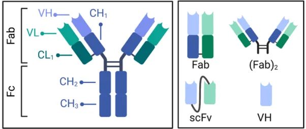
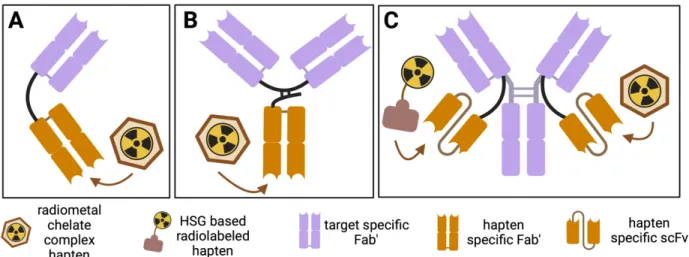
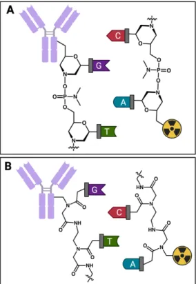
Structure of bispecific antibody (bsAb) and hapten molecule constructs for pretargeted nuclear medicine. In vivo biodistribution of pre-targeted [64Cu]Cu-NOTA-PEG3-Fc (16) in BxPC3 tumor-bearing nude mice with different doses of CB7-M5A.
INTRODUCTION
The objective included the characterization of the modified antibodies and guest radioligands to determine their potential for future in vivo studies. The performance of the proposed pre-targeting system was investigated in tumor-bearing mice for pre-targeted PET by evaluating the in vivo profile of various guest radioligands together with a cucurbit[7]uril-modified antibody.
LITERATURE
ANTIBODY BASED NUCLEAR MEDICINE
- Methodologies
- Clinical use in cancer treatment
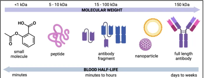
Due to the faster clearance of the antibody fragments from the blood pool, they could potentially lead to better tumor-to-background ratios. In addition, antibody fragments have shown higher penetration of the tumor tissue compared to full length.
PRETARGETED NUCLEAR MEDICINE
- Concept
- Methodologies
- Principles
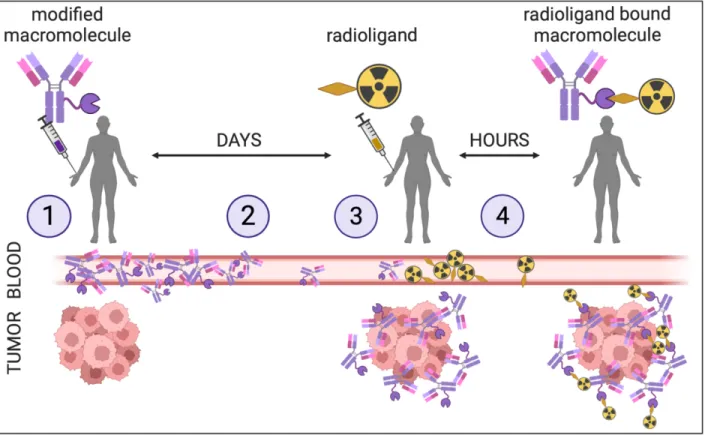
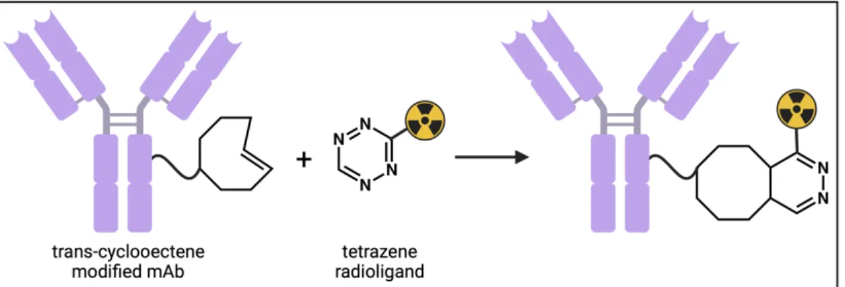
The administration of the modified molecule, such as a full-length antibody (1) and its accumulation at the target site over several days (2). The drawback and concern of the IEDDA pretargeting platform lies in the poor in vivo stability of the pretargeting components.
CUCURBITURIL AND GUEST MOLECULES AND THEIR INTERACTION
- Chemical and physical structure of the cucurbiturils
- Thermodynamics of complex formation
- CB[n]-guest complexes in biomedicine
- High affinity cucurbit[7]uril-guest pairs
- The promise of the CB[7]-guest chemistry
Moreover, the CB[n] cavity has low polarizability that does not promote the formation of van der Waals interactions between water molecules and the CB[n] cavity. It has been observed that with extremely high affinity guest molecules in the CB cavity[7].
![Figure 9. The chemical structure of the cucurbit[n]uril host molecule. Cucurbit[n]uril (A) and cucurbit[7]uril (B)](https://thumb-ap.123doks.com/thumbv2/123dok/10733035.0/40.918.116.813.234.565/figure-chemical-structure-cucurbit-uril-molecule-cucurbit-cucurbit.webp)
SPECIFIC AIMS
MATERIALS AND METHODS
DEVELOPMENT OF GUEST RADIOLIGANDS
- Synthesis of ferrocene radiolabeling precursors
- Synthesis of adamantane radiolabeling precursors
- Synthesis of fluorescein adamantane
- Radiosynthesis of 67/68 Ga-labeled radioligands
- Radiosynthesis of 64 Cu-labeled radioligands
- Distribution coefficient
- In vitro stability and plasma protein binding
- Cell internalization
- In vivo profile of radioligands in healthy mice
The chemical purity of the compounds was analyzed with a reversed-phase high-performance liquid chromatography (RP-HPLC) instrument using a C18 analytical column that detects a wavelength of 254 nm. The chemical purity of UV active compounds (10-12) and non-UV active compounds (7-9) was analyzed with an RP-HPLC instrument using a C18 analytical column detecting wavelengths of 254 nm or 1H-NMR respectively. The reaction solution was left at RT for 10 min before the labeling efficiency was measured by radio-HPLC.
The solution was incubated at 37 °C and 3-4 samples were taken between 30 min and 24 h after the start of incubation. Media, glycine solution, NaOH solution and PBS washes were collected in separate Eppendorf tubes and measured in a gamma counter to determine the percentage of unbound, membrane bound and internalized 16, 19 or [64Cu]CuCl2. Radioligand injections were performed as previously described for blood half-life experiments.

DEVELOPMENT OF CB7 MODIFIED ANTIBODY
- Synthesis of CB7 modified antibody
- Characterization of CB7 modified antibody
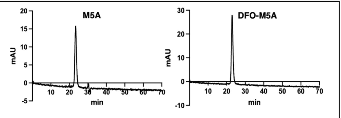
The blood half-life was calculated with a weighted average using equation 2, using the half-lives of the fast and slow phases (t1/2fast and t1/2slow) and their relative percentage (%fast and %slow) derived from the formed two-phase decay curve. To confirm that no aggregation or fragmentation of antibodies occurred during the CB7 modification procedure, CB7-M5A was run on a fast protein liquid chromatography (FPLC) instrument using size exclusion chromatography (SEC). For the Lindmo assay, increasing numbers of BxPC3 cells were incubated with a standard amount of radiolabeled antibody (950 Bq).
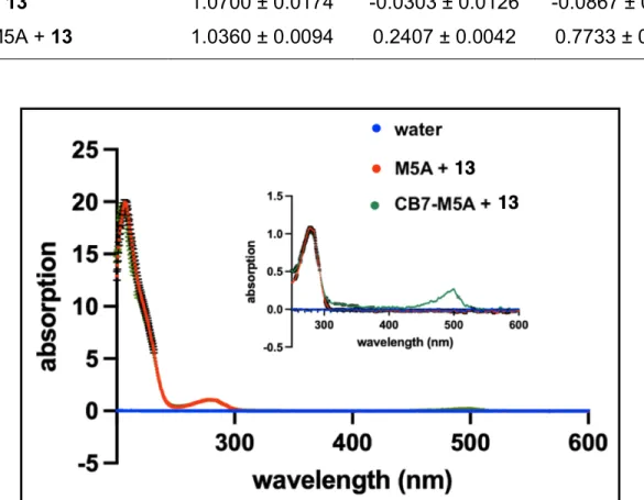
The total added activity to the bound activity of each sample was presented as a function of normalized cell concentration. Immunoreactivity was determined as the Y-intercept corresponding to the total activity to bound activity in a theoretically infinite number of cells. The absorbance of the collected eluate was measured at 280 nm and 495 nm with a UV-Vis spectrophotometer (Thermo Scientific NanoDrop).
![Figure 14. The reaction synthesis of the cucurbit[7]uril (CB7) modified hT84.66-M5A (CB7-M5A)](https://thumb-ap.123doks.com/thumbv2/123dok/10733035.0/67.918.112.817.363.558/figure-reaction-synthesis-cucurbit-uril-cb7-modified-ht84.webp)
DEVELOPMENT OF [ 89 Zr]Zr-DFO-M5A
- Synthesis of DFO modified antibody
- Characterization of DFO modified antibody
The radiochemical purity was determined by radio-TLC using the same method as for label monitoring. To confirm that no aggregation or fragmented antibody byproducts were formed during the DFO modification procedure, DFO-M5A was run on an FPLC instrument using SEC. The immunoreactivity of DFO-M5A was determined via Lindmo cellular binding assay with BxPC3 cell line using a [89Zr]Zr-DFO-M5A.
The number of conjugated DFO groups per M5A was analyzed by a Bruker Impact II quadrupole time-of-flight (QTOF, Bruker Daltonics, Massachusetts, USA) system with an electrospray ionization (ESI) source. The DFO-M5A and M5A reference samples in PBS7.4 were desalted with a PD10 desalting column prior to the experiment. The elution of the samples from the PD10 column was performed with water, resulting in a final concentration of 1.0 mg/mL.
CEA EXPRESSION
BIODISTIRBUTION AND IMAGING STUDIES IN XENOGRAFT MODELS
- In vivo profile of [ 89 Zr]Zr-DFO-M5A
- Pretargeting studies of ferrocene radioligands
- Pretargeting studies of adamantane radioligands
The biodistribution studies with the two earliest time points (4, 24 h) were performed only with a 0.67 nmol dose to obtain a more comprehensive in vivo profile of the antibody for a dosimetry analysis with one of the doses. The in vivo biodistribution of [89Zr]Zr-DFO-M5A was also studied in another mouse model of MIAPaCa-2 tumor bearing female nude mice which acted as the negative control: The mice (n=4) were treated with [89Zr]Zr injected. -DFO-M5A (0.67 nmol; 3.3 MBq) and imaged with a. PET scanner (15 min static scan) 72 hours after injection, followed by sacrificing them for in vivo biodistribution studies.
16 to obtain a comprehensive in vivo profile of one of the previously targeted ligands for a dosimetric study. Following the final imaging time point, mice were euthanized for in vivo biodistribution studies. All four cohorts were euthanized for in vivo biodistribution 4 h after the final injection of each cohort on 21
DOSIMETRY
RESULTS
GUEST RADIOLIGANDS AND THEIR CHARACTERIZATION
- The radiosyntheses of ferrocene and adamantane radioligands
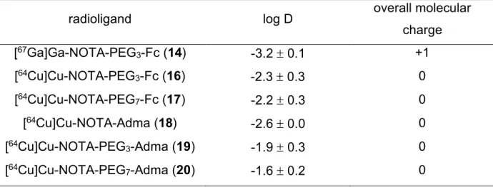
- Distribution coefficient
- In vitro stability and plasma protein binding
- Cell internalization of the radioligands
- In vivo characterization of the radioligands
All in all, the in vitro stability of the ferrocene radioligands was measured to be lower compared to the adamantane radioligands. The in vitro stability of the radioligands in PBS7.4 (A) and in bovine or human plasma (B) at 37 ºC as a function of time and their respective % plasma protein. Cell internalization of the gas radioligands. and [64Cu]CuCl2 of the total added activity as a function of time with BxPC3 cells at 37 ºC.
As with the partition coefficient values for the radioligands, it was expected that the charge of the compound and the length of the PEG linker would affect the blood half-life of the compound. However, as discussed earlier, based on the log D values of the ligands, longer PEG linkers produced less hydrophilic compounds. The maximum intensity projection PET images of the radioligand [64Cu]Cu-NOTA-PEG3-Fc (16) and.
CB7 MODIFIED ANTIBODY AND ITS CHARACTERIZATION
The absorbance at two wavelengths, 280 nm and 495 nm, was used to determine the number of CB7 moieties per M5A antibody. After the CB7 conjugation, the immunoreactivity of the CB7-M5A antibody was measured by the Lindmo assay to remain high (n=3). The immunoreactivity was calculated based on the y-intercept of the line formed based on the cell.
The total activity/bound activity of the cells is shown as the function of the normalized cell.
DFO MODIFIED ANTIBODY AND ITS CHARACTERIZATION
FPLC chromatograph of the unmodified M5A (left) and the purified DFO-M5A (right) with detection of absorption at 280 nm.
CEA EXPRESSION
BIODISTRIBUTION AND IMAGING STUDIES IN XENOGRAFT MODELS
- In vivo profile of [ 89 Zr]Zr-DFO-M5A
- Pretargeted ferrocene and adamantane radioligands
PET imaging showed excretion through the bladder and hepatobiliary system, which coupled with tumor location resulted in poor tumor visualization 2 h after radioligand injection in two of three mice at the 2 h time point. In the first set of experiments, the priming performance of second-generation Fc radioligands was compared using a delay of 72 hours. The location of the subcutaneous tumor in the right shoulder is highlighted with a red circle.
However, these tumor uptake values were all higher than those of the 0.67 nmol CB7-M5A dose used in the two previous pretargeting experiments. Scatter diagram of the blood and tumor uptake and the tumor-to-blood ratios of pretargeted [64Cu]Cu-NOTA-Adma (18), [64Cu]Cu-NOTA-PEG3-Adma (19) and. The PET imaging of the pre-targeted 18-20 resulted in successful delineation of the tumor mass at all investigated time points (Figure 38).
![Figure 25. Activity biodistribution of varying amounts (0.3; 0.7; 1.0; 2.3 nmol) of [ 89 Zr]Zr- Zr]Zr-DFO-M5A in BxPC3 tumor bearing nude mice](https://thumb-ap.123doks.com/thumbv2/123dok/10733035.0/92.918.116.812.609.829/figure-activity-biodistribution-varying-amounts-bxpc3-tumor-bearing.webp)
DOSIMETRY COMPARISON
The PET imaging of BxPC3 tumor-bearing nude mice infused with a first or a second dose of pretargeted 21 3 or 6 days p.i. of CB7-M5A resulted in the similar in vivo profile observed earlier in the in vivo biodistribution experiments. The dose-limiting organ for the use of the directly radiolabeled antibodies in humans is often red marrow and osteogenic cells.6 Importantly, the estimated dose received from pretargeted 16 and 19 to the red marrow is only 0.7 and 1 ,4 % respectively of the red marrow dose. estimated for the use of [89Zr]Zr-DFO-M5A. This was expected due to the longer presence of the pre-targeted 19 in the blood pool compared to 16.
![Table 8. The estimated organ dose values of an adult human male from a dose of [ 89 Zr]Zr- Zr]Zr-DFO-M5A, pretargeted [ 64 Cu]Cu-NOTA-PEG 3 -Fc (16) or pretargeted [ 64 Cu]Cu-NOTA-PEG 3 -Adma (19) (μSv/MBq)](https://thumb-ap.123doks.com/thumbv2/123dok/10733035.0/113.918.114.830.238.955/table-estimated-organ-values-adult-human-pretargeted-pretargeted.webp)
DISCUSSION
COMPARISON OF THE GUEST RADIOLIGANDS
The oxygen atoms of the ethylene glycol monomers make the PEG linker a hydrophilic component due to the hydrogen bonding of the oxygen atoms between water molecules. Positively or negatively charged compounds are often more hydrophilic than zero charged compounds due to the ion-dipole intermolecular interactions formed between the charged part of the compound and the polar water molecules. To further confirm and extend the trends observed in the lipophilicity of the different host radioligands, more compounds should ideally be added to the compound.
Establishing the stability of guest radioligands has been a major priority in understanding their potential as a radioligand component of a host-guest pretargeting strategy. Due to the extremely high in vitro stability of adamantane ligands, adamantane-based radioligands appear superior to ferrocene-based ligands. By potentially altering the degree of internalization of the guest radioligands, adjusting the zero charge of 16 and 19 to a positive one could increase the internalization of both compounds.
COMPARISON OF THE PRETARGETED RADIOLIGANDS IN VIVO
An additional factor that could have led to lower tumor uptake of the ferrocene radioligand was its compromised stability. The same trend of lower tumor uptake with ferrocene ligands was also observed with the gallium-68 labeled versions of the ligands, 15 and 21. With ferrocene and adamantane gas ligands, the blood half-life of the ligands decreased as the length of the PEG linker increased. increased.
Potentially higher signal from target to background could be achieved with extended delay times, as less radioligand would bind to the antibody still circulating in the blood pool. Few studies exploring longer lag times (5 days) have resulted in decreased tumor uptake due to poor long-term persistence of the default agents.28,72. Excitingly, the longer lag time also resulted in decreased presence of 21 in the blood group and thus increased tumor-to-blood ratio.
CONCLUSIONS AND FUTURE DIRECTIONS
CONCLUSIONS
LIMITATIONS
The shorter half-life of gallium-68 (t1/2=68 min) allowed for imaging only up to 4 h p.i., which was not enough time for the nontarget-bound radioligand to be excreted from the intestine so that to gain a high tumor-to-background ratio. Figure 40) The use of short-lived radionuclides, such as gallium-68 and fluorine-18 (t1/2=109 min) for pre-targeted PET is ideal from the point of view of radiation dose and safety, therefore further optimization of Labeled guest radioligands is needed with gallium-68. Potentially, directing excretion to occur through the kidneys by adjusting the molecular charge of the compounds could also increase the rate of excretion. Having a radioligand that would be excreted through the renal system would also allow a wider use of the approach and also target cancerous tissues located in the abdominal area.
FUTURE DIRECTIONS

![Figure 10. Binding of the guest molecule to the cavity of the cucurbit[n]uril host molecule](https://thumb-ap.123doks.com/thumbv2/123dok/10733035.0/42.918.157.766.112.514/figure-binding-guest-molecule-cavity-cucurbit-uril-molecule.webp)
