Development and Function of the Drosophila Giant Fiber Neural Circuit
By
Tyler Joseph Kennedy
Dissertation
Submitted to the Faculty of the Graduate School of Vanderbilt University In partial fulfillment of the requirements
for the degree of DOCTOR OF PHILOSOPHY
in
Biological Sciences May 10, 2019 Nashville, Tennessee
Approved:
Douglas McMahon, Ph.D.
Kendal Broadie, Ph.D.
David Miller, Ph.D.
Manuel Ascano, Jr., Ph.D.
Todd Graham, Ph.D.
ii
Dedication
This dissertation is dedicated to Amanda, Finnegan, Janning, Scott and Jorie, all of whom are quite simply the best.
iii
Acknowledgements
I first want to express my appreciation for the educational environment and wide range of resources Vanderbilt University provides. It has been a pleasure to spend time on this beautiful campus, interacting with the wonderful educators, students and staff that comprise this institution. The Biological Sciences Department is a very special part of Vanderbilt and I am grateful for the broad scientific exposure I received while here, especially the diversity of classes and seminars. Both department chairs during my degree, Dr. Charles Singleton and Dr. Douglas McMahon have been incredibly kind and helpful with all matters, especially in their support for the Graduate School Association activities. I would also like to thank Dr. Kathy Friedman and Dr. Donna Webb for all of their work as Director of Graduate Studies. They were always available to help with any issue relating to graduate work and unhesitating in offering guidance, support and kind words. I also cannot express enough appreciation for the help I received from the department’s front office. Alicia Goostree has been eternally patient and helped me with every problem that came up, big and small. Carol Wiley knows everything there is to know about grant submission and I cannot imagine having successfully completed mine without her. Leslie Maxwell was here when I began the program and helped me get settled into Nashville and navigate my way through beginning a PhD.
LaDonna Smith has my enduring thanks for helping me with my endless questions about funding, traveling for conferences, and finishing the PhD.
I want to give a special thanks to the Program in Developmental Biology. I credit this amazing organization with a great deal of my scientific maturation. Their weekly journal club has broadened my scientific knowledge, helped me understand how to critique a paper and shown me how to give high- quality presentations. PDB would not be possible without a number of dedicated individuals, but I want to especially thank Chris Wright for his impassioned dedication to this project and warm welcome to the program.
iv
My personal scientific research has benefitted from a huge number of people. I first would like to thank the innumerable individuals I called, emailed and spoke with over the years for sharing their advice and reagents. It is always humbling and amazing to realize how large the scientific community around the world is and to see how far people are willing to go to help a student with a strange science question.
Next, I would like to thank the incredible members of my committee. Dr. Douglas McMahon has been a wonderful chair and helped me keep my proposals organized and concise. Dr. David Miller, Dr. Manual Ascano Jr., and Dr. Todd Graham have been very supportive throughout the research process while also keeping a sharp eye on my work and helping me identify potential weaknesses in my arguments. I would like to thank Dr. Nick Reiter who has changed universities but was a highly valued member of my committee before he left. I also wish to express my sincere gratitude to Dr. Donna Webb, who is no longer with us. I was fortunate enough to take classes with Donna, rotate through her lab, and have her as a member of my committee. She was passionate about science and her students and was always available to help anyone who needed it. She gave extensive input on my dissertation proposal and was immensely helpful in the organization of my experiments. I still think about her often and miss having her delightful personality around the department.
Of key importance to all of the work that follows is the Broadie lab. I am grateful to Dr. Kendal Broadie for accepting me into his lab and providing me guidance for all of these years. He is a tireless, talented editor and his input on my work has made it immeasurably better. Our bi-monthly meetings have been filled with extensive discussions about research, ideas, techniques and his inside view of the academic and scientific worlds. I’ve learned a great deal from him and vastly improved my scientific abilities by working in his lab. The members of the Broadie laboratory have been an unending well of scientific knowledge and an incredible community. Meeting this group of people has changed me fundamentally and helped shape the scientist I am today. I will always be grateful for the conversations with, and input I received from Emma Rushton, Cheryl Gatto, Caleb Doll, James Sears, Neil Dani, Will
v
Parkinson, Mary Lynn Dear, Danielle Kopke, Dominic Vita, Randy Golovin and Qing Xia Chen. I would especially like to thank Emma for being my rotation advisor when I first joined the lab and introducing me to the world of Drosophila research. I also gained valuable lessons mentoring Moon Ryu, Ryan Moore and Demi Ajao. I had no idea how to train someone to become a scientist when I arrived at Vanderbilt, and I made a lot of mistakes along the way. I appreciate the patience my mentees had for me throughout the process. It has been an absolute joy to work in the lab with this wonderful collection of scientists and I will miss the spirited discussions during lab meeting and around the bench.
I also wish to acknowledge and thank my many mentors prior to arriving at Vanderbilt whose dedication to teaching helped me make it into and through a PhD program. Pam Durkee, my high school biology teacher was a powerful influence on my interest in the life sciences. She is a passionate and dedicated teacher and her devotion to her craft stimulated my interest in learning and sent me down a path towards biological research. I followed that path to the University of Redlands where I was given an excellent education by teachers who truly care about their students and the pursuit of knowledge. I especially want to thank Susan Blauth, Teresa Longin and David Soulsby, not only for leading fascinating classroom discussions, but also for mentoring me during my first true research projects and helping me learn how to read the scientific literature. After college, I took a job with Applied Speciation and Consulting, where I met two of my greatest mentors to date: Russ Gerads and Hakan Gürleyük. Working with Russ and Hakan changed the way I looked at science. They were willing to talk through anything, dissect the methods and findings of any paper and attempt any research project that came their way.
They instilled a healthy sense of skepticism and critical thinking in me, which I think are among the most important attributes a scientist can have.
Lastly, I could not have made it to this point without the unending support of my family. My parents, Janning and Scott, have always made my education their highest priority and encouraged me in everything I’ve done. My Mom was my first science teacher and is the source of my scientific curiosity.
vi
She always has a new science topic to discuss and I love hearing her insights on various findings. I couldn’t ask for a better role model in work or life. My Dad has always pushed me to be the best I can be and given endless encouragement to my pursuits. He has taken an interest in my work and asked detailed questions about my projects. His passion for life is infectious and he has helped keep a proper perspective throughout this program. My sister Jorie is one of my true inspirations and I am forever jealous of her talents. She is devoted to helping others, creating a better world and seeing beauty in everything. Having her in my life has reoriented my priorities and beliefs and I couldn’t ask for a better sister. The bulk of the work in supporting me through this PhD has fallen on my partner in life, Amanda Clayton. She is the most incredible and brilliant person I can imagine spending the beginning and end of every day with. Every time I have run up against obstacles, she has been unendingly positive and uplifting. I can always discuss my work with her and my writing has benefitted greatly from her sharp proofreading. She is always ready to celebrate successes and talk through disappointments. She has worked hard to make sure we enjoy everything life has to offer and ensures I do not spend all of my time working in. She has brought more joy to me than I thought possible and I cannot thank her enough for choosing to share her life with me.
And, finally, the newest member of my support team is Finnegan Kennedy. He has perhaps been the most disruptive person to my research program and has done everything he can to stop me from working on it so that I can play with him. Regardless, he brings pure unbridled happiness to my life and just seeing his little smile and hearing his little laugh is enough to prepare me for any trials the day may bring. Thank you, everyone, from the bottom of my heart.
vii
Acknowledgement of Funding
This work was supported by National Institutes of Health Grants MH084989 (to K.B.), and NS092250 and HD007502 (to T.K.).
Publications
Fragile X Mental Retardation Protein Restricts Small Dye Iontophoresis Entry into Central Neurons.
Kennedy T. and Broadie K. JNeuro. 2017 Sep 8; 0723—17.
Newly Identified Electrically Coupled Neurons Support Development of the Drosophila Giant Fiber Model Circuit. Kennedy T. and Broadie K. eNeuro. 2018 Nov 26; 0346-18.
viii
Table of Contents
Page
Dedication ... ii
Acknowledgements ... iii
Acknowledgement of Funding ... vii
Publications... vii
List of Figures and Tables ... xi
List of Abbreviations ... xiv
Chapter I: Introduction ... 1
Fragile X Syndrome ... 3
FMRP: RNA-Binding Functions ... 4
FMRP: Channel-Binding Functions ... 8
FMRP Response to Activity ... 10
Clinical Studies on FXS ... 12
The Drosophila Genetic Model System ... 16
Drosophila Giant Fiber Circuit ... 18
Neural Circuit Mapping ... 23
Mechanisms of Synapse Localization ... 27
Summary ... 30
II: Fragile X Mental Retardation Protein Restricts Small Dye Iontophoresis Entry into Central Neurons ... 32
Abstract ... 32
Significance Statement ... 33
ix
Introduction ... 33
Materials and Methods ... 35
Drosophila Genetics ... 35
Dye Iontophoresis ... 36
Dye Injection Manipulations ... 37
Confocal Imaging ... 37
Protein Quantification ... 38
Data Analyses ... 38
Results ... 39
FMRP Selectivity Limits Small Dye Iontophoresis into Neurons ... 39
FMRP-Dependent and Charge-Independent Dye Iontophoresis Defect ... 43
Increased Dye Iontophoresis Persists in the Absence of Gap Junctions ... 45
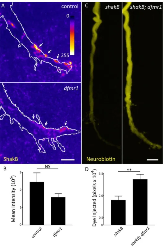
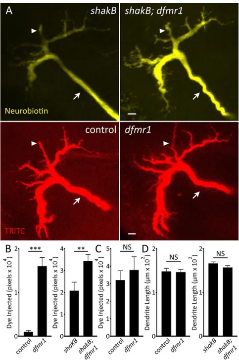
GFI Dendrites are Structurally Unaltered and Display Dye Loading Defect ... 48
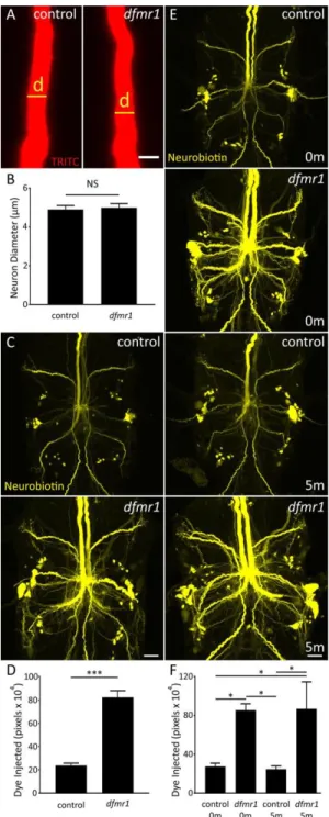
Intrinsic Neuronal Properties Unrelated to Dye Injection Defect ... 50
Ionic Manipulations Alter Dye Loading but do not Resolve dfmr1 Defect ... 54
Elevated Intracellular Dye Iontophoresis Rate in dfmr1 Null Neurons ... 57
Discussion... 60
III: Newly Identified Electrically Coupled Neurons Support Development of the Drosophila Giant Fiber Model Circuit ... 65
Abstract ... 65
Significance Statement ... 66
Introduction ... 66
Materials and Methods ... 68
Drosophila Genetics ... 68
Dye Iontophoresis ... 69
Confocal Imaging ... 69
Data Analyses ... 70
Results ... 71
The Giant Fiber Circuit Exhibits Extensive Dye-Coupled Connectivity ... 71
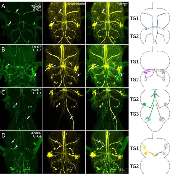
Transgenic Gal4 Drivers for Newly Identified Giant Fiber Coupled Neurons ... 72
Projection Architecture of GFC Neurons Within the Thoracic Ganglion ... 75
The Inframedial Bridge Connectivity Site of GFI-GFC Intersection ... 77
Shaking-B Gap Junction Synapses Between GFI and GFC Neurons ... 78
x
Pre- and Postsynaptic Polarity of Thoracic Ganglion GFC Neurons ... 80
GFC Requirements for the Development of GF Circuit Architecture ... 83
Discussion... 87
IV: Genetic Background Mutation Causes Hyper-Connectivity Within the GF Circuit ... 93
Abstract ... 93
Introduction ... 94
Materials and Methods ... 96
Drosophila Genetics ... 96
Dye Iontophoresis ... 96
Confocal Imaging ... 97
Western Blotting ... 98
Bulk Segregant Analysis and Whole Genome Sequencing ... 99
Data Analyses ... 99
Results ... 100
The dfmr150M Allele Causes Excess GFI Axonal Projections ... 100
Increased Axonal Projections Present During Early GFI Synaptogenesis ... 102
Overgrown Axonal Projections Contain Chemical Synaptic Machinery ... 104
Overgrown Axonal Projections Contain Electrical Synapses ... 107
GFI Synaptic Projections Target GFC Neurons to Cause Circuit Hyper-Connectivity ... 109
Background Mutation(s) in the FXS Disease Model Causes Synaptic Projections ... 113
Identifying Background Mutation(s) Causing Excess GFI Synaptic Projections ... 116
Discussion... 116
V: Conclusions and Future Directions ... 122
Dye Injection ... 122
Circuit Mapping the Giant Fiber System ... 131
Identification of Background Mutations in the dfmr150M Line ... 135
Further Experiments Not Included in Publications ... 138
Western Blot Screen for FMRP Targets ... 138
Neuron-Specific Protein Labeling ... 146
Conclusion ... 150
References ... 152
xi
List of Figures
Figure Page
1. FMRP’s Roles in the Neuron... 7
2. Map of the Known Giant Fiber Circuit ... 19
3. Gal4 Driven Expression of mCD8::GFP in the GFI ... 21
4. Modulators of Synaptic Formation During Development... 29
5. Selective Small Dye Iontophoresis is Increased in dfmr1 Null Neurons ... 41
6. Dye Iontophoresis Defect is FMRP Dependent and Charge Independent ... 44
7. Null dfmr1 Neurons Manifest Dye Injection Defect Without Gap Junctions ... 46
8. Specific Small Dye Iontophoresis is Increased in dfmr1 GFI Dendritic Arbors ... 49
9. Dye Injection Defect is not Related to Multiple GFI Neuron Properties... 51
10. K+ Channel Block Reduces Dye Loading Without Correcting dfmr1 Defect ... 55
11. Dye Iontophoresis Rate is Highly Elevated in dfmr1 Null Mutant Neurons ... 59
12. Giant Fiber Interneuron Dye Injection Reveals Coupled Neurons ... 72
13. Transgenic Gal4 Drivers for the Newly Identified GFC Neurons ... 74
14. The GFI Interacts with the GFC Neurons at the Inframedial Bridge ... 79
15. GFCs Form Electrical Synapses with the GFI at the Inframedial Bridge ... 81
16. Pre- and Postsynaptic Polarity of the Newly Identified GFC Neurons ... 84
17. GFC Neurons Support GF Circuit Architectural Development ... 86
18. Presynaptic Projections From the Giant Fiber Interneuron Terminal Bend ... 101
19. Supernumerary GFI Axonal Projections in dfmr150M Null Mutants ... 103
20. GFI Axonal Projection Overgrowth Begins Early in Synaptogenesis ... 105
21. The GFI Axonal Projections Contain Chemical Synapse Markers ... 108
xii
22. The GFI Axonal Projections Contain Electrical Synapse Markers ... 110
23. GFI Axonal Projections Synapse Within the GF Circuit on GFC Neurons ... 112
24. GFI Synaptic Projections Caused by dfmr150M Background Mutations ... 115
25. Potential Mechanisms of Differential Dye Loading ... 124
26. Brain Size is Increased in dfmr150M Animals ... 128
27. LY Dye Loading into Sucrose Droplets... 130
28. dfmr150M Causes Overgrowth in Dendrites ... 137
29. Csw Western Blot ... 142
30. The GLEAM Technique for Cell-Specific Labeling of Endogenously Expressed Proteins ... 147
31. RNA Editing Mechanism to Cell-Specifically Label Proteins ... 149
xiii
List of Tables
Table Page
1. A Summary of Clinical Trials Performed for FXS. ... 15
2. Summary of Circuit Mapping Methods ... 25
3. Transgenic Gal4 Driver Lines for the Giant Fiber Circuit ... 73
4. Known FMRP Targets for Western Blot Screen ... 139
5. Alternative Neuronal Targets Tested in FMRP Western Blot Screen... 139
6. Initial Results of FMRP Target Western Blot Screen. ... 145
xiv
List of Abbreviations
4-AP 4-Aminopyridine
9-AC 9-Anthracenecarboxylic Acid
ACh Acetylcholine
AG Abdominal Ganglia
Ag Silver
AIS Axon Initial Segment
AMPA α-amino-3-hydroxy-5-methyl-4-isoxazolepropionic Acid APF After Puparium Formation
ASD Autism Spectrum Disorder ATR All-Trans Retinal
Bap Bagpipe
BC1 Bran Cytoplasmic RNA 1 BCA Bicinchoninic Acid
Ben Bendless
BK Big Potassium
BrdU Bromodeoxyuridine
Brp Bruchpilot
BSA Bulk Segregant Analysis BSA Bovine Serum Albumin
Bsn Bassoon
CAM Cell Adhesion Molecule
CaMKII Calcium/Calmodulin-Dependent Protein Kinase Type II
xv CC Cervical Connective
Ced-6 Cell Death Protein 6
Chmp4 Charged Multivesicular Body Protein 4A
ChR Channelrhodopsin
Cl Chloride
CNS Central Nervous System CNV Copy Number Variant
CRISPR Clustered Regularly Interspersed Palindromic Repeats
Csw Corkscrew
DAG Diacylglycerol DenMark Dendritic Marker
dfmr1 Drosophila Fragile X Mental Retardation 1 DgkK Diacylglycerol Kinase, Kappa
DLM Dorsal Longitudinal Muscle DLMn Dorsal Longitudinal Motor Neuron eIF4F Eukaryotic Initiation Factor Complex 4F
EM Electron Microscopy
ERK Extracellular-Signal Regulated Kinases
FIB/SEM Focused Ion Beam/Scanning Electron Microscopy
Flp Flippase
FMR1 Fragile X Mental Retardation 1 FMRP Fragile X Mental Retardation Protein
FRAP Fluorescence Recovery After Photobleaching FRT Flp Recombination Target
xvi
FXPOI Fragile X-Associated Primary Ovarian Insufficiency
FXR1 Fragile X Mental Retardation Syndrome-Related Protein 1 FXR2 Fragile X Mental Retardation Syndrome-Related Protein 2 FXS Fragile X Syndrome
FXTAS Fragile X-Associated Tremor/Ataxia Syndrome
Fz Frizzled
GCI Giant Commissural Interneuron
GF Giant Fiber
GFC Giant Fiber Coupled GFI Giant Fiber Interneuron GFP Green Fluorescent Protein
Gig Gigas
GLEAM Gal4 Limited Enchainment Activated Marker GOF Gain of Function
GRASP GFP Reconstruction Across Synaptic Partners Hid Head Involution Defective
HITS-CLIP High-Throughput Sequencing of RNA Isolated by Crosslinking Immunprecipitation
hnRNP Heterogeneous Nuclear Ribonucleoprotein IB Inframedial Bridge
iBLINC in vivo Biotin Labeling of Intercellular Contacts IHC Immunohistochemistry
Irk2 Inwardly Rectifying Potassium Channel 2 KAc Potassium Acetate
xvii KCl Potassium Chloride
KH K Homology
LTD Long Term Depression LTP Long Term Potentiation
LY Lucifer Yellow
MAP1B Microtubule Associated Protein 1B
MAPseq Multiplexed Analysis of Projections by Sequencing
MB Mushroom Body
MCFO Multicolor FlpOut
mGluR Metabotropic Glutamate Receptor MiMIC Minos Mediated Integration Cassette MIP Maximum Intensity Projection MMP Matrix Metalloprotease
mTOR Mammalian Target of Rapamycin
Na Sodium
NATF Native and Tissue Specific Fluorescence
NB Neurobiotin
Nrg Neuroglian
Nrx Neurexin
NS Not Significant
PA Phosphatidic Acid
PBS Phosphate Buffered Saline POI Protein of Interest
PSD-95 Postsynaptic Density 95
xviii PSI Peripherally Synapsing Interneuron PTEN Phosphatase and Tensin Homolog
Pum Pumilio
Rac Ras-Related C3 Botulinum Toxin Substrate RBP RNA Binding Protein
RGG Arginine-Glycine-Glycine
Rho Ras Homolog
RNAi RNA Interference ROI Region of Interest
RSD Relative Standard Deviation SEM Standard Error of the Mean Sema-1a Semaphorin-1a
ShakB Shaking B
Shaw Shaker Cognate W
Shh Sonic Hedgehog
SNP Single-Nucleotide Polymorphism spGal4 Split-Gal4
STaR Synaptic Tagging with Recombination
Stau Staufen
Syb Synaptobrevin
SynCAM Synaptic Cell Adhesion Molecule
Syt Synaptotagmin
TDE 2, 2’ Thiodiethanol
TEA Tetraethylammonium
xix
TG Thoracic Ganglia
TRITC Tetramethylrhodamine Isothiocyanate Trp Transient Receptor Potential
TTM Tergotrochanteral Muscle TTMn Tergotrochanteral Motor Neuron UAS Upstream Activating Sequence UPR Unfolded Protein Response UTR Untranslated Region VNC Ventral Nerve Cord
WB Western Blot
WGA Wheat Germ Agglutinin
WGS Whole Genome Sequencing
Wnt Wingless/Integrated
1
Chapter I
Overview of Dissertation
The goal of this dissertation was to study Fragile X Mental Retardation Protein’s (FMRP) roles in neural circuit development and to advance our understanding of Fragile X syndrome (FXS), the human disorder caused by loss of this protein. FXS causes intellectual disability, autism spectrum disorder (ASD) and other neurological impairments, and is therefore a useful model for uncovering treatments for a wide variety of neuropathies (Harris et al., 2008). I performed my work in the Drosophila Giant Fiber (GF) neural circuit, which coordinates the escape reflex in response to threatening stimuli (Allen et al., 2006; Boerner and Godenschwege, 2011). More specifically, I used the central Giant Fiber Interneuron (GFI) as a model for neuron development and function due to its large size and amenability to manipulation. My initial experiments were intended to expand on research showing loss of FMRP causes overgrown synaptic connectivity with reduced maturity (Comery et al., 1997; Irwin et al., 2001; Zhang et al., 2001). FMRP is canonically known as an RNA-binding protein and my aim was to identify new target RNAs related to FXS connectivity defects, as well as to study other RNA-binding proteins which may partner with FMRP, such as Pumilio (Pum) and Staufen (Stau; Brown et al., 2001; Darnell et al., 2001, 2011; Dubnau et al., 2003;
Vessey et al., 2006; Olesnicky et al., 2012; Pai et al., 2013).
The GFI proved an amenable model neuron and I was able to identify a synaptic overgrowth phenotype in mutants null for the FMRP gene, Drosophila fragile x mental retardation 1 (dfmr1; Wan et al., 2000; Zhang et al., 2001). Pursuing this finding, I asked whether the overgrowth results from increased connections to normal partners or new connections to inappropriate partners. In the process of investigating this using dye injection to determine the GFI synaptic partners, I found that dfmr1 null neurons take up far more dye than paired controls, a puzzling phenotype that has never been reported
2
before. This finding was the focus of my first paper, included in this dissertation as Chapter 2 (Kennedy and Broadie, 2017). Another outcome of these dye filling experiments was the identification of several dye-coupled members of the GF circuit which had been previously unreported. A recently published library of Gal4 transgenic drivers, many of which provide near single-neuron resolution, allowed a fine- scale mapping of this expanded GF circuit (Brand and Perrimon, 1993; Jenett et al., 2012; Tirian and Dickson, 2017). The identification of these neurons was the subject of my second paper, included in this dissertation as Chapter 3 (Kennedy and Broadie, 2018).
While publishing these two papers, I continued to study the connectivity overgrowth in the GFI, looking for FMRP targets or RNA-binding proteins that might modulate the phenotype. However, a series of control experiments eventually uncovered a surprising result: the phenotype was not caused by loss of FMRP, but instead by a background mutation in the dfmr1 null line (dfmr150M; Zhang et al., 2001). As this phenotype is central to an important aspect of developmental neurobiology, namely how a neuron regulates synaptic connectivity, I pursued the mutation’s identity using whole genome sequencing (WGS).
This work is ongoing but is expected to identify a gene that controls synaptogenesis. This work is described in Chapter 4 of this dissertation and is currently being finalized for publication.
While this dissertation work has deviated significantly from the original aims, it has remained true to the broader intent of better understanding neurodevelopment and neuropathies. I discovered a new phenotype caused by loss of FMRP, which may underlie some of the symptoms of the FXS disease state.
The expanded GF neural circuit described here characterizes numerous interconnected neurons, which may prove useful for more complex neurodevelopmental studies than have been possible before. And finally, the work on the synaptogenic gene mutation found in the background of the dfmr150M allele not only may open up a new set of targets with which to study synaptic growth and regulation, but also provides a cautionary tale on the background mutations that can accumulate in commonly used genetic lines. The remainder of this introduction provides background on the extensive literature on FXS, the GF
3
neural circuit, and synapse localization mechanisms. I then conclude with an outline of the remaining chapters.
Fragile X Syndrome
FXS is caused by the loss or inactivation of the fragile X mental retardation 1 (FMR1) gene and occurs in approximately 1:7,000 males and 1:11,000 females (Hunter et al., 2014). FXS is the leading known genetic cause of ASD and intellectual disability, and subsets of patients also present with attention deficits, anxiety, hyperactivity, childhood seizures, depression, elongated facial features, large ears and macroorchidism (Bailey et al., 2008). The FMR1 gene was first cloned in 1991 and has since been the subject of enormous study (Dietrich et al., 1991; Willemsen and Kooy, 2017). The majority of FXS cases are caused by a CGG triplicate expansion in the 3’ untranslated region (UTR) of the gene (Fu et al., 1991).
Unaffected members of the population have 50 or fewer of these repeats, while those with 200 or more manifest the disorder, due to hypermethylation of the CGG repeats leading to gene silencing (Oberlé et al., 1991). Intermediate numbers of repeats are considered a “pre-mutation condition,” and are associated with disabilities, such as Fragile X-Associated Primary Ovarian Insufficiency (FXPOI) and Fragile X-Associated Tremor/Ataxia Syndrome (FXTAS, Jacquemont et al., 2007; Hunter et al., 2008). Several point mutations and deletions have also been uncovered that cause FXS by disrupting functional domains in the protein (Feng et al., 1997a; Myrick et al., 2015).
Within neurons, FMRP localizes primarily to the cell body where it resides in the cytosol and traffics to and from the nucleus (Feng et al., 1997b; Doll and Broadie, 2015; He and Ge, 2017). FMRP is also observed in large protein/RNA granules trafficking along dendrites and into synaptic terminals (Fig.
1, Kanai et al., 2004; Antar et al., 2005). Axonal growth cones contain FMRP and some research suggests FMRP is present in mature axons as well, though this remains contentious (Antar et al., 2006; Price et al., 2006; Akins et al., 2017). FMRP expression in Glia has been observed to also play a role in neural
4
development, but this work is still in the early stages of research (Higashimori et al., 2013, 2016; Connor et al., 2017). The primary model organisms used for FXS research are mice and Drosophila. Zebrafish and Xenopus FXS models exist, but they are less well studied, with fewer than 10 total publications for each at the close of 2018. C. elegans interestingly does not appear to contain an FMR1 homolog, suggesting FMRP arose after brain evolution and specialization were well underway (Shtang et al., 1999).
The enormous amount of research into FMRP function can be categorized in different ways. For this dissertation, I have summarized the research in four categories; 1) FMRP as an RNA-binding protein, 2) FMRP as a channel-binding protein, 3) activity-dependent FMRP roles, and 4) clinical FXS treatment.
FMRP: RNA-Binding Functions
FMRP was originally characterized as an mRNA-binding protein and is currently recognized to have at least three RNA-binding domains: two heterogeneous nuclear ribonucleoprotein (hnRNP) K Homology (KH) domains and an Arginine-Glycine-Glycine (RGG) box (Ashley et al., 1993; Siomi et al., 1993, 1994;
Darnell et al., 2005a). There is also reported to be a less well understood RNA-binding domain at the FMRP N-terminus (Adinolfi et al., 2003; Zalfa et al., 2005). The initial discovery that FMRP binds mRNA set off a search to determine target transcript(s), with the hope that misregulated expression could be corrected in clinical treatments. Unfortunately, multiple studies using a range of techniques have shown FMRP binds a long list of RNAs, with one early study showing up to 4% of brain RNAs are bound (Ashley et al., 1993).
More recent studies have identified upwards of 900 putative mRNA targets (Brown et al., 2001; Darnell et al., 2011; Ascano et al., 2013). While overwhelming, these target lists provided candidates that could be studied in model organisms missing the FMR1 gene. The initial model established was the mouse FMR1 knockout, generated in 1994 by a Dutch-Belgian consortium (The Dutch-Belgian Fragile X Consorthium et al., 1994). A Drosophila FXS model was later developed in 2000 and has been instrumental in studying the disease (Wan et al., 2000; Zhang et al., 2001). Humans and mice both have two FMRP paralogs, Fragile X
5
Mental Retardation Syndrome-Related Protein 1 and 2 (FXR1 and 2), which interact with FMRP and can also bind RNA (Tamanini et al., 1997; Spencer et al., 2006; Darnell et al., 2009). Drosophila has only the single dfmr1 gene and no known paralogs (Siomi et al., 1995; Zhang et al., 1995; Wan et al., 2000). The genetics of FMRP are further complicated by splicing: humans have at least five coding isoforms of the FMR1 gene, while flies have six, not including a large number of UTR isoforms (Flybase.org, 12/3/18;
NCBI.NLM.NIH.gov 12/3/18). The roles of these many isoform are largely unknown.
Over the course of nearly three decades of research on FMRP RNA-binding capabilities, three major results have emerged. First, the vast majority of target genes studied show increased expression when FMRP is lost, indicating FMRP acts primarily as a translational repressor (Brown et al., 2001; Darnell et al., 2001, 2005a; Phan et al., 2011; Niere et al., 2012; Gkogkas et al., 2014). However, exceptions exist including a list of very long RNAs, the postsynaptic scaffold Postsynaptic Density Protein 95 (PSD-95) and Diacylglycerol Kinase, Kappa (DgkK), all of which are cases of FMRP promoting expression as a translational activator (Zalfa et al., 2007; Bechara et al., 2009; Tabet et al., 2016; Greenblatt and Spradling, 2018).
Reduced DgkK expression in FMR1 nulls has been proposed as the actual culprit in the loss of translational repression, as it acts in the Diacylglycerol (DAG) to Phosphatidic Acid (PA) conversion pathway thought to control general protein translation (McMahon and Rosbash, 2016; Tabet et al., 2016). Importantly, this argument suggests DgkK RNA is the primary FMRP target, and all others are false positives. However, this contradicts observations that FMRP binds the ribosome and can impede its procession along RNA, which has been suggested to be one mechanism of FMRP translational repression (Fig. 1, Darnell et al., 2011).
Moreover, a wide range of independent studies have shown FMRP binding transcript targets (Kim et al., 2009; Yan and Denman, 2011; Braat et al., 2015).
The second result that has emerged is that FMRP uses a complex method to discriminate mRNA targets, which has thus far eluded researchers (Veneri et al., 2004; Darnell et al., 2005b). No agreed consensus binding sequence exists, although several motifs have been found, including UGGA, ACUK and
6
GAC (K = G or U). However, these motifs are not well agreed upon, nor complex enough to specifically delineate FMRP targets. Some work suggests high numbers of these motifs recruit sufficient FMRP to mediate its role in repression (Ascano et al., 2013; Ray et al., 2013; Suhl et al., 2014; Anderson et al., 2016).
G-quartets and uridine tracts have also been suggested as guiding sequences, but again these structures are not specific to FMRP targets or present in all targets (Chen et al., 2003; Dolzhanskaya et al., 2003;
Ramos et al., 2003). One difficulty in these experiments is that the list of FMRP targets is still indeterminate, thus preventing robust study. A consensus has also not been reached on the RNA region bound by FMRP, with groups separately finding either the UTR or the coding sequence is the primary target (Brown et al., 1998; Sung et al., 2003; Darnell et al., 2011; Ascano et al., 2013; Zhang et al., 2014b;
Anderson et al., 2016). The lack of a target sequence has created speculation that FMRP uses other mechanisms to regulate transcripts. There is work showing FMRP acts via RNA interference (RNAi) machinery, and other forms of untranslated RNA, such as Brain Cytoplasmic RNA 1 (BC1), to block translation (Zalfa et al., 2003; Jin et al., 2004). Partner RNA-binding proteins (RBPs) such as Pum and Stau have been suggested as well, and have been shown to interact with FMRP at genetic, cellular and behavioral levels, but not yet in molecular terms (Barbee et al., 2006; Elvira et al., 2006; Wells, 2006;
Bolduc et al., 2008; Sialana et al., 2016; Zhang et al., 2017). It is known that FMRP coexists with several RBPs in RNA granules, mobile conglomerations of RBPs, RNA, ribosomes and scaffolds which bind motor proteins and are trafficked along the neuronal cytoskeleton (Fig. 1; Anderson and Kedersha, 2006; Thomas et al., 2011).
The third result that has emerged is that restoring many presumptive FMRP targets corrects FXS phenotypes. For example, corrected expression of Microtubule Associated Protein 1B (MAP1B, Drosophila Futsch), Charged multivesicular body protein 4A (Chmp4, Drosophila Shrub), matrix metalloproteases (MMPs), adenylyl cyclase, DAG kinase and others have ameliorated a range of phenotypes in mouse and Drosophila FXS disease models (Zhang et al., 2001; Lu et al., 2004; Siller and Broadie, 2011; Tabet et al.,
7
2016; Sethna et al., 2017; Vita and Broadie, 2017). Frequently studied phenotypes include brain morphology, synaptic architecture, calcium signaling, long term potentiation and depression (LTP and LTD), learning and memory, anxiety, repetitive behavior, hyperactivity and seizures (Irwin et al., 2001; Nimchinsky et al., 2001; Bear et al., 2004;
Bolduc et al., 2008; Gatto and Broadie, 2008; Spencer et al., 2011; Gholizadeh et al., 2014;
Koga et al., 2015; Ruby et al., 2015; Doll and Broadie, 2016;
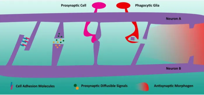
Tian et al., 2017). Published studies often correct one FMRP target and find restoration of a subset of the known phenotypes. It is difficult to reconcile how so many different proteins can seemingly treat FXS in model organisms, but two possibilities might explain these results. First, targets may operate within overlapping pathways, and correcting one target may modify or stabilize multiple targets leading to the finding that multiple proteins can rescue the same phenotype (Bear et al., 2004; Sharma et al., 2010; Tabet et al., 2016). Second, the wide variety of disruptions seen in FXS means researchers could be rescuing some phenotypes with their experiment, but not others (Strumbos et al., 2010; Portera-Cailliau, 2011). In Figure 1. FMRP’s Roles in the Neuron
FMRP (blue) travels in RNA granules with other RNA binding proteins (green, yellow, purple, dark red). These complexes are carried along the microtubule cytoskeleton attached to motor proteins such as kinesin (black). By stalling ribosomes (red) along mRNA, FMRP can repress translation. FMRP also regulates ion channels, such as Slack (orange), increasing their ion conductivity.
8
this case, the many FXS rescues in the literature are only partial, correcting a subset of the disrupted pathways.
It is tempting to link all of the FXS symptoms together, suggesting, for example, that altered neuronal activity is due to overelaborated synaptic connectivity and is the root of weakened memory, seizures and hyperactivity; but it is worth remembering that FXS patients do not have homogenous symptom profiles (Bailey et al., 2008). Correcting one misregulated pathway may partially or completely fix one FXS phenotype, while leaving others uncorrected, and the total correction of the disorder may require repair of many separate pathways. The range of FXS severity suggests a third possibility to explain the disparate findings noted above: FXS is caused by the interaction of FMRP loss and other background mutations. This idea is supported by findings that different mouse strains display different FMR1 null phenotypes (Dobkin et al., 2000; Spencer et al., 2011). This could complicate the analysis of some of the rescue experiments mentioned above: clean genetic backgrounds in the mice and flies carrying the rescue construct could unintentionally mask background mutations, reducing phenotypes and emulating rescue (Burgess, 2011; Chandler et al., 2013). Overall, the disparate findings across FXS research may be reconcilable by attempting to place the many “verified” FMRP targets in common pathways and identifying core proteins responsible for specific phenotypes. Furthermore, background effects must be carefully accounted and controlled for to either remove modifiers or identify and bring them into the FXS disease model as potential therapeutic targets themselves.
FMRP: Channel-Binding Functions
Compared to well established RNA-binding roles the FMRP channel-binding function is a relatively new discovery, and the full implications have not yet been determined. FMRP has been shown to regulate expression levels of ion channels by RNA-binding mechanisms, but in 2010 it was found that FMRP also has a direct protein-protein interaction with the sodium-activated potassium channel Slack (Fig. 1, Brown
9
et al., 2010; Strumbos et al., 2010; Lee et al., 2011; Zhang et al., 2012). Later research showed that Big Potassium (BK) calcium-activated potassium channels also directly interact with FMRP (Deng et al., 2013;
Zhang et al., 2014a). FMRP interaction with Slack causes an increase in the open state of the channel, helping to hyperpolarize neurons through increased net conductance. Therefore, FMRP loss may leave neurons more sensitive to stimulation, which is a phenotype often reported in FXS disease models (Zhang et al., 2012; Contractor et al., 2015). FMRP is also known to increase BK channel conductance, though researchers believe this is accomplished by increasing the sensitivity to calcium (Deng et al., 2013; Zhang et al., 2014a). BK channels shape action potentials by releasing potassium in response to high calcium levels (Contet et al., 2016). At presynaptic terminals they help end neurotransmitter release by terminating action potentials and repolarizing the neuron. The loss of FMRP causes action potential broadening as the BK channels are slower to respond to calcium influx, and thus delay repolarization (Deng et al., 2013). Interestingly, there is little to no strong evidence showing FMRP exists in mature axon terminals, indicating the interactions may be with somatic or dendritic BK channels, though several experiments have suggested this is not the case (Deng et al., 2013). Future studies may require enhanced imaging techniques to reveal FMRP at presynaptic sites or further examination of the FMRP-BK channel interaction in other neuronal compartments (Bock and Stuart, 2016). Lastly, FMRP has been shown to interact with the neuronal N-type voltage gated calcium channel, Cav2.2 (Ferron et al., 2014). Again, this interaction is shown to be direct, rather than RNA mediated; however, in contrast to the potassium channels, it appears FMRP does not regulate the biophysical properties of Cav2.2. Instead, FMRP participates in escorting the channel to the proteasome for degradation. When FMRP is lost Cav2.2 degradation is diminished and levels increase. As Cav2.2 regulates synaptic vesicle release, one consequence of FMRP loss is increased vesicle exocytosis, which could be one source of the hyperexcitability seen in FXS models and patients (Turner et al., 1993; Bailey et al., 2008).
10
The FMRP-channel interaction raises the question of what role this function plays in the FXS disease state. A recent paper suggests loss of the channel interaction is responsible for intellectual disability and seizures in FXS patients. The authors identified a novel mutation in the FMRP coding sequence in an FXS patient that disrupts the segment of the protein responsible for binding to BK channels. Importantly, this FMRP single nucleotide polymorphism (SNP), R138Q, retains RNA binding capabilities thus suggesting channel binding is a critical part of FMRP function (Myrick et al., 2015).
Separately, another human FMRP SNP, I367N, has been identified that blocks FMRP-ribosome interaction but does not impair channel binding. The patient with this mutation also displayed severe intellectual disability, seizures and other typical FXS symptoms. This suggests FMRP’s regulation of both RNA and channels is required to avoid neurological disorders (De Boulle et al., 1993; Feng et al., 1997a). It remains to be seen whether there is a functional association between the FMRP RNA-binding and channel-binding roles. One possibility is that these roles exist in separate compartments (pre- vs. postsynaptic), and separately impact cognitive development and function. Alternatively, these two functions may be linked, and FMRP presence on channels positions it to sense activity and alter RNA translation in response (Ferron, 2016). Such an interaction would be well in line with research on FMRP responses to neuronal activity.
FMRP Response to Activity
Over time a consensus has emerged that the primary function of FMRP is to regulate protein translation in response to neuronal activity, acting as an on-demand dispenser of new synaptic proteins (Zalfa et al., 2003; Weiler et al., 2004; Price et al., 2006). As noted above, this seems at odds with the vast bulk of FMRP concentration being in the soma rather than in neuronal processes or at synapses (Tang et al., 2001; Doll and Broadie, 2015). In addition, FMRP binds a wide array of disparate transcripts, and the majority of these target mRNAs are not localized to synapses or known to be involved in activity-
11
dependent processes. Moreover, several studies have identified roles for FMRP in the neuronal soma and nucleus, albeit these studies are in the distinct minority (Shamay-Ramot et al., 2015; Filippini et al., 2017;
Guo et al., 2017; Zhou et al., 2017). It remains to be seen whether FMRP’s critical neurodevelopmental functions take place at the soma or if this is simply a loading station for FMRP to pick up mRNAs and carry them into processes for activity-dependent translation.
The metabotropic glutamate receptor (mGluR) theory posed in 2004 is the most studied mechanism of FMRP activity-dependent translation (Bear et al., 2004). mGluRs respond to specific activity patterns by enacting a protein synthesis dependent form of LTD which leads to the removal of excitatory α-amino-3-hydroxy-5-methyl-4-isoxazolepropionic acid (AMPA) glutamate receptors from the postsynaptic membrane (Snyder et al., 2001; Xiao et al., 2001). Removal of AMPA receptors weakens, and can lead to removal of, synapses (Schikorski and Stevens, 1997; Nusser et al., 1998). According to the mGluR theory, the translation of the proteins necessary for AMPA removal is regulated by FMRP. The theory suggests activity triggers liberation of RNAs from FMRP control, permitting their translation and beginning the process of synaptic degradation. Importantly, FMRP represses its own mRNA, which is included in the liberated targets (Brown et al., 1998). The newly synthesized FMRP begins to repress the mRNAs again and terminates AMPA receptor removal, which is termed end-product inhibition. When FMRP is absent, the process becomes uncontrolled and excessive synaptic removal results, presumably leading to the immature synapses seen in human patients (Hinton et al., 1991). It seems likely other proteins are capable of at least partially regulating this process, otherwise no synapses would form in FMRP’s absence. Studies have also suggested FMRP is an intermediary between mGluR signaling and mammalian Target of Rapamycin (mTOR) and Extracellular-Signal Regulated Kinases (ERK). These proteins control complex signaling pathways, including regulating RNA translation via members of the translation machinery such as eukaryotic initiation factor complex 4F (eIF4F; Sharma et al., 2010; Sawicka et al., 2016;
Ehyai et al., 2018). Both pathways have been shown to be hyperactivated when FMRP is lost, and their
12
inhibition can correct FXS model phenotypes (Wang et al., 2012; Richter et al., 2015; Sawicka et al., 2016).
The overarching mGluR theory was supported by several studies in mouse and fly models, which show genetic and pharmacological approaches to reduce mGluR activity can correct FXS phenotypes (McBride et al., 2005; Yan et al., 2005; Pan and Broadie, 2007; Pan et al., 2008). However, clinical trials designed to transition this theory into a human treatment have thus far been unsuccessful. The mGluR antagonist Mavoglurant (AFQ056) failed to provide significant improvement in FXS patients (Jeste and Geschwind, 2016). This result is not necessarily a repudiation of the mGluR theory (see below), but does mirror a string of failed clinical trials, suggesting the many roles of FMRP will not be so easily corrected (Erickson et al., 2017).
How does FMRP respond to activity? The protein is regulated by several phosphorylation sites, including the well-studied S500 site (S499 in mice, S406 in Drosophila; Ceman et al., 2003; Bartley et al., 2016). Dephosphorylation of S500 permits translational activation and is accomplished through protein phosphatase 2A, which can be activated by stimulated membrane receptors (Narayanan et al., 2007).
Phosphorylation of S500 is believed to trigger translational repression by FMRP (Coffee et al., 2012). This function is controlled by the kinases Casein Kinase II and p70 S6 kinase 1, and possibly others (Narayanan et al., 2008; Bhattacharya et al., 2012; Bartley et al., 2016). Recent research suggests sumoylation also contributes to FMRP activation (Khayachi et al., 2018). Each of these identified interactions, while complicating the picture of what FMRP does and how it functions, have provided potential therapeutic targets for future FXS treatment drug development.
Clinical Studies on FXS
The complex interactions of FMRP identified thus far have suggested numerous targets that might be susceptible to pharmacological treatment, resulting in many FXS clinical trials (Darnell and Klann, 2013). Targets have included mGluR antagonists (e.g. Mavoglurant, Basimglurant), GABA receptor
13
agonists (e.g. Arbaclofen, Riluzole), MMP inhibitors (e.g. Minocycline), and ERK/mTOR pathway modulators (e.g. Lovastatin, Metformin; Bilousova et al., 2009; Erickson et al., 2011; Siller and Broadie, 2011; Osterweil et al., 2013; Ligsay and Hagerman, 2016; Gantois et al., 2017). While some FXS patients have responded positively to treatments, often based on caregiver assessments, overall these trials have been largely unsuccessful in providing statistical improvements in various parameters (Table 1, Davenport et al., 2016; Erickson et al., 2017). It is unclear why successful treatments in model organisms have not yet been replicated in human patients, though several proposals exist. Model organisms are limited in their ability to replicate human-specific disease traits. Seizures, some forms of memory, and hyperactivity can be assessed fairly well in FXS models, but social and mental disorders such as ASD and intellectual disability are much harder to score. Thus some of the most crucial parts of the disorders scientists are attempting to correct are relatively inaccessible to their research (Watson and Platt, 2012; Sestan and State, 2018).
It can also be difficult to evaluate whether a drug has been effective in treating FXS. As noted above, FMRP loss impinges on many pathways and causes a wide range of symptoms in FXS patients. If these are mediated by separate pathways, distinct measurements may be needed for each symptom.
Further, some of these symptoms are part of broad categories. Within intellectual disability should spatial learning or associative learning be monitored? For autistic children, which social or repetitive behavior tests should be applied? As one cannot tell which outcomes the drugs might improve, it is difficult to know what to assay when designing trials (Berry-Kravis et al., 2013). Another difficulty is determining when to treat. For ethical reasons, very few clinical trials have been conducted on children under the age of 5, but it could be that those are the only treatable timepoints for FXS (Caldwell et al., 2004; Jacquemont et al., 2014). FMRP levels peak very early in development suggesting a specific role in brain formation (Lu et al., 2004; Tessier and Broadie, 2008; Pacey et al., 2013). Much of the current FXS model research is conducted using rescue treatments that are present from conception. Because of this, it is unclear whether FMRP
14
roles are developmental, during adult function or both, leaving drug developers with the question of when treatment needs to be applied to maximally correct the disorder. Can pharmaceuticals applied to adults correct FXS, or must the treatment be given in utero? Some studies have addressed this timing issue;
however, they are split on the answer with some suggesting correction is possible in adults (e.g. Michalon et al., 2012; Dolan et al., 2013; Sun et al., 2016), and others showing intervention must come during development (Doll and Broadie, 2016).
Perhaps a more promising approach than drug treatment is the emerging possibility of genomic editing, which has the potential to clip away the excess FMR1 CGG repeats or correct SNPs to treat the disorder. Such treatment would have to overcome numerous technical and ethical challenges, but is making rapid progress (Park et al., 2015; Yrigollen and Davidson, 2019). Despite a single protein being the root cause of FXS, research into treating the disorder has been stymied by the extraordinary degree of complexity surrounding this protein. FMRP not only interacts with potentially hundreds of RNAs, but also directly regulates channels, and perhaps sits within the inner circle of extremely complex signal transduction pathways. Still, the field has made extraordinary gains in identifying FMRP roles in the brain and has come to understand much about autism and intellectual disability along the way. Many new drugs are in the pipeline which target newly identified FMRP pathways and researchers are optimistic that we are nearing a viable treatment despite past disappointments.
15
Drug Mechanism of Action Test Subject
Age
Clinical Trial Status
Donepezil Acetylcholine esterase inhibitor 12+ Complete
Ampalex AMPA receptor positive allosteric modulator 18-50 Complete
Arbaclofen GABA receptor agonist 5-50 Complete
Ganaxolone GABA receptor positive allosteric modulator 13-22 Complete
Trofinetide Insulin like growth factor analog 12-45 Complete
Basimglurant mGluR5 negative allosteric modulator 5-50 Complete
Oxytocin Oxytocin receptor agonist 13-29 Complete
Aripiprazole Serotonin receptor agonist, Dopamine receptor
agonist/antagonist 5-35 Complete
Metadoxine Serotonin receptor antagonist, GABA transaminase
inhibitor 6-40 Complete
Zoloft Serotonin transporter inhibitor (SSRI) 2-6 Complete
Riluzole Sodium channel inhibitor 18+ Complete
ZYN002 Cannabinoid receptor agonist 3-18 Ongoing
Gaboxadol GABA receptor agonist 13-22 Ongoing
AZD7325 GABA receptor positive allosteric modulator 18-50 Ongoing Acamprosate GABA receptor positive allosteric modulator, NMDA
receptor antagonist 5-55 Ongoing
Mavoglurant mGluR5 antagonist 3-45 Ongoing
Minocycline MMP9 inhibitor 3.5-55 Ongoing
Metformin MMP9 inhibitor, ERK signaling inhibitor 10-17 Ongoing
Lovastatin ERK signaling inhibitor 6-55 Ongoing
BPN14770 Phosphodiesterase allosteric modulator 18-45 Ongoing
Epigallocatechin
Gallate (ECGC) Undetermined 6-60 Ongoing
Vitamin C, E Undetermined 1-18 Ongoing
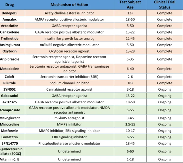
Table 1. A Summary of Clinical Trials Performed for FXS.
Clinical trials have been run for numerous drugs that target pathways associated with FMRP. Most studies have not resulted in beneficial outcomes for patients and have been abandoned. Some of the ongoing trials have shown promising results but are still in early stages. Pharmaceutical treatment has been attempted as early as 2 years of age to correct developmental disruptions. Data compiled from www.clinicaltrials.gov (accessed 2/1/19).
16
The Drosophila Genetic Model System
This dissertation research was performed using the Drosophila genetic model, which is simple enough to assay single genes in single neurons, yet complex enough to have intricate brain organization and sophisticated behavioral outputs. The ~10-day lifecycle and large populations of Drosophila permit a wide range of genetic manipulations. The embryo develops for one day in the egg before hatching and entering the larval stage. The larvae go through three developmental stages, termed instars, with the first two instars lasting one day each and the third instar lasting two days. The larval stage is followed by a four-day pupal stage after which the adult ecloses and becomes sexually mature within eight hours.
Drosophila genetics are relatively simple, with ~14,000 protein-coding genes on 4 chromosomes. Despite having fewer proteins than humans, Drosophila has homologs for ~75% of human disease causing genes (Reiter et al., 2001). One result of this slim genome is far fewer paralogs for each gene. For targets of FMRP, the reduced paralogs make for easier manipulations. MMPs for example are represented by 2 genes in the fly, 24 in mice and 23 in humans (Page-McCaw et al., 2007; Dear et al., 2017). Similarly, Drosophila have 8 gap junction genes, part of the innexin family, while humans have gap junctions from two families: 3 genes for the closely related pannexins and 21 genes in the connexin family (Stebbings et al., 2002; Söhl and Willecke, 2003; Baranova et al., 2004). These simplifications help in manipulations of the nervous system such as, for example, determining and removing gap junctions expressed in a neuron of interest.
The Drosophila genome is amenable to a myriad of manipulations. Mutagenesis was originally carried out by chemical or physical assault on DNA but was later simplified by the discovery of P-element transposons that can be inserted randomly into the genome (Spradling and Rubin, 1982; Price, 2005).
These transposons can be imprecisely excised to create small mutations, such as the dfmr150M mutation used in this work (Rorth et al., 1998; Zhang et al., 2001; Kennedy and Broadie, 2017). P-elements have a preference for 5’ insertion in genes and typically avoid introns (Bellen et al., 2004; Aleksic et al., 2009).
17
Other transposons, such as Minos elements, are helping to probe more of the genome (Venken et al., 2011). Of course, the Clustered Regularly Interspersed Palindromic Repeats (CRISPR) technique has obviated these strategies, making targeted Drosophila genome editing precise and routine (Jinek et al., 2012; Gratz et al., 2013). Naturally, many mutations block viability or fertility, and balancer chromosomes are used to circumvent this problem (Casso et al., 1999). A lethal or infertile mutation of interest is maintained in a heterozygous state over a balancer which has its own lethal mutation. As neither chromosome can propagate as a homozygote, the mutation of interest in maintained over generations.
Dominant markers make it easy to select for or against the balancer in genetic crosses. Balancer inversions block genetic recombination by interfering with chromosome alignment, thus preventing the shedding of deleterious mutations.
Another powerful Drosophila resource is the binary Gal4/Upstream Activating Sequence (UAS) system, a yeast transcription factor/enhancer element pair that allows all manner of transgenic manipulations (Brand and Perrimon, 1993). The Gal4 sequence downstream of an enhancer region of interest drives expression in target cells (Mlodzik and Hiromi, 1992; Jenett et al., 2012). The UAS responder is linked to a protein coding or RNAi sequence, both of which are available for a wide range of genes (Dietzl et al., 2007; Ni et al., 2009). Endogenous genes, with or without tagged fluorophores, enable studies of rescue, localization and function (Zhang et al., 2002; Christiansen et al., 2011; Chen et al., 2014).
Exogenous genes allow structural studies (membrane green fluorescent proteins (GFPs)), functional manipulations (channelrhodopsins (ChRs)), and calcium recordings (GCaMP), permitting exquisite cellular experiments (Nagel et al., 2003; Pfeiffer et al., 2010; Akerboom et al., 2012). Recently, two groups have made an enormous effort to randomly insert small Gal4 enhancer elements into individual fly lines and screen UAS-gfp expression patterns across the brain (Jenett et al., 2012; Tirian and Dickson, 2017). This has resulted in a collection of Gal4 drivers that can express in small subsets of neurons, even single cells, opening new avenues for circuit mapping, developmental studies and neuron-neuron interactions. This
18
technology was further expanded by creating split-Gal4 (spGal4) versions of many of these lines. spGal4s operate by expressing the DNA binding half and RNA polymerase recruiting half of Gal4 under separate enhancers, requiring an overlapping expression pattern to form a complete Gal4, thus greatly reducing the neurons targeted (Luan et al., 2006; Pfeiffer et al., 2010).
In terms of neurodevelopment, the Drosophila brain provides an excellent model for research.
Drosophila have ~100,000 neurons, which is much more tractable than the ~70 million found in mice (Herculano-Houzel et al., 2006; Chiang et al., 2011). Even with relatively few neurons, flies demonstrate a diverse behavioral repertoire controlled by fairly complex, but accessible, neural circuits (O’Kane, 2011;
Kennedy and Broadie, 2018; Namiki et al., 2018). The high density of the central nervous system (CNS) means flies still must solve complex developmental problems such as axon pathfinding and target selection in a crowded arena, providing an accessible way to study such processes at high resolution.
Moreover, rapid development makes the Drosophila brain amenable to live imaging of neuron formation and maturation. Drosophila is even beginning to challenge C. elegans, with a full mapping of all of the brain neurons and their synapses in the works (Zheng et al., 2017). Hence Drosophila provides a particularly attractive model for neurodevelopmental research and neurological disease modeling. Within the Drosophila nervous system, one of the best models for studying individual neuron formation and function is the Giant Fiber Circuit.
Drosophila Giant Fiber Circuit
The Drosophila GF neural circuit, first described by Maxwell Power in 1948, is an escape reflex arc like those found in a range of invertebrates, from squid to cockroach (Hodgkin and Huxley, 1939; Power, 1948; Keegan and Comer, 1993). The circuit rapidly carries information about threatening stimuli from sensory modalities to muscles, enabling a quick escape behavior (Pézier et al., 2014). GF neurons have been used extensively to study neurodevelopment and model human neuropathies including FXS,
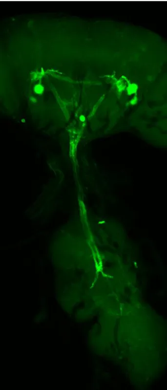
19 Figure 2. Map of the Known Giant Fiber Circuit
This cartoon represents the known neurons of the GF circuit prior to the work described in this dissertation. The Giant Commissural Interneuron (GCI, blue) interconnects the paired Giant Fiber Interneurons (GFI, green) in the Central Nervous System (CNS). The GFI axons descend into the Ventral Nerve Cord (VNC), which is composed of three Thoracic Ganglia (TG1-3) and an Abdominal Ganglion (AG). The GFI axons synapse on the Peripherally Synapsing Interneuron (PSI, orange) at the Inframedial Bride (IB), and the Tergotrochanteral Motor neuron (TTMn, red) along large axonal bends. All of these neurons are bilaterally symmetric, but only the GFIs are shown as such here for clarity. Not pictured are sensory neurons upstream of the GFI/GCI which activate the circuit in response to threatening stimuli, and the DLMns which control the wings.
20
tauopathies and L1 syndrome (Tanouye et al., 1981; Martinez et al., 2007; Kudumala et al., 2013; Lee and Godenschwege, 2014; Orr et al., 2014; Kadas et al., 2019). These studies take advantage of the known synaptic connectivity, established electrophysiology recording paradigms, and large neuron size. The GF circuit also contains numerous genetically tractable interneurons which permit the study of neuron- neuron interactions at single cell resolution.
The GF circuit architecture begins with visual, auditory and mechanical sensory organs collecting information on threatening stimuli, which is then passed to the central brain GFIs, the paired central interneurons which conduct responses downstream (Fig. 2, Allen et al., 2006; Pézier et al., 2014). The GFIs are interconnected by the Giant Commissural Interneurons (GCI) through mixed chemical/electrical synapses, which are believed to create a balanced response on both sides of the body. Each GFI projects an axon into the ventral nerve cord (VNC) where they come together at the posterior end of thoracic ganglion segment 1 (TG1) to form the Inframedial Bridge (IB). The axons then move latterly apart to terminate in large bends. These bends are the sites of synaptic output to the Tergotrochanteral motor neuron (TTMn), which controls the jump reflex of the central legs. The IB is a site of multiple small extensions from the GFI, and the location of mixed chemical and electrical synapses on the Peripherally Synapsing Interneuron (PSI). The PSI connects through a cholinergic synapse to the Dorsal Longitudinal Motor neurons (DLMns), which control the wing depressing muscles (Mejia et al., 2013).
Several characteristics of the GF circuit make it appealing for studying neurodevelopment, including the large diameter neurons (Allen et al., 1998). As flies lack the myelin sheathing found in vertebrates, they use large neuron calibers to increase transmission speed. The GFI neurons are so large relative to the other Drosophila neurons that they can be individually recognized under a 10x microscope objective. This size offers two benefits. First, the GF neurons are easily targeted and manipulable, allowing access via glass electrodes which can deliver dye or other molecules. Dye injections rapidly label the entire neuron, with no requirement of transgenic labels such as GFP. As the GFI contains electrical synapses,
21
small tracers can also be injected to map connected neurons (Boerner and Godenschwege, 2011). Second, the size of the GF neurons makes characterization and quantification much easier, allowing for rapid and confident assessment of diameters, synaptic sites, and structural abnormalities (Mejia et al., 2010; Kudumala et al., 2013; Borgen et al., 2017). These neurons are also genetically tractable, and highly specific Gal4 drivers are available to manipulate each neuron in the GF circuit. Early drivers (such as A307 and C17) were less specific, but still enabled excellent studies on GF circuit development (Phelan et al., 1996; Allen et al., 1999). The Janelia Farms and Vienna Drosophila Resource Center collectives have since generated new drivers, which provide near single neuron resolution for most members of this circuit (Fig. 3; Jenett et al., 2012; Tirian and Dickson, 2017). The motor outputs of the GF circuit also allow electrophysiology characterization. A simple recording can be taken from the two muscle targets of the circuit, providing information on how the circuit develops and functions (Tanouye and Wyman, 1980; Augustin et al., 2011).
Figure 3. Gal4 Driven Expression of mCD8::GFP in the GFI The 91H05-Gal4 driver expresses strongly and specifically in the GCI and GFI. The GCI runs horizontally across the CNS to interconnect the GFI. The large GFI cell bodies form dendrites in the CNS and send axons into the VNC, where they briefly contact at the IB and then diverge to form the characteristic bends.