A Chronology of
Middle Missouri Plains Village Sites
By Craig M. Johnson
with contributions by
Stanley A. Ahler, Herbert Haas, and Georges Bonani
Smithsonian Institution Scholarly Press
s m i t h s o n i a n c o n t r i b u t i o n s t o p a l e o b i o l o g y • n u m b e r 9 5
New Materials of
Masiakasaurus knopfleri
Sampson, Carrano, and Forster, 2001, and Implications for the Morphology of the Noasauridae
(Theropoda: Ceratosauria)
Matthew T. Carrano, Mark A. Loewen,
and Joseph J. W. Sertich
SerieS PublicationS of the SmithSonian inStitution Emphasis upon publication as a means of “diffusing knowledge” was expressed by the first Secretary of the Smithsonian. In his formal plan for the Institution, Joseph Henry outlined a program that included the following statement: “It is proposed to publish a series of reports, giving an account of the new discoveries in science, and of the changes made from year to year in all branches of knowledge.” This theme of basic research has been adhered to through the years by thousands of titles issued in series publications under the Smithsonian imprint, com- mencing with Smithsonian Contributions to Knowledge in 1848 and continuing with the following active series:
Smithsonian Contributions to Anthropology Smithsonian Contributions to Botany
Smithsonian Contributions to History and Technology Smithsonian Contributions to the Marine Sciences Smithsonian Contributions to Museum Conservation Smithsonian Contributions to Paleobiology
Smithsonian Contributions to Zoology
In these series, the Institution publishes small papers and full-scale monographs that report on the research and collections of its various museums and bureaus. The Smithsonian Contributions Series are distributed via mailing lists to libraries, universities, and similar institu- tions throughout the world.
Manuscripts submitted for series publication are received by the Smithsonian Institution Scholarly Press from authors with direct affilia- tion with the various Smithsonian museums or bureaus and are subject to peer review and review for compliance with manuscript preparation guidelines. General requirements for manuscript preparation are on the inside back cover of printed volumes. For detailed submissions require- ments and to review the “Manuscript Preparation and Style Guide for Authors,” visit the Submissions page at www.scholarlypress.si.edu.
New Materials of
Masiakasaurus knopfleri
Sampson, Carrano, and Forster, 2001, and Implications for the Morphology of the Noasauridae
(Theropoda: Ceratosauria)
Matthew T. Carrano, Mark A. Loewen, and Joseph J. W. Sertich
washington d.c.
2011
ABSTRACT
Carrano, Matthew T., Mark A. Loewen, and Joseph J. W. Sertich. New Materials of Masiakasaurus knopfleri Sampson, Carrano, and Forster, 2001, and Implications for the Morphology of the Noasau- ridae (Theropoda: Ceratosauria). Smithsonian Contributions to Paleobiology, number 95, viii + 53 pages, 26 figures, 3 tables, 2011. — Osteology of the noasaurid theropod Masiakasaurus knopfleri Sampson et al., 2001, is now two-thirds complete. We describe Masiakasaurus knopfleri in detail on the basis of examination of new specimens and emphasis on previously unknown elements. The skull is anteroposteriorly long but low in height, unlike the foreshortened abelisaurid condition. Premaxil- lary teeth are procumbent, like those of the dentary. Frontal bones are flat and unornamented, but the lacrimal and postorbital exhibit surface texturing. The braincase resembles that of abelisaurids but is more highly pneumatized. The neck is curved anteriorly but horizontal posteriorly, and it transitions to the trunk without significant proportional changes. Centrum pneumaticity appears confined to the neck and anterior trunk. The sacrum includes six vertebrae, and the expanded transverse processes of caudal vertebrae may articulate with caudal ribs. The scapulocoracoid is large and broad. The ilium is both anteroposteriorly long and dorsoventrally deep, and it bears pegs for articulation with sockets on the pubis and ischium, as in other ceratosaurs. The nearly complete pes shows no particular locomotor specializations and allows reinterpretation of the “raptorial” pedal ungual of Noasaurus as a manual element. These new specimens also illuminate the morphology of other noasaurids, especially those from the Lameta Formation.
In addition to Madagascar, noasaurids are known from Europe, India, South America, and Africa, spanning at least Aptian–Albian through Maastrichtian time. The new materials of Masiakasaurus increase character resolution within Abelisauroidea, identifying many formerly equivocal features as synapomorphies of the nodes Noasauridae, Abelisauridae, or Abelisauroidea. Unfortunately, the frag- mentary nature of nearly all other noasaurids obviates any meaningful ingroup resolution, and as a result no particular evolutionary or biogeographic scenarios for the clade can presently be supported (or rejected) with confidence.
Cover images: Skeletal reconstruction of skull (left) and life restoration of head (right) of the theropod Masiakasaurus knopfleri. Skull image by Mark Loewen; head restoration by Lukas Panzarin.
Published by SMITHSONIAN INSTITUTION SCHOLARLY PRESS P.O. Box 37012, MRC 957
Washington, D.C. 20013-7012 www.scholarlypress.si.edu
Text and images in this publication may be protected by copyright and other restrictions or owned by indi- viduals and entities other than, and in addition to, the Smithsonian Institution. Fair use of copyrighted mate- rial includes the use of protected materials for personal, educational, or noncommercial purposes. Users must cite author and source of content, must not alter or modify content, and must comply with all other terms or restrictions that may be applicable. Users are responsible for securing permission from a rights holder for any other use.
Library of Congress Cataloging-in-Publication Data Carrano, Matthew T.
New materials of Masiakasaurus knopfleri Sampson, Carrano, and Forster, 2001, and implications for the morphology of the Noasauridae (Theropoda: Ceratosauria) / Matthew T. Carrano, Mark A.
Loewen, and Joseph J. W. Sertich.
p. cm. — (Smithsonian contributions to paleobiology, ISSN 0081-0266 ; no. 95) Includes bibliographical references.
1. Masiakasaurus. 2. Noasauridae—Morphology. 3. Paleontology—Cretaceous. I. Loewen, Mark A. II. Sertich, Joseph J. W. III. Title.
QE862.S3C276 2011
567.912—dc22 2010042359
ISSN: 0081- 0266 (print) / 1943- 6688 (online)
The paper used in this publication meets the minimum requirements of the American National Standard for Permanence of Paper for Printed Library Materials Z39.48–1992.
Contents
LIST OF FIGURES v
LIST OF TABLES vii
INTRODUCTION 1
Institutional Abbreviations 2
Acknowledgments 2
SYSTEMATIC PALEONTOLOGY 3
DISCUSSION 34
Noasaurid Morphology 34
Comparative Anatomy 35
Relationships within noasauridae 37
Evolutionary Implications for ceratosauria 38
Noasaurids 38
Character Distributions 38
CONCLUSIONS 39
APPENDIX 41
REFERENCES 51
Figures
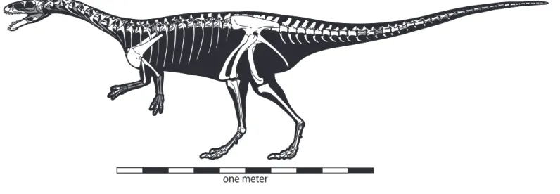
1. Composite skeletal reconstruction of Masiakasaurus knopfleri 2
2. Left premaxilla of M. knopfleri 4
3. Lacrimal and postorbital of M. knopfleri 5
4. Left frontal of M. knopfleri 6
5. Right quadrate of M. knopfleri 7
6. Braincase of M. knopfleri 8
7. Braincase of M. knopfleri 9
8. Left dentary of M. knopfleri 10
9. Left angular of M. knopfleri 10
10. Right prearticular and articular of M. knopfleri 11
11. Reconstructed skull of M. knopfleri 12
12. Cervical and anterior dorsal vertebrae of M. knopfleri 14
13. Morphological changes in the cervical and anterior dorsal
vertebrae of M. knopfleri 17
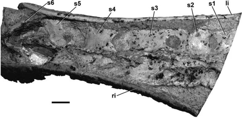
14. Sixth dorsal vertebra of M. knopfleri 19
15. Sacrum of M. knopfleri 20
16. Middle caudal vertebra of M. knopfleri 21
17. Right sixth cervical rib of M. knopfleri 22
18. Right scapulocoracoid of M. knopfleri 24
19. Left humerus of M. knopfleri 25
20. Manual phalanges of M. knopfleri 28
21. Left ilium of M. knopfleri 29
22. Ventral pelvic elements of M. knopfleri 30
23. Right fibula of M. knopfleri 32
24. Right astragalus of M. knopfleri 33
25. Metatarsals of M. knopfleri 33
26. Associated individuals of M. knopfleri 34
Tables
1. Measurements of associated individuals of Masiakasaurus
knopfleri from locality MAD05-42 26
2. Isolated noasaurid specimens from the Lameta Formation 36
A.1. Complete list of Masiakasaurus knopfleri specimens from the Upper Cretaceous Maevarano Formation of Madagascar
collected through 2007 42
New Materials of Masiakasaurus knopfleri Sampson, Carrano, and Forster, 2001, and Implications for the Morphology of the
Noasauridae (Theropoda: Ceratosauria)
IntroductIon
Masiakasaurus knopfleri was first described from disassociated elements discovered in the Upper Cretaceous Maevarano Formation of northwestern Madagascar (Sampson et al., 2001). Representing nearly 40% of the skeleton, these remains were identified as pertaining to a small-bodied abelisauroid, which implied a greater morphological diversity within Ceratosauria than had been depicted by the better-known Ceratosaurus, Elaphrosaurus, and Abelisau- ridae. Masiakasaurus bore specific resemblances to otherwise poorly understood forms such as Noasaurus and Laevisuchus, but little more could be said about its relationships at that time.
A more detailed study of Masiakasaurus by Carrano et al. (2002) elaborated on these similarities and proposed that the three latter taxa—Masiakasaurus, Noasaurus, and Laevisuchus—might pertain to a single clade, Noasauridae, al- beit with equivocal phylogenetic support. A secondary result was an increase in the evidence supporting the dissolution of Ceratosauria sensu Gauthier (1986), in favor of a more basal placement for Coelophysoidea within Theropoda. This hypothesis has since received further support (Rauhut, 2002; Wilson et al., 2003; Sereno et al., 2004), although it is not universally accepted (e.g., Tykoski and Rowe, 2004; Allain et al., 2007).
A recent phylogeny of Ceratosauria (Carrano and Sampson, 2008) provided further evidence for a monophyletic Noasauridae, which also included Velocis‑
aurus and Genusaurus but not Deltadromeus or Elaphrosaurus, as has been suggested (Sereno et al., 2004; Canale et al., 2009). Noasaurids have now been recognized in South America, Africa, Madagascar, India, and Europe, and in deposits as ancient as Aptian, implying a history reaching back at least into the earliest Cretaceous (Carrano and Sampson, 2008). Jurassic ceratosaurs appear to be more primitive, for example Ceratosaurus, Elaphrosaurus, and
Matthew T. Carrano, Curator of Dinosauria, Department of Paleobiology, National Museum of Natural History, Smithsonian Institution, P.O. Box 37012, MRC 121, Washington, D.C.
20013‑7012, USA; Mark A. Loewen, Depart‑
ment of Geology and Geophysics, University of Utah, 1390 East Presidents Circle, Salt Lake City, Utah 84112‑0050, USA; and Joseph J. W.
Sertich, Department of Anatomical Sciences, Stony Brook University, Stony Brook, New York 11794‑8081, USA. Correspondence: M. T.
Carrano, [email protected].
Manuscript received 2 February 2009; accepted 9 June 2010.
2 • S M I T H S O N I A N C O N T R I B U T I O N S T O P A L E O B I O L O G Y
Limusaurus (Xu et al., 2009), but also include indetermi- nate abelisauroids (Rauhut, 2005).
Unfortunately, most noasaurid species are repre- sented by extremely fragmentary remains (e.g., Novas et al., 2004), and as a result several important aspects of the noasaurid skeleton remain poorly known, particularly the skull and forearm. This makes it difficult to resolve many important character states within Ceratosauria. In par- ticular, it remains equivocal whether numerous distinctive
“ceratosaur” features are primitive for Ceratosauria or Abelisauroidea, or instead characterize only more highly nested clades.
Here we describe abundant new materials of Ma‑
siakasaurus that help resolve several of these problems (a complete list is given in the Appendix). Most of the skull is now known, along with nearly the entire vertebral series and hind limb, and additional bones from the forelimb.
Overall, approximately 65% of the skeletal elements of Masiakasaurus have been found (Figure 1). These new specimens, some of which derive from associated individu- als, permit a much more accurate reconstruction of Ma‑
siakasaurus and allow greater insight into the anatomical specializations of noasaurids more generally.
InstItutIonal abbrevIatIons
The following institution names are abbreviated for subsequent mention in the text.
FMNH Field Museum of Natural History, Chicago
GSI Geological Survey of India, Kolkata UA Université d’Antananarivo, Madagascar
acknowledgments
We are very thankful for the efforts of participants in the 2001, 2003, 2005, and 2007 Mahajanga Basin Project expeditions (funded by the National Science Foundation and the National Geographic Society), which yielded these new discoveries of Masiakasaurus, as well as participants in previous expeditions for the original finds. D. Krause (Stony Brook University) and S. Sampson (University of Utah) have been particularly supportive of our research and encouraged us to continue as new materials were un- earthed, and C. Forster (George Washington University) was especially helpful with access to specimens. We thank J. Groenke and V. Heisey (Stony Brook University) for their expert preparation of the fossils, often on short no- tice, and W. Simpson (Field Museum) for curatorial work.
The manuscript was significantly improved thanks to thor- ough reviews by M. Lamanna and C. Forster. Figure 6 was skillfully rendered by M. Parrish (Department of Paleobiol- ogy, National Museum of Natural History). Translations of Accarie et al. (1995), Bonaparte and Novas (1985), and Janensch (1925) are available from the Polyglot Paleon- tologist web site (http://www.paleoglot.org).
FIGURE 1. Composite skeletal reconstruction of Masiakasaurus knopfleri in left lateral view, showing recovered (white) and missing (gray) elements. Scale bar = 1 m.
one meter
SyStematIc Paleontology Dinosauria owen, 1842
saurischia Seeley, 1888 TheropoDa marsh, 1881 ceraTosauria marsh, 1884
abelisauroiDea (Bonaparte and novas, 1985) Bonaparte, 1991
noasauriDae Bonaparte and Powell, 1980 type taxon. Noasaurus leali Bonaparte and Powell, 1980.
included taxa. Compsosuchus solus Huene and Matley, 1933; Genusaurus sisteronis Accarie et al., 1995; Jubbulpuria tenuis Huene and Matley, 1933; Lae‑
visuchus indicus Huene and Matley, 1933; Masiakasaurus knopfleri Sampson et al., 2001; Ornithomimoides? bara‑
simlensis Huene and Matley, 1933; Velocisaurus unicus Bonaparte, 1991. Note that not all of these are valid, al- though they do represent noasaurids, and that the clade Noasauridae also includes unnamed taxa from Niger (Sereno et al., 2004; Sereno and Brusatte, 2008) and Ar- gentina (Brissón Egli and Apesteguía, 2008).
geographic Range. Europe (France), South America (Argentina), India, Madagascar, and Africa (Niger).
temporal Range. Cretaceous, Aptian–
Albian through Maastrichtian (minimum).
Masiakasaurus knopfleri Sampson, carrano, and Forster, 2001
Figures 1–20
holotype. UA 8680, a right dentary with sev- eral teeth (Sampson et al., 2001).
Referred specimens. See Appendix for complete list.
Localities. Thirty distinct localities near the village of Berivotra, along Route National 4 in Mahajanga Province (Boeny Region), northwestern Madagascar (see Appendix for complete list).
age. Maastrichtian, Late Cretaceous (Rogers and Hartman, 1998; Rogers et al., 2000, 2007).
strata. Anembalemba and Masorobe mem- bers, Maevarano Formation.
description.
Skull and Lower Jaw
Important new materials from the skull of Masiakasau‑
rus provide a far better understanding of the general mor- phology of the skull. Only the nasal, squamosal, parietal, jugal, quadratojugal, and palate remain unknown, and whereas only three elements from the lower jaw were pre- viously known, only the surangular is now lacking.
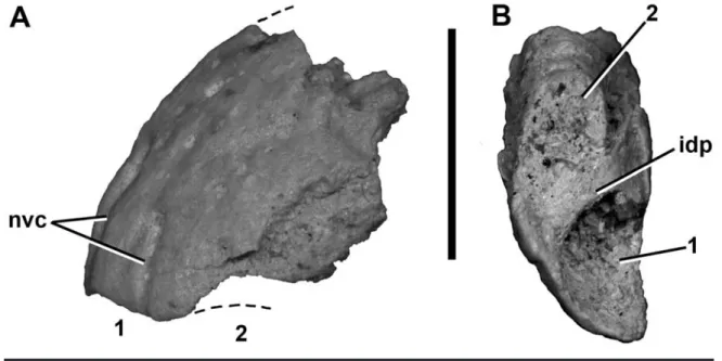
Premaxilla: A partial left premaxilla (FMNH PR 2453, Figure 2) reveals that this element mirrors some of the unusual morphologies of the dentary (Sampson et al., 2001; Carrano et al., 2002). This fragment preserves two anterior alveoli along with the adjacent body of the bone;
the narial margin and maxillary contact are absent. How- ever, if the ventral margin of the preserved premaxilla is held horizontally, these two alveoli are anteroventrally oriented (Figure 2A). Thus, the first and second premaxil- lary teeth (missing in this specimen) complemented the an- terodorsal orientation of the anterior dentary teeth. This had been suggested previously on the basis of the antero- ventral orientation of the first maxillary alveolus (FMNH PR 2183; Carrano et al., 2002) and is confirmed here, but the premaxillary teeth appear to be more strongly angled than the first maxillary tooth.
In ventral view, the alveoli are large, oval, and sepa- rated by a thin interdental plate (Figure 2B). The medial surface is flat and largely featureless, and presumably articulated against the contralateral element. The lateral surface is broadly convex and exhibits a number of small foramina that open into neurovascular channels directed toward the ventral margin of the bone (Figure 2A).
Lacrimal: One nearly complete lacrimal is known (FMNH PR 2473; Figure 3A–D), preserving most of the ventral ramus. The lateral surface shows a high degree of texturing and numerous vessel traces, as in abelisaurids (Figure 3B), but is otherwise flat, with sinuous anterior and posterior margins. The presence of an orbital flange cannot be definitively ascertained, but the preserved or- bital curvature is consistent with an eye that filled most of the orbital fenestra, as is typical for small theropods. As in most theropods, the dorsal and ventral moieties of the lac- rimal antorbital fossa are separated on the external bone surface; however, unlike nearly all other theropods except Torvosaurus, this separation is incomplete.
Anteriorly, the ventral portion of the antorbital fossa is well developed but shallow, whereas the dorsal portion houses a large opening into the nasolacrimal canal, as in
4 • S M I T H S O N I A N C O N T R I B U T I O N S T O P A L E O B I O L O G Y
Ceratosaurus and abelisaurids (Figure 3A; e.g., Sampson et al., 1998; Sampson and Witmer, 2007). Only the ven- tral margin of this foramen can be seen; it connected to a large internal chamber, the lacrimal pneumatic recess, which is visible through the broken dorsal surface of the bone. Also in anterior view, a vertical ridge separates the lateral components of the antorbital fossa from the shal- low medial vacuity.
Posteriorly, a large, teardrop-shaped medial nasal pneumatic fossa (Sampson and Witmer, 2007) is visible at the level of the mediolateral widening of the lacrimal (Fig- ure 3C). It sits nearly centered within the concave anterior orbital wall and leads to a foramen that enters the body of the bone. The medial surface of the lacrimal is marked by a thin ridge that connects dorsally with a triangular concave area (Figure 3D). It is not clear whether this rep- resents the contact surface for the ventral process of the prefrontal or the medial surface of a fused prefrontal.
Postorbital: Like the lacrimal, the postorbital (Figure 3E–G) exhibits some of the characteristic textur- ing seen in abelisaurids. The lateral surface of the ventral ramus bears numerous vessel traces and small tubercles, which continue onto the anterior ramus (Figure 3E). A thickened and roughened brow ridge, also bearing ves- sel traces, emerges where these two rami join. The ventral ramus curves slightly anteriorly as it descends to its distal point, but lacks a flange that enters the orbital fenestra, as
in many abelisaurids. The posterior, or squamosal, ramus is a shorter, flat, blunt triangle of bone that would have lodged within a slotted facet on the squamosal.
The medial surface of the ventral ramus (Figure 3F) is marked by a distinct curving ridge that separates the orbital and lateral temporal fossae, as in many theropods including abelisaurids such as Carnotaurus and Majun‑
gasaurus (Sampson and Witmer, 2007). Dorsally, this ridge terminates at an elongate fossa for the laterosphe- noid head. The dorsal margin of this fossa is adjacent to a very dorsoventrally thin, medially facing articular surface, probably for the parietal. The more expansive frontal ar- ticulation would have been present along the thin antero- lateral and anterior margins, and is only partly preserved in the present specimens. Finally, the posterodorsal corner of the orbital fenestra houses a shallow, elliptical fossa containing a small foramen. In Majungasaurus, a simi- lar foramen is present but without a corresponding fossa (Sampson and Witmer, 2007).
The dorsal surface of the postorbital is triangular, broad anteriorly along the frontal but tapering to a point posteriorly where it articulates with the squamosal (Figure 3G). This surface is most markedly textured along the lat- eral edge. The medial margin is smooth along much of its length, where it forms the anterolateral corner and lateral margin of the upper temporal fenestra. The parietal con- tact is straight and may have been a lappet joint.
FIGURE 2. Left premaxilla (FMNH PR 2453) of Masiakasaurus knopfleri in lateral (A) and ventral (B) views. Abbre- viations: idp = interdental plate; nvc = neurovascular channel; 1, 2 = alveoli. Scale bar = 1 cm. (Anterior is down in B.)
Frontal: The frontal (Figure 4) is a very dorso- ventrally thin, gently curved bone that entirely lacks the texturing or thickening seen in many abelisaurids (e.g., Majungasaurus; Sampson and Witmer, 2007). Instead, it more closely resembles the frontals of other small-bodied, primitive theropods such as Coelophysis in its proportions
(quadrangular in dorsal view) and in the large orbital com- ponent along its lateral edge. It is also quite long relative to its width; it exhibits none of the anterior shortening seen in many larger or more derived theropods.
The anterior fifth of the lateral edge is occupied by a triangular articulation that leads into a conical socket for FIGURE 3. Lacrimal (A–D) and postorbital (E–G) of Masiakasaurus knopfleri. Partial right lacrimal (FMNH PR 2473) in anterior (A), lateral (B), posterior (C), and medial (D) views. Right postorbital (FMNH PR 2456) in lateral (E), medial (F), and dorsal (G) views. Abbreviations:
aofe = antorbital fenestra; aofo = antorbital fossa; ar = anterior ramus; lsc = laterosphenoid contact; ltf = lateral temporal fenestra; ltfo = lateral temporal fossa; mr = medial ridge; mv = medial vacuity; nlc = opening into nasolacrimal canal; npf = nasal pneumatic fossa; of = orbital fenestra;
ofo = orbital fossa; pac = parietal contact; sqr = squamosal ramus; utf = upper temporal fenestra; vr = ventral ramus. Scale bar = 1 cm. (Anterior is up in G.)
6 • S M I T H S O N I A N C O N T R I B U T I O N S T O P A L E O B I O L O G Y
the prefrontal (Figure 4C). The depth of this socket sug- gests that it may have allowed for limited invasion of the frontal by pneumatic structures from the lacrimal, as in Majungasaurus (Sampson and Witmer, 2007). The poste- rior fifth bears a small, flat, triangular facet for the anterior ramus of the postorbital. Between these two articulations, the lateral edge of the frontal is sharp, subtends an arc of approximately 70°, and forms the dorsal rim of the orbit.
The anterior edge is also sharp, forming the underside of a broad lappet joint for the nasal that covers approxi- mately 10% of the dorsal surface (Figure 4A). This edge is relatively straight and meets the midline at an angle of about 20°. The medial surface is a nearly flat and weakly striated facet for the opposing frontal (Figure 4D). It exhib- its almost no curvature, and tapers in thickness both ante- riorly and posteriorly from its thickest point, dorsal to the posterior half of the orbit. Although broken in both speci- mens, the posterior frontal appears to have been thicker and more rounded than the anterior. It arcs ventrally to form the anteromedial wall of the upper temporal fossa, the border of which is marked by a distinct rim. This fossa occupies about one-fifth of the surface of the frontal.
Ventrally, the lateral portion of the frontal is mark- edly concave and forms the roof of the orbital fossa (Figure 4B). It is demarcated from the medial portion by a strongly curving ridge that thins mediolaterally from anterior to
posterior. This medial moiety is flat and narrows between its expanded anterior and posterior portions. The anterior expansion contains a small, elliptical fossa for the olfac- tory lobe of the brain. Posteriorly, the expansion marks the location of the cerebral hemisphere, which is followed by a deeper concavity. A groove runs along the medial edge of the curved ridge anteriorly from the cerebral fossa.
Quadrate: A partial right quadrate (Figure 5;
FMNH PR 2496) is one of few elements that can be di- rectly compared with material in other noasaurids (i.e., Noasaurus). It has a fairly straight shaft, as in most abelis- aurids, but unlike the strongly posteriorly concave condi- tion seen in Noasaurus.
There is no evidence of a foramen on the posterior surface, either in the body of the bone or along the qua- dratojugal contact (Figure 5B). However, the body is suf- ficiently damaged that the presence of a small foramen or fossa (as in Majungasaurus; Sampson and Witmer, 2007) cannot be ruled out. The quadratojugal contact is very thin, and wraps around onto the posterior surface along the ventralmost portion of the bone (Figure 5A), as in Car‑
notaurus, Abelisaurus, and other abelisaurids.
The quadrate condyles are similar in width but mark- edly asymmetrical in anteroposterior length, with the me- dial condyle length nearly twice that of the lateral. The two condyles are oriented at an acute angle to one another FIGURE 4. Left frontal (FMNH PR 2475) of Masiakasaurus knopfleri in dorsal (A), ventral (B), lateral (C), and medial (D) views. Abbrevia- tions: chf = cerebral hemisphere fossa; mfrc = midline frontal contact; nac = nasal contact; of = orbital fenestra; ofo = orbital fossa; olf = olfactory lobe fossa; poc = postorbital contact; prfc = prefrontal contact; utfo = upper temporal fossa. Scale bar = 1 cm.
(Figure 5D). This is comparable to the condition in No‑
asaurus, Ilokelesia, and Majungasaurus. The intercondylar sulcus is only about two-thirds of the width of either con- dyle, and is oriented approximately 45° anteromedially.
On the medial side of the quadrate, the pterygoid articular flange is only partly preserved (Figure 5C). Its
ventral extent reaches nearly to the base of the quadrate, unlike the condition in some theropods (e.g., Afrovenator, Baryonyx) where up to one-third of the quadrate shaft ex- tends ventral to the flange. There does not appear to be a ventral shelf on the pterygoid ramus, as seen in Majungas‑
aurus (Sampson and Witmer, 2007).
Braincase: FMNH PR 2457 (Figures 6, 7) is a partial braincase that comprises the basioccipital, basi- sphenoid, and left exoccipital-opisthotic. It is evidently from a subadult individual because the right exoccipital–
opisthotic has separated from the remaining braincase el- ements part way along their natural sutural planes. The braincase is broken just anterior to the prootic pendant, transecting the hypophyseal fossa. This affords an anterior view of the fossa along with the foramina for the entrance of cranial nerve VI.
As seen in posterior view, the foramen magnum is large (Figure 6A). A wide, shallow furrow in the dorsal surface of the occipital condyle indicates the path of the spinal cord. The condyle itself is semilunate; its dorsolat- eral edges are formed by the exoccipitals but the remain- der is composed of the basioccipital. The exoccipital gives rise to a narrow ridge along the edge of the foramen mag- num, ventrolateral to which are found the exits for cranial nerves XI and XII. A median vertical ridge descends from the base of the condyle and is flanked laterally by gently concave areas that probably represent a ventral extension of the paracondylar recesses. The basioccipital–basisphe- noid contact is incomplete, but appears to have been hori- zontal (transverse) as in abelisaurids, rather than oblique as in most other theropods.
When observed in lateral view (Figures 6B, 7A), the braincase is extensively pneumatized by numerous fossae, many of which are also present in the braincases of the basal tetanurans Eustreptospondylus and Piatnitzkysaurus (Rauhut, 2004; Sadleir et al., 2008), as well as the abelisau- rid Majungasaurus (Sampson and Witmer, 2007). These in- clude a large triangular fossa that comprises the columellar recess and the foramen ovale. The anterior tympanic recess is oval and located posteroventral to the prootic pendant.
Smaller fossae are present both dorsal and posterodorsal to this recess, separated from it by an oblique ridge.
The prootic pendant has a short, ventrally directed ala. Its anteroventral edges are much less distinct than its posterior ones, similar to Majungasaurus (Sampson and Witmer, 2007) and Carnotaurus, but unlike Sinrap‑
tor (Currie and Zhao, 1993 [1994]) in which the ala is strongly demarcated around its entire margin. The ala it- self floored a fossa, bounded by a dorsally situated ridge.
Ventral to the ala, the canal for the cerebral carotid artery FIGURE 5. Right quadrate (FMNH PR 2496) of Masiakasaurus
knopfleri in posterolateral (A), posterior (B), medial (C), and ventral (D) views. Abbreviations: ics = intercondylar sulcus; lc = lateral con- dyle; mc = medial condyle; ptf = pterygoid flange; qjc = quadratoju- gal contact. Scale bar = 1 cm.
8 • S M I T H S O N I A N C O N T R I B U T I O N S T O P A L E O B I O L O G Y
passes horizontally into the anterior tympanic recess and toward the exit for the internal carotid artery. The large, oval trigeminal foramen lies dorsal to the prootic pen- dant, along the broken anterior margin of the braincase.
Although the margins of the foramen are incomplete as a result of breakage, the dorsal edge appears to be com- plete and the ventral edge exhibits a partial partition; thus we interpret that the exit for cranial nerve V was incom- pletely segregated into distinct openings for the ophthal- mic (V1) andmaxillary (V2) plus mandibular (V3) nerves, as in abelisaurids (Sampson and Witmer, 2007; Carrano and Sampson, 2008). The groove for the middle cerebral vein is visible along the dorsal margin of the trigeminal foramen. The exit for cranial nerve VII is posteroventral to this, separated from it by a low ridge. Further posterodor- sally, the otosphenoidal crest (crista prootica) bounds the
stapedial groove and forms the anterodorsal margin of the columellar recess/foramen ovale. This large fossa would have housed the exits for cranial nerves IX and X; the margins of these exits are broken, but are more intact on the endocranial surface.
Much of the ventral surface has been damaged, but a deep basisphenoidal recess is present, including the rem- nant of a laminar median partition. The recess appears to have been teardrop-shaped, as in abelisaurids, rather than oval, as is typical of other theropods. Anterior to it lies the base of the cultriform recess, which is transversely narrow and would have invaginated into the (now lost) ventral portion of the basisphenoid.
Some of the internal structures of the braincase can be seen, including foramina for the passage of cranial nerves and cerebral vasculature, as well as portions of the inner FIGURE 6. Braincase (FMNH PR 2457) of Masiakasaurus knopfleri in posterior (A) and left lateral (B) views. Abbreviations: atr = anterior tympanic recess; bocc = basioccipital; bsp = basisphenoid; ccac = cerebral carotid artery canal; cr/fo = columellar recess/foramen ovale; ex/op = exoccipital/opisthotic; df = dorsal furrow; fm = foramen magnum; mcvg = middle cerebral vein groove; oc = occipital condyle; otsc = otosphe- noidal crest; pcr = paracondylar recess; prp = prootic pendant; sg = stapedial groove; V2-3, VII, XI, XII = exits for respective cranial nerves. Scale bar = 1 cm.
ear and semicircular canals (Figure 7B). Details on these will be reported elsewhere (e.g., Sipla, 2007).
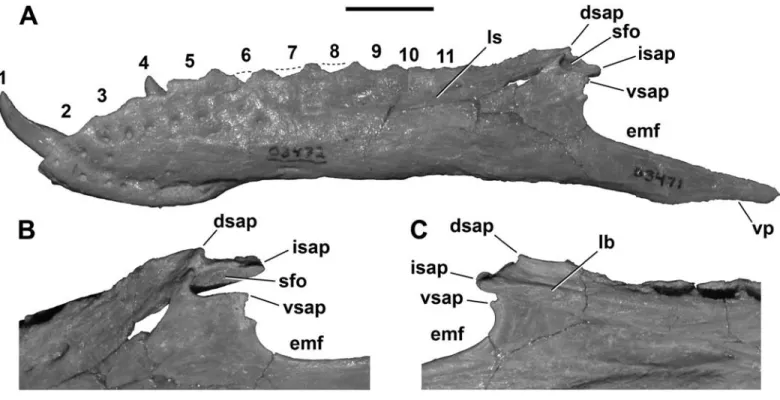
Dentary: A complete left dentary (Figure 8A;
FMNH PR 2471) shows—for the first time—the true pro- portions of this bone, which are somewhat more elongate than had been reconstructed originally (Sampson et al., 2001; Carrano et al., 2002). Specifically, the ventral pro- cess of the dentary is not shortened as in abelisaurids, but extends posteriorly far past the surangular contacts.
The articulations with the postdentary bones are also better preserved than in previously described specimens and reveal more detail about the unusual morphology of the intramandibular articulations. The posterior region of the dentary is very similar to that of both Carnotaurus and Majungasaurus in exhibiting four distinct processes, three
of which form a dorsally placed “socket” for reception of a prong from the surangular (Figure 8B). These three dorsal processes are arrayed in close proximity to one another:
the short dorsal and ventral surangular processes mark the lateral margins of this socket; the moderate-sized interme- diate surangular process forms the medial wall (Sampson and Witmer, 2007). The proportions of these three pro- cesses are more heterogeneous in Masiakasaurus than in Majungasaurus, and the fossa is narrower and deeper.
In medial view, the intermediate surangular process lies at the terminus of a longitudinal buttress that is ven- tral to the paradental plates and forms the dorsal limit of the splenial articulation (Figure 8C). This articulation is particularly pronounced toward its anterior end, at about midlength along the dentary. Along the ventral dentary, FIGURE 7. Braincase (FMNH PR 2457) of Masiakasaurus knopfleri in left lateral (A) and left medial (B) views. Abbreviations: atr = anterior tympanic recess; cr/fo = columellar recess/foramen ovale; ic = foramen for internal carotid artery; mcvg = middle cerebral vein groove; oc = oc- cipital condyle; prp = prootic pendant; scc = region housing semicircular canals; sg = stapedial groove; V2-3, VII, IX, X, XI, XII = foramina for respective cranial nerves. Scale bar = 1 cm.
1 0 • S M I T H S O N I A N C O N T R I B U T I O N S T O P A L E O B I O L O G Y
the splenial ridge is more elongate and farther from the ventral margin of the bone than in Majungasaurus (Samp- son and Witmer, 2007). As in Majungasaurus, a single neurovascular foramen is visible at the base of the first al- veolus on the medial side, although this is correspondingly more posteriorly placed in Masiakasaurus.
The neurovascular foramina on the lateral surface of the dentary are relatively large and arrayed serially within the longitudinal sulcus (Figure 8A). They become more el- liptical posteriorly. Unusually, these foramina also extend down along the dorsoventral depth of the first alveolus.
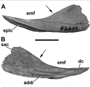
Angular: Two nearly complete left angulars (FMNH PR 2455, UA 9147) throw suspicion on the iden- tification of FMNH PR 2166, previously described as a right angular (Carrano et al., 2002). The identities of FMNH PR 2166 and its parent taxon remain unclear.
The angular of Masiakasaurus is triangular in me- diolateral view and broadly curved along its dorsal and ventral edges (Figure 9). In this view, the dorsal curve suggests an anteroposteriorly elongate external mandibu- lar fenestra, the posterior terminus of which is indicated by a subtle change in curvature (Figure 9A). The poste- rior edge of the element is blunt but incomplete at the
FIGURE 8. Left dentary (FMNH PR 2471) of Masiakasaurus knopfleri in lateral view (A). Enlargement of intramandibular joint in posterolat- eral (B) and medial (C) views. Abbreviations: dsap = dorsal surangular process; emf = external mandibular fenestra; isap = internal surangular process; lb = longitudinal buttress; ls = longitudinal sulcus; sfo = surangular fossa [“socket”]; vp = ventral process of dentary; vsap = ventral surangular process; 1–11 = tooth positions/alveoli. Scale bar in A = 1 cm.
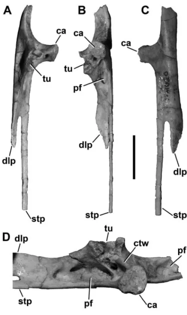
FIGURE 9. Left angular (UA 9147) of Masiakasaurus knopfleri in lateral (A) and medial (B) views. Abbreviations: addi = insertion for adductor musculature; dc = dentary contact; emf = external man- dibular fenestra; sac = surangular contact; splc = splenial contact;
arrow marks posterior extent of emf. Scale bar = 1 cm.
posteroventral corner, where it presumably had a long, thin extension along the ventral mandible as in other the- ropods. The anterior end tapers to a slender point. A long, convexo-concave facet along the anteroventral edge rep- resents the splenial articulation, above which the angular is slightly textured and thickened. This facet is longer and more laterally exposed than in Majungasaurus (FMNH PR 2100). A shallow fossa is also present near the pos- terodorsal corner, presumably for the anterior portion of the M. pterygoideus ventralis (Holliday, 2009).
Medially, the posterior part of the angular bears a long, dorsally facing, oval fossa that marks the insertion site of the adductor musculature (Figure 9B). This fossa lies adjacent to a faint contact surface along the vertical face of the posterior angular for the surangular. A flat, dorsally facing facet sits on the dorsomedial surface of the anterior prong, marking the dentary contact. This is unlike the condition in Majungasaurus, where these two bones were not in direct articulation (Sampson and Wit- mer, 2007). The prearticular would have articulated along the convex ventral margin of the bone.
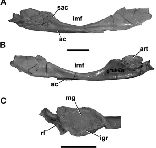
Prearticular: The prearticular resembles that of most basal theropods in overall shape (Figure 10). In
mediolateral view, it is much longer than tall, gently curved along its ventral edge, and more strongly curved dorsally, where it forms the ventral border of the internal mandibu- lar fenestra (Figure 10A,B). This curvature is shallow, con- trasting the stronger arc seen in the foreshortened jaws of the abelisaurids Majungasaurus and Carnotaurus.
Anteriorly and posteriorly, the prearticular expands to approximately equal dorsoventral depths. The anterior flange is incompletely known, but a short portion of con- vex curvature along its ventral edge suggests that it may have had a shape similar to Majungasaurus (Sampson and Witmer, 2007). The posterior flange articulates against and is partly ventral to the lateral portion of the articular.
This flange is complete in UA 9149, where it tapers pos- teroventrally to a long, finger-like projection with a con- cave dorsal surface for the articular.
The prearticular curves ventrolaterally to create a concavity (Figure 10A). Below this, the ventral edge forms a flat, posteromedially twisted facet for the angular. The lateral surface is marked by thickenings along the ven- tral and dorsal edges, which bound a narrow furrow that runs from the articular contact to the anterior flange. A roughened patch at the posterodorsal edge of the internal
FIGURE 10. Right prearticular and articu- lar (UA 9166) of Masiakasaurus knopfleri in lateral (A) and medial (B) views. Dorsal view (C) of mandibular glenoid. Abbrevia- tions: ac = angular contact; art = articular;
igr = inerglenoid ridge; imf = internal man- dibular fenestra; mg = medial glenoid fossa;
rf = retroarticular fossa; sac = surangular contact. Scale bars = 1 cm.
1 2 • S M I T H S O N I A N C O N T R I B U T I O N S T O P A L E O B I O L O G Y
mandibular fenestra represents the contact surface for the surangular.
Articular: The single right articular (UA 9166) is articulated with the corresponding prearticular but is somewhat damaged, obscuring several of its surfaces.
However, most of the medial glenoid fossa is intact, and presents a deeply concave trough for the medial quadrate condyle that is oriented approximately 30° anteromedial to the midline axis (Figure 10C). The medial edge of the fossa is tall, and the lateral edge is marked by a low in- terglenoid ridge. The surangular would have contacted the angular lateral to this point, forming most of the lateral glenoid fossa for articulation with the lateral quadrate condyle. A concave fragment lying posterior to the me- dial glenoid fossa represents the anteriormost portion of the dorsally facing retroarticular fossa, the lateral side of which would have been formed by the surangular.
In medial view, a small, convex portion of the articu- lar is exposed dorsal to the prearticular, but the remainder is covered (Figure 10B). The lateral side is damaged and much is missing, including the lateral glenoid fossa and the area of the chorda tympani foramen.
Hyoid: A long, slender, curved element associated with FMNH PR 2481 is identified as a ceratobranchial.
The articulation with the basihyal has two facets that meet at a 120° angle. The shaft tapers adjacent to the articula- tion, but remains uniformly narrow throughout most of
the rest of its length. The distal end is not preserved. The lateral surface is convex, but a thin concavity runs along the medial side. Overall it is similar to the same element in Carnotaurus (Bonaparte et al., 1990) and Majungasaurus.
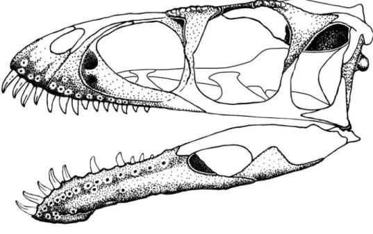
General skull morphology: With these new ele- ments we have attempted to reconstruct the entire skull and lower jaw (Figure 11). We have restored the skull to be relatively long and low based on the primitively pro- portioned maxilla and frontal. Nonetheless, an accurate length:height ratio cannot yet be provided. In profile the skull of Masiakasaurus superficially resembles those of other small theropods, including Coelophysis, Compsog‑
nathus, and Ornitholestes; these similarities are a result of the relatively large orbit and long snout rather than any particular phylogenetic affinity. The most unusual features of the skull are confined to the anterior portions of the jaws. In dorsal view, the skull likely had a similarly plesio- morphic shape, tapering to a narrow snout from its widest point near the anterior edge of the frontal; posteriorly, the sides of the skull were approximately parallel. The lower jaw is closer to the abelisaurid condition than to that of other theropods, particularly in the morphology of the in- tramandibular joint.
Axial Column
Numerous vertebrae from all parts of the column greatly extend our knowledge of axial column morphology
FIGURE 11. Reconstructed skull of Masiakasaurus knopfleri in left lateral view. Known elements (or parts thereof) are stippled; unknown elements are outlined.
in Masiakasaurus and clarify comparisons with Noasau‑
rus and Laevisuchus (Huene and Matley, 1933; Bonaparte and Powell, 1980; Carrano et al., 2002). Most of the cervical series is represented; only the atlas is missing.
Among the dorsals, only portions of the midtrunk remain unknown or unidentified. All sacrals and portions of all of the major caudal regions are also represented.
Although individual variations make precise iden- tification of position uncertain for some specimens, the relatively complete cervical and dorsal series of Carnotau‑
rus (Bonaparte, 1991), Elaphrosaurus (Janensch, 1925), Majungasaurus (O’Connor, 2007), and Spinostropheus (Sereno et al., 2004) provide instructive comparative ma- terials. We can also clarify the positions of some previously identified cervicals (Carrano et al., 2002). The pre-caudal vertebral formula of Masiakasaurus matches well with that of Carnotaurus, which possesses 10 cervicals, 12 free dorsals, and 6 sacrals (including 2 dorsosacrals and 2 cau- dosacrals). The terminology of Wilson (1999) regarding most laminae and fossae is followed here.
Cervical vertebrae: The axis (Figure 12A–D) is known from two specimens (FMNH PR 2462, 2466), and in both cases the axial centrum and atlantal intercentrum are coossified. Two isolated atlantal intercentra (FMNH PR 2477, 2630) show a morphology consistent with these fused examples.
In lateral view, the axis bears a small but distinct oval parapophysis; the long axis is oriented slightly posteroven- trally (Figure 12A). A large, horizontally oval pneumatic foramen lies posterior to the parapophysis and leads into a sizable internal chamber. A second, smaller foramen is present more posteriorly in FMNH PR 2462. Unlike in Majungasaurus, this foramen is not located exclusively posterior to the diapophysis but also partly ventromedial to it (O’Connor, 2007). The outer edges of the foramen are smooth, showing no evidence of a surrounding pneu- matic fossa. The chamber is separated from its opposite by a median septum; it is further subdivided in FMNH PR 2466. The neurocentral suture can still be seen although it is clearly tightly closed in both specimens. The intercen- trum projects horizontally anterior to the parapophysis;
it is not upturned as in Sinraptor (Currie and Zhao, 1993 [1994]). The odontoid projects farther anteriorly than the intercentrum and bears a shallow lateral fossa.
On the neural arch, a distinct oval facet marks the articular surface of the prezygapophysis. The pendant di- apophysis is located directly posteroventrally and is an- gled slightly posteriorly. On its posteroventral surface, a small infradiapophyseal fossa is demarcated by anterior and posterior laminae. The diapophysis is connected to
the epipophysis and postzygapophysis by a pronounced postzygodiapophyseal lamina. This lamina forms the anterodorsal edge of the infrapostzygapophyseal fossa, which houses either a distinct foramen or a deep fossa.
The postzygapophysis is quadrangular, and is consider- ably surpassed by the dorsoventrally flat epipophysis. In lateral view, the neural spine is broadly arched, closer in shape to that of Carnotaurus than Majungasaurus, and hangs slightly posteriorly over the centrum.
Ventrally, the axis is nearly flat, with a very faint midline ridge but no keel. A similar transverse ridge connects the parapophyses. The centrum is somewhat spool-shaped, and its width is subequal to that of the at- lantal intercentrum.
Anteriorly, the intercentrum presents a wide, reniform, concave articular facet for the atlantal centrum (Figure 12B). It sits ventral to the prominent, D-shaped odontoid, which has a nearly flat dorsal surface, flanked laterally by slight ridges that would have floored the neural canal. The odontoid is rounded anteriorly. In this view the diapophy- ses curve ventrolaterally, whereas the prezygapophyses face dorsolaterally. In posterior view the epipophyses are nearly as wide as the postzygapophyses (Figure 12C), al- though they do not project laterally as in Majungasaurus (O’Connor, 2007).
The third cervical vertebra (C3) is characterized by anterior and posterior centrum faces that are dorsoven- trally offset and not parallel in lateral view. A small, round pneumatic foramen pierces the centrum posterodorsal to the parapophysis, which is broken in the only known specimen (UA 9121). On the arch, a strong posterior centrodiapophyseal lamina runs from the diapophysis to the posterior end of the neurocentral suture, separating the elongate infradiapophyseal fossa ventrally from the expansive infrapostzygapophyseal fossa dorsally. A lon- ger postzygodiapophyseal lamina is discriminated from the diapophysis by a kink in its trajectory. The infrapre- zygapophyseal fossa is shallow and faces anteriorly. The prezygapophyseal–epipophyseal lamina is pronounced, and exhibits a small rise and thickening at its midlength.
The short diapophysis hangs nearly vertically.
Anteriorly, the centrum face is flat and approximately rectangular. More dorsally, the neural canal is circular and approximately 80% as large as the centrum face. The small, triangular infraprezygapophyseal fossa is separated from the more medially placed anterior peduncular fossa by an additional lamina. The prezygapophyses are widely sepa- rated from the midline, where they flank a deep prespinal fossa. The posterior centrum face is concave and D-shaped.
The postzygapophyses are angled laterally approximately
1 4 • S M I T H S O N I A N C O N T R I B U T I O N S T O P A L E O B I O L O G Y
30°. The deep postspinal fossa houses a pair of large sub- sidiary foramina, and is separated from the triangular pe- duncular fossae ventral to it by thin laminae.
In dorsal view, the arch is rectangular, with two V-shaped incisions marking the prespinal and postspinal fossae between their respective zygapophyses. A distinct dorsal fossa is visible adjacent to the prezygapophyseal–
epipophyseal lamina, near the base of the prezygapoph- ysis. The neural spine is very thin transversely, located above the midlength of the centrum, and extends only a few millimeters above the flat dorsal surface. The ventral surface of the centrum is nearly flat.
The fourth cervical vertebra (C4; Figure 12E–H) more strongly resembles the only known cervical of Noasaurus than any of those previously described, especially in its elongate proportions (Carrano et al., 2002). It is gener- ally similar to C3, although the centrum faces are more strongly offset, and a second pneumatic foramen is pres- ent on the centrum (Figure 12E). This smaller foramen is located posterior to the diapophysis, and on one specimen (UA 9106) has a scalloped posteroventral margin mark- ing the path of associated soft-tissues. In this specimen, a portion of the cervical rib is still articulated, but not fused, with the left diapophysis.
The proportions of the laminae and fossae are similar to those of C3. Here, the infrapostzygapophyseal fossa is partitioned into a large dorsal chamber, which commu- nicates with the interior of the arch, and a smaller, blind posteroventral fossa. The infradiapophyseal fossa has an uneven surface, and may connect to the infrapostzyg- apophyseal fossa on the right side. The neural spine is thicker and more rectangular than in C3. In C4 the prezygapophyseal–epipophyseal lamina undulates such that the zygapophyses are more dorsally positioned than is the midpoint of this lamina. The epipophysis is long and acuminate, extending posteriorly well past the postzyg- apophysis and the posterior centrum face. The neural spine is half as long anteroposteriorly as the centrum, and bears a sloping anterior edge but a more vertical posterior one (Figure 12E, H).
Anteriorly, the centrum face is reniform, and the pe- duncular fossae are more conical and larger than in C3 (Figure 12F). The prezygapophyses extend laterally only as far as the diapophysis, which has a shallower infrapre- zygapophyseal fossa. The prespinal fossa is narrower and deeper than in the preceding vertebra. The posterior cen- trum face is rounder than the anterior face. In dorsal view, the prezygapophyses are oblate, and the postzygapophyses are visible laterally, ventral to the acuminate epipophyses (Figure 12H). The prezygapophyseal–epipophyseal lamina is straight. Ventrally, the centrum is more elongate than in C3; the anterior and posterior edges are twice as wide as the midcentrum.
The fifth cervical vertebra (C5) is generally similar to the preceding elements, but the prezygapophyseal–epi- pophyseal lamina is less distinct at its midlength, repre- sented by an undulating curve rather than a marked ridge.
This change is related to the emergent elevation of the prezygapophysis relative to the dorsal arch surface. The neural spine is shorter anteroposteriorly and more poste- riorly positioned over the neural arch; the epipophysis is more downturned.
The anterior centrum face is more rounded than in C3 and C4, and the prezygapophyses are more horizon- tally oriented and spaced farther apart laterally. The infra- prezygapophyseal fossa is very small and shallow, and is contiguous with a fossa on the medial portion of the cervi- cal rib in FMNH PR 2465. The shallow prespinal fossa is wide and more open dorsally. Posteriorly, the peduncular fossae are very shallow.
In the only known sixth cervical vertebra (C6; Figure 12I–L; FMNH PR 2628), the centrum faces remain off- set from one another and are vertically inclined (Figure 12I). A single pneumatic foramen is present, adjacent to the left parapophysis, but faint traces of a pair of pneu- matic fossae are evident on both sides of the centrum.
The prezygapophysis is now elevated to the same height as the neural spine, and the infrapostzygapophyseal fossa is large but less pronounced than in preceding vertebrae.
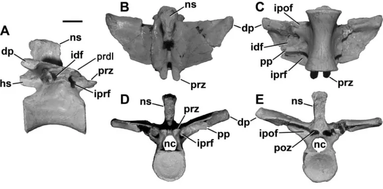
The prezygapophyseal–epipophyseal lamina dips ventrally FIGURE 12. Cervical (A–O) and anterior dorsal (M–P) vertebrae of Masiakasaurus knopfleri in left lateral (A, E, I, M), anterior (B, F, J, N), posterior (C, G, K, O), and dorsal (D, H, L, P) views. A–D, axis (FMNH PR 2466); E–H, fourth cervical vertebra (UA 9106); I–L, sixth cervical vertebra (FMNH PR 2481); M–P, first dorsal vertebra (FMNH PR 2837). Abbreviations: ai = atlantal intercentrum; dp = diapophysis; epi = epipophysis; hs = hyposphene; idf = infradiapophyseal fossa; ipof = infrapostzygapophyseal fossa; iprf = infraprezygapophyseal fossa; ncs = neu- rocentral suture; ns = neural spine; od = odontoid; pcdl = posterior centrodiapophyseal lamina; pdf = peduncular fossa; pf = pneumatic foramen or fossa; podl = postzygodiapophyseal lamina; poz = postzygapophysis; pp = parapophysis; prepl = prezygapophyseal-epipophyseal lamina; prz
= prezygapophysis. Scale bars = 1 cm. (Anterior is up in D, H, L, and P.)
1 6 • S M I T H S O N I A N C O N T R I B U T I O N S T O P A L E O B I O L O G Y
before rising to meet the very small epipophysis, and lacks any thickening at midlength. The neural spine is shorter anteroposteriorly than in C3–C5.
Anteriorly, the infraprezygapophyseal fossa is deeper and extends along the length of the extended diapophysis, whereas the peduncular fossa is very shallow (Figure 12J).
The prezygapophyses are angled at 45°. As seen in poste- rior view, the postspinal fossa is very deep but narrow, and the peduncular fossa is small but also deep (Figure 12K).
The posterior centrum face is more transversely oval than in preceding vertebrae.
The prezygapophyseal–epipophyseal lamina is more distinct in dorsal view than in more anterior cervicals, and is slightly concave along its course (Figure 12L). A U-shaped margin leads to the prespinal fossa; the post- spinal fossa sits at the base of a V-shaped embayment. A pair of large foramina is evident on the dorsal surface, just posteromedial to the prezygapophyses. In ventral view, the parapophyses are connected by a thin transverse ridge, and the infradiapophyseal fossa appears deep and multi-chambered.
The previously illustrated Laevisuchus vertebra (GSI K27/696; Carrano et al., 2002: fig. 7B) is very similar to this specimen in proportions, and we intepret it as C6 based on the offset of the centrum faces and the relative positions of the neural spine and neural arch processes.
The seventh and eighth cervical vertebrae (C7, C8) lack any offset between the centrum faces but are other- wise similar to C6. In these elements the epipophysis is slender and does not extend posteriorly past the postzyg- apophysis; the neural spine is small and blocky. Note that three specimens (FMNH PR 2140, 2139, and 2141) were originally assigned to three successive positions within the neck (Carrano et al., 2002). Based on comparison with newly discovered material, we now identify FMNH PR 2139 as C7, FMNH PR 2141 as C8, and FMNH PR 2140 as C10 (see Appendix). C7 and C8 have, therefore, been previously described in detail (Carrano et al., 2002).
In lateral view, the ninth cervical vertebra (C9) dif- fers from the two preceding elements in that it exhibits a slightly backswept neural arch. The distance between the prezygapophysis and postzygapophysis is reduced such that the prezygapophyseal–epipophyseal lamina is folded.
The epipophysis is very small. The postzygodiapophyseal lamina exhibits a distinct kink along its trajectory.
Relative to more anterior elements, in C9 the di- apophysis extends more directly laterally and bears a well-defined infraprezygapophyseal fossa that is very deep medially. Anteriorly, the peduncular fossa is absent, and the neural canal is smaller relative to the centrum face.
The prespinal fossa is very shallow and fully opened an- teriorly. Posteriorly, the peduncular fossa is much smaller than the postspinal fossa, and the infrapostzygapophyseal fossa exhibits deep foramina.
The tenth cervical vertebra (C10) lies at or near the cervicodorsal transition, although this can be difficult to define in theropods. Its unusually long proportions in- dicate that Masiakasaurus was characterized by antero- posteriorly lengthened centra throughout the presacral vertebral column. This is unlike the condition in most the- ropods, where the cervicodorsal transition is marked by one or two anteroposteriorly short vertebrae.
Vertebra C10 retains a small, anteriorly placed pneu- matic foramen, but lacks any evidence of a more posterior foramen or fossa. The parapophysis is elevated just dorsal to the ventral margin of the centrum. Unlike all other cer- vicals (and dorsals), the prezygapophysis is oriented nearly vertically, such that its posterior margin is approximately even with the anterior edge of the neural spine. In addi- tion, the postzygodiapophyseal lamina is not concave but bears an outward kink close to its dorsal terminus.
In anterior view, the diapophysis is more elevated than in C9, with a more expansive anterior face and as- sociated infraprezygapophyseal fossa. As seen in posterior view, the postzygapophysis is elevated to the level of the neural spine. As in C8 and C9, the dorsal surface of the arch bears a pneumatic foramen that faces the area of the prezygapophysis. The arch is visibly set back from the anterior border of the centrum in dorsal view.
Trends within the cervical series: The entire cervi- cal series of Masiakasaurus is highly pneumatized. This pneumaticity includes: (1) two fossae on the centrum, the anterior of which consistently forms a foramen; (2) com- munications between the centrum and arch pneumaticity via the neurocentral suture; (3) three deeply invaginated fossae ventral to the transverse process on the neural arch, each with one or more foramina; and (4) peduncu- lar fossae and foramina. Within the series, pneumaticity increases posteriorly; examples include the appearance of peduncular fossae subsequent to C4 and the presence of foramina on the dorsal surface of the arch starting on C6.
It is likely that the pneumaticity of the anterior centrum fossae is directly related to cervical rib pneumaticity (see below). Similarly, the dorsal arch fossae of C6–C10 might bear some relation to the presence of a fossa alongside the prezygapophyseal–epipophyseal lamina in C3 and C4.
The anterior half of the cervical series articulates along a gentle curve in its neutral position, but the last half is nearly straight because of the lack of offset between the anterior and posterior centrum faces. The epipophyses
are more pronounced anteriorly as well, and the neural spine is longer, although still small. Pronounced left-right asymmetry is common with respect to both the presence and degree of development of pneumatic structures.
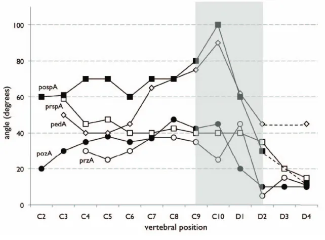
Although the absence of significant centrum length changes obscures some aspects of the cervicodorsal transi- tion in Masiakasaurus, this transition is nonetheless evident in other morphological features. For example, the trans- verse distance between both the pre- and postzygapophy- ses declines rapidly from C9–D2, and both articulations become more horizontally oriented (Figure 13). A the same time, the prezygapophysis and its peduncle become verti- cally oriented in lateral view, and then abruptly lower again.
Dorsal vertebrae: Compared with the most pos- terior cervicals, the only known first dorsal vertebra (D1;
Figure 12M–P; FMNH PR 2837) has a more equilateral
profile overall. The centrum remains proportionally long (length:height = 2.0), although the ventral margin is more strongly arched (and this arch has a more posteriorly posi- tioned apex) than in the posterior cervicals. The parapophy- sis is D-shaped and situated just ventral to the neurocentral suture, which remains visible but not patent (Figure 12M).
A large, elliptical pneumatic foramen lies directly poste- rior to the parapophysis and opens into a chamber within the centrum. Marked, longitudinally oriented striations are present along the rims of the centrum faces.
The arch laminae are well developed (Figure 12M).
An extensive pneumatic fossa occupies most of the large infraprezygapophyseal fossa on the left side, whereas the right is perforated by a large foramen; on both sides a smaller, round foramen is present in the posterodorsal corner of the fossa. The infradiapophyseal fossa is small
FIGURE 13. Morphological changes in the cervical and anterior dorsal vertebrae of Masiakasaurus knopfleri. Dashed lines represent interpolations across missing data; gray area indicates the region of the cervicodorsal transition. Abbre- viations: pedA (diamonds) = angle between floor of neural canal and axis of prezygapophyseal peduncle in lateral view;
pospA (solid squares) = angle between left and right edges of postspinal fossa in dorsal view; pozA (solid circles) = angle of vertical inclination of postzygapophysis in posterior view; prspA (open squares) = angle between left and right edges of prespinal fossa in dorsal view; przA (open circles) = angle of vertical inclination of prezygapophysis in anterior view.