Chapter 3
Mutation Candidates for Human CCR1
In conjunction with experimental data from our collaborators at Berlex, this chapter will be submitted as a communication to Nature Chemical Biology.
Abstract
The goal of the work presented in this chapter is to predict point mutation candidates in hCCR1 that would improve the binding of a low affinity small molecule antagonist. Such a study will not only validate the predicted antagonist binding site in hCCR1 but would also lay the groundwork for using this structure to optimize binding affinities of lead compounds. This analysis involves comparison of the binding site of a strong and weak affinity antagonist to hCCR1, with the goal of designing receptor
mutants that markedly improve the binding of the low-affinity antagonist. We dock to our validated hCCR1 structure two antagonists with completely different (3 orders of
magnitude) known binding affinities. We then identify the functional group responsible for the binding differential, and choose two residues in the binding cavity (TM5 residues Leu260 and Val263) that could enhance the binding energy for the low affinity binder, for computational point mutation. Systematic changes are made from the non-polar Leu and Val to a series of polar residues (Asn, Gln, Ser, Thr, and Tyr) using a side chain rotamer placement program with accurate energy functions. In most cases we calculate a large improvement in binding energy, and have suggested these mutations to our
experimental collaborators at Berlex (experiments are underway).
Computational Methods
Force Fields
Protein receptor calculations used the DREIDING Force Field (DFF)1 with CHARM22 charges2. The Cell Multipole Method (CMM)3 in MPSim4 was used to calculate all non-bond interactions.
Jaguar v4.0 was used to perform Hartree-Fock (HF) level quantum mechanical calculations to extract electrostatic potential (ESP) fitted charges on the isolated ligands5. These ligand charges have been used for docking calculations.
All solvent (water) calculations used the Analytical Volume Generalized Born (AVGB) approximation to the Poisson-Boltzmann (PB) continuum solvation model6.
The DFF along with the charge equilibration (QEq)7 method was utilized to describe lipids.
Generation of Mutants
The binding sites for C5 (a strong affinity antagonist with Ki = 10 nM) and C6 (a weak antagonist with Ki > 10,000 nM) were predicted using the HierDock method8-10 followed by side chain rotamer optimization of the residues in the binding site. The weak binder has a polar secondary amine group added to the ligand and the environment of this functional group is hydrophobic in the protein. After comparison of the binding sites for these two compounds, we also calculated the contribution of each residue in the 5 Å
binding cavity to the ligand interaction energy. This showed that Leu260 (TM6) and Val263 (TM6) were the two positions where mutation to a polar residue could make a hydrogen bond with the ligand. Therefore we generated single mutants using the
mutation builder Python script cbMuts (written by Caglar Tanrikulu). This program calls SCREAM11 for side chain replacement, then performs minimization on the resulting mutant structure using MPSim. SCREAM uses a library of 1,478 rotamers with accurate energy functions that calculates ligand-protein interactions along with protein-protein interactions to side chain placement. This method has been tested on many co-crystal structures for globular proteins. The binding energies are recalculated using DFF energy functions in HierDock, and binding site residues within 6 Å reassigned with another round of SCREAM.
Results and Discussion
Antagonist Binding Site
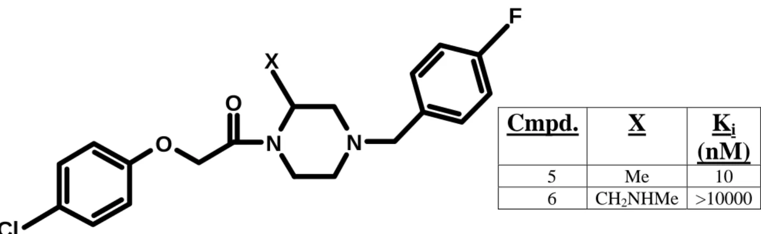
Figure 3-1 shows the structures of the two proprietary antagonists which were docked to hCCR1. The binding region is identical to that presented in Chapter 2, and is detailed for each ligand in Figures 3-2 and 3-3. The pKa of the tertiary amine substituent in C6 was calculated to be 9.38, and so the protonated species was docked. This had the effect of creating a large hydrophobic pocket around TM6, and so we searched for residues in the 5 Å binding site that were favorable in the C5 cavity, but clashing in the C6 binding mode.
I. Predicted Binding Site for C5 (Ki = 10 nM)
The 5 Å binding cavity of C5 involves contributions from TMs 2, 3, 4, 6, and 7 (Figure 3-2). Of the polar residues, Tyr113 (TM3), Ser110 (TM3), and Tyr291 (TM7) all anchor the ligand in this site. The major vdW contributor to the binding energy is Tyr113 (TM3), which stacks beneath the ligand piperazine ring. Ser110 (TM3) hydrogen bonds to the oxygen atom adjacent to the ester carbonyl at a larger than ideal distance of 4.74 Å.
The fluorine substituted aromatic interacts with Tyr291 (TM7) and weakly (yet favorably) with Glu287 (TM7). This indicates only a mild perturbation of the Lys94 (TM2) to Glu287 (TM7) salt bridge in the apo protein, affirming our view of C5 as a strong affinity antagonist. Although the chlorine substituted aromatic seems to hydrogen
bond with the side chain of Ser172 (TM4), the other TM4 residues shown (Gly168, Leu169, and Phe171) also contribute favorably to both the vdW and Coulombic components of the binding energy.
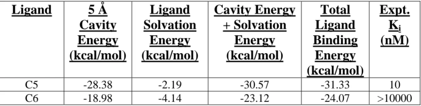
All of these interactions with the ligand were also seen in the binding cavities of a similar class of antagonists (C1-C4 ligands discussed in Chapter 2). It is encouraging to note that the calculated binding energy is 98% recovered upon summation of the ligand solvation and cavity energies (Table 3-1).
II. Predicted Binding Site for C6 (Ki > 10,000 nM) and C5-C6 Binding Differential
The 5 Å binding cavity of C6 involves contributions from TMs 3, 4, 6, and 7 as shown in Figure 3-3. Compared to C5, the contribution of Tyr113 (TM3) is less in this cavity, as the ligand piperazine ring is tilted slightly away from the side chain. The interaction of the fluorine substituted aromatic ring with Tyr291 (TM7) is stronger (2.94 Å), and the chlorine substituted aromatic ring now hydrogen bonds very weakly with Gly168 (TM4). This latter interaction is in contrast to the hydrogen bonding to Ser172 (TM4) we observed in the case of C5.
However, this site exhibits two repulsive hydrophobic interactions, namely with Leu260 (TM5) and Val263 (TM5). In light of the fact that the only difference between these two antagonists is a secondary amine substituent in C6 where the methyl group was in C5, the calculated repulsive energy can be accounted for by analyzing the local
hydrophobic environment around these residues. Figure 3-4 clearly shows these bulky hydrophobic side chains sterically clashing with the equally bulky non-polar alkyl
substituent in C6. In Figure 3-5 we see the same view for C5, where the packing is actually favorable.
Addition of the cavity and solvation energies yields 96% of the calculated binding energy, although only 79% of this is actually from the cavity. It is instructive to note that C6 exhibits a lower binding energy due to the desolvation penalty. The solvation energies in Table 3-1 show that this term for C6 is twice that of C5, which is due to the
stabilization gained through hydrogen bonding when the basic amino group is hydrated (as this compound is charged).
hCCR1 Mutant Design
An overlay of the binding modes of C5 and C6 is shown in Figure 3-6. The binding mode is in fact the same, and we can clearly see that the functional group change (–Me in C5 to –CH2NHMe in C6) is pointing toward TM6. As described above, the difference in the calculated binding energies between these two antagonists stems from the large repulsive interactions with both Leu260 (TM6) and Val263 (TM6) for C6.
Analysis of the apo receptor structure shows that these residues are in fact not directly participating in stabilizing inter-helical interactions. Therefore, we chose to mutate these two non-polar residues to the polar amino acids: Asn, Gln, Ser, Thr, and Tyr. We did not investigate charged polar mutations (Asp, Glu, and His) due to the practical difficulty of introducing these residues in the middle of TM domains.
Table 3-2 shows the entire spectrum of hCCR1 mutants generated, and the resulting binding energy calculated after using HierDock to re-dock C6 into the mutant
receptor. The values in blue indicate that the mutation resulted in a more favorable docked conformation than what was observed in the wild type. Conversely, the residues highlighted in red exhibit more repulsive interactions, and make ligand binding worse.
This would be expected for non-polar mutations (Ala and Gly), which would not
significantly change the character of the local environment. As a gauge of the reliability of our method, we used the same protocol to “mutate” Leu260 into Leu, and Val263 into Val, and calculated the new binding energy. In both cases, the result was within 0.02 kcal/mol, ensuring that our procedure was indeed valid for this study.
BE Calculations for Mutant hCCR1-C6 complexes
I. Leu260Gln
Figure 3-7 shows the interaction between the Leu260Gln mutant and C6. In the apo protein, this residue would make two hydrogen bonds with Val263 and Asn256. The local environment of this replacement is a mix of polar (Ser262, Thr258, and Asn256) and non-polar residues (Leu257, Ile259, Ile261, Val263, and Phe265), which could be a potential issue for mutant expression. Decomposition of the interaction energy of the residues in the binding site (within 6Å) with the ligand shows an increase of 5.3 kcal/mol in Coulombic energy by Gln260, to yield a total interaction energy of -31.05 kcal/mol.
Based on this data, we predict that this mutation would bring the binding affinity of C6 back up to the level of C5.
II. Leu260Asn
The interaction between the Leu260Gln mutant and C6 is shown in Figure 3-8. In the apo protein, this residue would make two hydrogen bonds with Asn256, and one hydrogen bond with Phe264. Decomposition of the interaction energy of the residues in the binding site (within 6Å) with the ligand shows an increase of 5.2 kcal/mol in
Coulombic energy by Asn260, to yield a total interaction energy of -30.83 kcal/mol.
Based on this data, we predict that this mutation would enhance the binding of C6.
III. Val263Gln
Figure 3-9 shows the interaction of this mutant with C6. The mutation does not appear to disrupt any apo protein contacts, nor does it create new hydrogen bonds within the structure. However, the Gln side chain is about the same size as the wild type Val.
There is an increase of 5.5 kcal/mol in the binding energy, indicating that the hydrogen bond to the amine functionality in C6 is indeed a strong interaction, and that this mutation should also enhance binding.
IV. Val263Ser
In Figure 3-10 we observe that the Val263Ser mutation hydrogen bonds with the backbone of Leu260, while not creating any repulsive interactions. There is a hydrogen bond with C6, although it is weaker here than in the other mutants. Furthemore, the Ser
side chain is quite a bit smaller than the wild type Val. Accordingly, the binding energy becomes more favorable by 3.8 kcal/mol, a smaller increment than the mutants discussed thus far.
V. Val263Thr
Figure 3-11 shows that replacement of Val263 by Thr263 is essentially the same as the Ser mutation. The backbone hydrogen bond with Leu260 changes from 1.91 to 2.33 Å. In the same manner, there are no repulsive interactions created by introducing this mutation. The Thr side chain is also larger than Ser. Analysis of the binding energy shows that 3.4 kcal/mol is gained in this mutant.
VI. Val263Tyr
Figure 3-12 shows the interaction of the Tyr mutant with C6. There is a weak hydrogen bond (5.55 Å) with the amine substituent, and a similar backbone interaction with Leu260 at a larger distance than seen in the other mutants. 3.2 kcal/mol of
interaction energy is gained in this mutation.
Table 3-3 shows the different hCCR1 mutants that have been proposed to our experimental collaborators.
Conclusions
Two antagonists with order of magnitude different binding affinities were docked to our validated hCCR1 structure. The binding differential was ascertained both
quantitatively (repulsive terms in the cavity energy) and schematically (steric clashes in the binding site), in an effort to design mutants that would bind more favorably to the weaker binder. Two non-polar residues in TM6 (Leu260 and Val263) were selected for mutation, and exhibited much stronger ionic coupling to the ligand after being mutated to Gln and Asn. Mutations from Leu to Leu and Val to Val returned the initial rotamer conformation and ligand binding energy, assuring us that lower binding energies seen in most mutants were in fact reflective of more stable complexes. The proposed mutations and corresponding favorable change in BE are Leu260Gln (5.3 kcal/mol), Leu260Asn (5.2 kcal/mol), Val263Gln (5.5 kcal/mol), Val263Ser (3.8 kcal/mol), Val263Thr (3.4 kcal/mol), and Val263Tyr (3.2 kcal/mol).
References
[1] Mayo, S. L., Olafson, B. D., and Goddard, W. A. III (1990) J. Phys. Chem. 94, 8897-8909.
[2] MacKerell, A. D., Bashford, D., Bellott, M., Dunbrack, R. L., Evanseck, J. D., Field, M. J., Fischer, S., Gao, J., Guo, H., Ha, S., Joseph-McCarthy, D., Kuchnir, L., Kuczera, K., Lau, F. T. K., Mattos, C., Michnick, S., Ngo, T., Nguyen, D. T., Prodhom, B., Reiher, W. E., Roux, B., Schlenkrich, M., Smith, J. C., Stote, R., Straub, J., Watanabe, M., Wiorkiewicz-Kuczera, J., Yin, D., and Karplus, M.
(1998) J. Phys. Chem. B 102, 3586-3616.
[3] Ding, H. Q., Karasawa, N., and Goddard, W. A. III (1992) Chem. Phys. Lett. 97, 4309-4315.
[4] Lim, K-T., Brunett, S., Iotov, M., McClurg, R. B., Vaidehi, N., Dasgupta, S., Taylor, S., and Goddard, W. A. III (1997) J. Comput. Chem. 18, 501-521.
[5] Trabanino, R. J. (2004) Ph.D. thesis, California Institute of Technology.
[6] Zamanakos, G. (2001) Ph.D. thesis, California Institute of Technology.
[7] Rappé, A. K., and Goddard, W. A. III (1991) J. Phys. Chem. 95, 3358-3363.
[8] Vaidehi, N., Floriano, W. B., Trabanino, R., Hall, S. E., Freddolino, P., Choi, E.
J., Zamanakos, G., and Goddard, W. A. III (2002) Proc. Natl. Acad. Sci. USA 99, 12622-12627.
[9] Kekenes-Huskey, P. M., Vaidehi, N., Floriano, W. B., and Goddard, W. A. III (2003) J. Chem. Phys. B 107, 11549-11557.
[10] Datta, D., Vaidehi, N., Zhang, D., and Goddard, W. A. III (2004) Prot. Sci. 13, 2693-2705.
[11] Kam, V. W. T., Vaidehi, N., and Goddard, W. A. III (2005) unpublished results.
Table 3-1: Correlation between calculated binding energies (BE) and experimental
binding affinities (Ki) for docked hCCR1 ligands.
Ligand 5 Å
Cavity Energy (kcal/mol)
Ligand Solvation
Energy (kcal/mol)
Cavity Energy + Solvation
Energy (kcal/mol)
Total Ligand Binding
Energy (kcal/mol)
Expt.
Ki
(nM)
C5 -28.38 -2.19 -30.57 -31.33 10
C6 -18.98 -4.14 -23.12 -24.07 >10000
Table 3-2: Binding energies (kcal/mol) for C5 docked to mutant hCCR1 receptor.
L V S Q N T Y G A
L260X -24.09 N/A -26.68 -31.05 -30.83 -26.79 -26.01 -22.97 -22.96 V263X N/A -24.08 -29.30 -31.19 -24.57 -28.97 -28.19 -24.14 -23.64
Table 3-3: Planned hCCR1 mutations (* indicates mutant is energetically more stable than wild type receptor).
S Q N T Y
L260X * *
V263X * * * *
Figure 3-1: Structural template for proprietary antagonists.
Template for two hCCR1 antagonists with distinct binding affinities (see table above).
Data courtesy of Berlex AG.
O
O
N N
F
Cl
X
Cmpd. X K
i(nM)
5 Me 10
6 CH2NHMe >10000
Figure 3-2: Interactions in the C5 binding cavity (top).
Major contributions from Tyr113, Ser110, and Tyr291. Each binding site residue shown here interacts favorably with C5 (shown in magenta).
4.59 Å
4.74 Å
3.44 Å
TM1 TM2
TM3
TM4
TM5
TM6
TM7
T86 (2) L109 (3) S110 (3)
S172 (4)
F171 (4) L169 (4)
G168 (4)
Y113 (3)
Y291 (7) E287 (7)
T286 (7) A290 (7)
V263 (6) L260 (6)
Figure 3-3: Interactions in the C6 binding cavity (top).
4.95 Å
5.30 Å
2.94 Å
TM7 TM2 TM3
TM4
TM5
TM6
Y291 (7) E287 (7)
V283 (7) T286 (7) A290 (7)
V263 (6) L260 (6)
F264 (6)
Y113 (3) L109 (3) S110 (3)
L117 (3) G168 (4)
F171 (4) S172 (4)
Y114 (3) L169 (4)
Figure 3-4: Repulsive packing in the C6 binding cavity (top).
Leu260 (TM6)
Val263 (TM6) C6
Figure 3-5: Packing between C5 and TM4’s L260 and V263 (side).
Val263 (TM6)
Leu260 (TM6)
C5
Figure 3-6: Overlay of the C5 and C6 binding modes (top).
C5
C6
TM1 TM2
TM3 TM4
TM5
TM6
TM7
Figure 3-7: Leu260 to Gln260 mutation in the C6 binding cavity (top).
2.12 Å
Gln260 (TM6) Val263
(TM6)
Asn256 (TM6)
3.01 Å
4.20 Å
Figure 3-8: Leu260 to Asn260 mutation in the C6 binding cavity (side).
Asn256 (TM6)
Phe264 (TM6)
Asn260 (TM6) 3.43 Å
3.68 Å 2.67 Å
1.98 Å
Figure 3-9: Val263 to Gln263 mutation in the C6 binding cavity (top).
Gln263 (TM6)
2.41 Å
Figure 3-10: Val263 to Ser263 mutation in the C6 binding cavity (side).
Ser263 (TM6)
Leu260 (TM6)
4.41 Å 1.91 Å
Figure 3-11: Val263 to Thr263 mutation in the C6 binding cavity (side).
4.66 Å 2.33 Å
Ser263 (TM6)
Leu260 (TM6)
Figure 3-12: Val263 to Tyr263 mutation in the C6 binding cavity (top).
Leu260 (TM6) Leu260
(TM6)
5.62 Å 5.55 Å