137
CHAPTER 6: New Approaches to Photochemical Cleavage of Peptide and Protein Backbones
∗
6.1 ABSTRACT
A strategy for photochemical cleavage of peptide and protein backbones is described, which is based on a selenide-mediated cleavage of a backbone ester moiety. Studies in model systems establish the viability of the chemistry and suggest the method could be a valuable tool for chemical biology studies of proteins. Also described are two alternative strategies based on a “caged” aniline and the (2-nitrophenyl)ethyl (NPE) protecting group.
6.2 INTRODUCTION
We describe here new strategies for photochemical cleavage of peptide and protein backbones and model studies intended to evaluate the viability of the novel chemistry involved. Strategies for preparing photoresponsive biomolecules are finding increasing use in chemical biology.1, 2 Some time ago we introduced a strategy for photochemically initiating backbone cleavage of a protein, employing the unnatural amino acid 2- nitrophenylglycine (Npg, Figure 6.1).3 After incorporation of Npg into a protein or peptide, the well-known o-nitrobenzyl “deprotection” of the peptide bond nitrogen results in cleavage of the protein backbone, a site-specific, nitrobenzyl-induced, photochemical proteolysis (SNIPP). Other strategies for photochemical cleavage of protein and peptide
∗ This work was done in collaboration with Dr. Amy L. Eastwood and Dr. Niki M. Zacharias and is adapted from: Eastwood, A. L.; Blum, A. P.; Zacharias, N. M.; Dougherty, D. A., A selenide‐based approach to photochemical cleavage of peptide and protein backbones at engineered backbone esters. The Journal of Organic Chemistry 2009, 74, (23), 9241‐44. Copyright 2009 American Chemical Society.
138
backbones have recently appeared. Imperiali and Kron have both employed o-nitro-β- phenylalanine as a linker between two protein or peptide fragments,4, 5 while Muir has employed an expanded nitrobenzyl linker.6 Otaka and co-workers introduced a novel system based on a nitrobenzyl-caged phenol and the “trimethyl lock” motif, which promoted intramolecular cleavage of the backbone amide after the phenol was decaged.7 Schultz and co-workers demonstrated a novel cleavage mediated by 2- nitrophenylalanine.8
All of these strategies have advantages and disadvantages. The Npg approach has been employed in several contexts3, 9, 10 and is compatible with both solid phase peptide synthesis (SPPS) and in vivo incorporation into full proteins via nonsense suppression.
However, the photochemical efficiency of the cleavage is not high, with a ~50% cleavage yield after four hours of photolysis.3 Also, the incorporation into proteins of the relatively crowded, β-branched residue is often not efficient. Similarly low photoefficiency is seen with 2-nitrophenylalanine.8 The other strategies can show more efficient cleavage of peptides but are not compatible with in vivo incorporation into proteins.
Described herein are alternative strategies for the photochemical cleavage of peptide and protein backbones. The first strategy is based on a caged selenide. Model studies establish that this strategy is effective, with up to 72% backbone cleavage of depsipeptides. Other strategies are also described which are based on a nitroveratryloxycarbonyl (NVOC)-protected aniline and the photochemistry of (2- nitrophenyl)ethyl (NPE) derivatives.
139 6.3 RESULTS
6.3.1 Caged selenide strategy
In our first strategy the key cleavage reaction is based on a novel, intramolecular SN2 reaction, shown schematically in Figure 6.1. Selenide is one of the most potent nucleophiles known, and at physiological pH a selenol (pKa ~5-6) should be predominantly in the selenide form. The essential reaction, SN2 cleavage of an ester carbon with a carboxylate as the leaving group, has ample precedent.11-15 Formation of the ester leaving group requires incorporation of an α-hydroxy acid (rather than an α- amino acid), but such backbone esters can be efficiently incorporated into peptides by SPPS16, 17 and into proteins by nonsense suppression.10, 18-22 In addition, selenium- containing natural amino acids such as selenocysteine and selenomethionine are well- known to be efficiently incorporated into proteins. Finally, “caging” the selenide with an o-nitrobenzyl group allows the process to be initiated photochemically.
Figure 6.1. Npg and the second-generation SNIPP unnatural α-hydroxy acids, 1 and 2.
HN O
HN
R
O XSe
O R O2N
HN O
HN
R
O XSe–
O R
HN
OH
HN
R O
O R
h! +
HO CO2H Se
HO CO2H Se
1 2
X Se
O2N O2N
H2N CO2H
Npg O2N
140
The process proposed in Figure 6.1, however, raises many questions. Like all SN2 reactions, the selenide-induced ester displacement is sensitive to steric effects. As such, the reaction is typically applied to methyl esters, although under optimal conditions and with heating, ethyl, benzyl, and even isopropyl esters along with many lactones are reactive. Certainly, the α-carbon of the α-hydroxy acid that is incorporated will be sterically crowded. The question is whether the intramolecularity of the process, perhaps aided by the inductive effect of the neighboring amide carbonyl, will overcome the steric burden. Caged selenides are not common, and so there is the question of the efficiency of the photochemical step. In addition, selenides are sensitive to oxidation, more so for aliphatic than aromatic (selenophenol) derivatives. On the other hand, an aliphatic selenide might be expected to be the stronger nucleophile, but an aryl selenide has fewer rotatable bonds that need to be restricted in the cyclization reaction.
Given these chemical uncertainties, it seemed prudent to first evaluate the viability of the chemistry proposed in Figure 6.1 before proceeding with chemical biology studies. In the present work we evaluate two structures that are meant to provide such a test. We describe the synthesis and characterization of aliphatic (1) and aromatic (2) variants of the design, along with mechanistic characterization.
Synthesis of enantiopure 1 (Scheme 6.1), as devised and executed by Dr. Amy L.
Eastwood23 and Dr. Niki M. Zacharias,24 began with conversion of S-(−)-tetrahydro-2- furoic acid to the ring-opened bromide 4 as previously described.25, 26 Conversion to the diselenide, and ester hydrolysis then produced 6.27, 28 Acid diselenide 6 was directly reduced with sodium borohydride and the product alkylated with o-nitrobenzyl bromide to give the target compound 1.29
141
Scheme 6.1. Synthesis of selenide α-hydroxy acid 1. This route was devised and executed by Dr.
Amy L. Eastwood23 and Dr. Niki M. Zacharias.24
Scheme 6.2. Synthesis of selenide α-hydroxy acid 2.
The synthesis of 2 (Scheme 6.2) began with the known reduction of 2- nitrophenylpyruvic acid by (+)-β-chlorodiisopinocampheylborane (“Ipc2BCl”) to yield 7 in 94% ee.30 The selenocyanate was prepared by a modification of the standard sequence, and the nitrobenzyl group was introduced by reductive alkylation. The bulky t- butyl protecting groups were installed to discourage intramolecular cyclization, which was seen when 7 was subjected to reducing conditions, as well as to improve the
O OH
O
O OEt
O EtOH, toluene
80 !C (61% yield)
HO OEt
O
HO OEt
O Br
3 4 5
CH2Cl2 0 !C (71% yield)
Se
THF, H2O (97% yield)
HO OH
O 6
Se
75 oC (96% yield)
1 1. NaBH4
NaOH, EtOH 0 !C to RT
HO OH
O Se O2N
NO2 Br
H2SO4 Me2BBr, TEA Se (s), NaBH4
2
2
HCl
NaOH 0 !C to RT (85% yield) 2.
O2N
O OH
O
O2N
HO OH
O
O2N
O O
O
H2N
O O
O
NCSe
O O
O
Se
O O
O O2N
Se
HO OH
O O2N
TFA, CH2Cl2 Ipc2BCl
TEA, THF
NaBH4, Pd-C MeOH, THF
1. NaNO2, AcOH 2. KSeCN, H2O
1. NaBH4, EtOH, THF
2. Br
NO2 cyclohexane
THF (18% yield)
O CCl3 NH
BF3-Et2O -42 !C to RT
(89% yield)
0 !C to RT (90% yield)
0 !C (50% yield)
0 !C to RT
(70% yield) (96% yield)
2
7 8 9
10 11
142
solubility and ease of purification of subsequent compounds in the sequence. Alternative synthetic routes were also attempted and are described in Appendix 3.
To evaluate whether the proposed cleavage mechanism was viable, studies in model systems were performed. Depsipeptides 12 and 14 were chosen for synthetic accessibility and because they introduce a UV chromophore into the carboxylate cleavage product. They were prepared through standard solution-phase coupling procedures, employing PyBop/N-methylmorpholine and DCC/DMAP for the hydroxy-peptide- and depsipeptide-forming reactions, respectively (Scheme 6.3).
Scheme 6.3. Synthesis of depsipeptides 12 and 14. Depsipeptide 12 was prepared by Dr. Amy L.
Eastwood.23
Mass spectrometric analysis of initial photolysis studies indicated the formation of the mechanistically revealing selenacyclopentane (15, 16) and the appropriate carboxylic acid, but also suggested several complicating side reactions. Both 12 and 14 produced
Se
HO OH
O O2N
2
Se
HO HN
O O2N
O O
O 13 O
Se
O HN
O O2N
O O
O O
14 H O
N O O
NH PyBop
N-methylmorpholine di-tert-butyl-L-glutamate
DMF (90% yield)
DCC, DMAP
t-Boc-L-tryptophan-OH CH2Cl2 (40% yield) Se
HO OH
O O2N
1
Se
HO HN
O O2N
O O
O O
Se
O HN
O O2N
O O
O O 12 H O
N O O PyBop
N-methylmorpholine di-tert-butyl-L-glutamate
CH2Cl2 (41% yield)
DCC, DMAP
t-Boc-L-phenylalanine-OH CH2Cl2 (63% yield)
143
characteristic isotope pattern for a structure with two selenium atoms. In addition, the aliphatic variant 12 showed m/z ratios consistent with a depsipeptide containing dehydronorvaline (i.e., an allyl side chain), presumably formed by elimination of the selenoxide produced by air oxidation. These undesirable reactions could be suppressed by running the photolyses in the presence of excess dithiothreitol (DTT),31 which was expected to discourage both dimerization and oxidation.
Scheme 6.4. Depsipeptide cleavage reactions.
For aryl selenide 14 an unanticipated side product, identified by mass spectrometry, was the depsipeptide in which the original nitrobenzylselenyl group had been replaced by a hydrogen atom. While this work was in progress, Kitahara and co- workers reported that short wavelength photolysis of arylalkyl selenides can lead to C-Se bond cleavage.32 As suggested by that study, we found that changing the photolysis wavelength from >300 nm (pyrex filter) to >330 nm (uranium glass filter) eliminated this side reaction.
Se
O O
HN O2N
O O
O O HN
O O
O
NH
O HN Se OH
HN O
O
O
NH
O ON +
+ O
O
O O O
HN
O O O
O O HN
O O
O
Se
HN OH
O H
N O
O O O
O
O O Se
ON
O +
+ 1. h! , DTT (100 eq.)
ACN, 20 mM phos. buffer (pH 7.6) 2. 70 oC
O2N
12
14
15
16 1. h! , DTT (10 eq.)
ACN, H2O (pH 8.0)
144
Of course, the novel process of Figure 6.1 cannot be considered validated without unambiguous confirmation that the selenacyclopentanes 15 and 16 were formed. Using the knowledge gained from the preliminary studies, preparative-scale photolyses were performed (Scheme 6.4). For both depsipeptides the selenacyclopentane product could be isolated after irradiation, providing clear support for the proposed scheme; mass spectrometry and NMR spectroscopy confirm product identities. We find that cleavage is more efficient with the aryl selenide (14) than the alkyl (12), in that isolated yields of the selenacyclopentane are consistently higher (72% vs. 29%). In addition, obtaining significant yields of the selenacyclopentane from the aliphatic system (12) required heating the photolysis mixture to ~70 °C, which was not necessary in the case of aryl selenide 14 under optimal photolysis conditions. These observations, coupled with the lack of complications due to olefin formation, suggest that aryl selenide 2 may be the better system to incorporate for subsequent protein and peptide studies.
6.3.1.1 Chemical biology studies of proteolysis by 1 and 2
In preparation for nonsense suppression experiments, the key α-hydroxy acids 1 and 2 were each activated as a cyanomethyl ester and subsequently transesterified by published protocols33 to yield acylated suppressor tRNA as needed for the following experiments.
6.3.1.2 In vitro studies of proteolysis by 1 and 2
Studies were conducted to determine whether 1 or 2 could promote proteolysis of a full- length protein in vitro. These studies used in vitro nonsense suppression methodology to incorporate 1 and 2 into the α1 subunit of the muscle-type nicotinic acetylcholine receptor (nAChR) at residue Met243 (located in the M2 transmembrane helix). In these
145
studies, the α1 subunit of the nAChR contains a hemmagglutinin epitope (in the M3-M4 cytoplasmic loop) to facilitate Western blotting. It was envisioned that cleavage of the nAChR protein backbone at Met243 would yield cleavage fragments that are sufficiently different in molecular weight from the full-length protein to be easily separated by polyacrylamide gel electrophoresis (PAGE) and subsequently visualized by Western blotting. We chose rabbit reticulocyte and wheat germ lysate as our in vitro translation systems. Rabbit reticulocyte is generally the preferred system for in vitro nonsense suppression, because it tends to yield higher quantities of protein than other systems, but the deep red color of this lysate (owing to its high heme content) was anticipated to complicate experiments requiring irradiation with UV light. Wheat germ lysate generally gives lower protein yields, but it is translucent and therefore more amenable to our experiments. Both systems successfully incorporated 1, but not 2 into the nAChR.
While it is unclear why proteins containing 2 did not express, it is likely that the side chain of this unnatural amino acid was too large or otherwise incompatible with the ribosomes of these translation systems. Proteins containing 1 were susceptible to base (concentrated NH4OH) cleavage of the backbone ester, but irradiation of the samples never resulted in the appearance of cleavage products when visualized by Western blotting. Attempts to optimize photolysis conditions (including heating the solutions before or after irradiation and increasing the pH of the media) were unsuccessful.
Additional in vitro studies (also unsuccessful) are described in the theses of Dr. Amy L Eastwood23 and Dr. Niki M. Zacharias.24
146
6.3.1.3 Nonsense suppression experiments with 1 and 2 in Xenopus oocytes
Nonsense suppression experiments were also conducted in Xenopus oocytes. These studies were based on experiments designed to evaluate the effectiveness of Npg, the original unnatural amino acid shown to promote photochemical cleavage of protein backbones. In these experiments, Npg was expressed in the N-terminal domain of the Drosophila Shaker B K+ channel at residues Leu47 and Pro64.3
The Shaker B (ShB) channel is a voltage-gated ion channel that is comprised of four identical subunits. Each subunit consists of six transmembrane regions, a short intracellular C-terminus and a long intracellular N-terminus (Figure 6.2). The pore loop resides between the fifth and sixth transmembrane regions. The first twenty amino acids of the N-terminus form a structural domain or “ball” that is known to inactivate the channel on a millisecond timescale by plugging the channel’s pore (through “ball and chain” or “N-type” inactivation).34 The ball domain is tethered to the remainder of the protein by a “chain” sequence of ~60 amino acids. Channel inactivation occurs when any of the four balls of the homotetrameric protein plug the channel pore. Deletion of the ball and chain regions (residues 6-46) results in the well-characterized ShakerIR (ShIR; where IR stands for “inactivation domain removed”) channel that does not inactivate on a millisecond timescale.35-37
147
Figure 6.2. Depiction of the topology of the Shaker B K+ channel and the location of sites used to incorporate Npg. Figure is adapted from England et al.3
Npg was inserted at Leu47 and Pro64 in the N-terminal ball and chain domain of ShB.3 Irradiation of the mutant channels lead to cleavage of the inactivation ball (essentially converting the ShB protein into ShIR) and a corresponding reduction in the channel inactivation as measured by two-electrode voltage-clamp electrophysiology.
Thus cleavage of the inactivation ball of the ShB protein provides a convenient phenotype for evaluating the effectiveness of 1 and 2 in promoting protein backbone cleavage.
Negative control experiments led us to question the fidelity of nonsense suppression experiments at Leu47 and Pro64 using the amber THG73 suppressor tRNA.
These controls included injection of THG73 without an appended amino acid to look for misacylation or “reaminoacylation” − a situation in which the suppressor tRNA is charged with a natural amino acid by an endogenous aminoacyl-tRNA synthesase, which enables incorporation of the natural amino acid at the mutation site. From these controls it was clear that misacylation was likely to be occurring, suggesting that natural amino acids might be incorporated at Leu47 and Pro64 during nonsense suppression
148
experiments. For this reason, we turned to frameshift nonsense suppression, because the suppressor tRNA used in this methodology is more orthogonal with the Xenopus ooctye translational system.38, 39
Compounds 1 and 2 were successfully incorporated into the ShB protein expressed in Xenopus ooctyes using frameshift nonsense suppression methodology as indicated by the larger currents (10-fold higher) seen for suppression with the unnatural residues relative to control (misacylation) experiments. Unfortunately, irradiation of ShB proteins expressing 1 or 2 did not give the ShIR phenotype, suggesting that, unlike Npg, α-hydroxy acids 1 and 2 could not promote backbone cleavage in these systems. Several attempts were made to optimize the conditions, including increasing the pH of the ND96 recording solution to ensure deprotonation of the selenol (to yield a the more reactive selenide), prolonged irradiation times (5 min to several hrs) with different light sources (a 1000 W Hg/Xe arc lamp and a 288 W Hg lamp), but we were never able to see evidence of backbone cleavage by either compound. Thus, despite their success in cleaving protein backbones of depsipeptide model systems, 1 and 2 have not been shown to be capable of promoting backbone cleavage in vitro or in vivo.
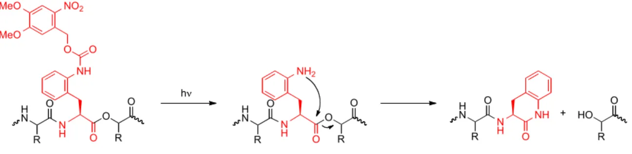
6.3.2 Strategies based on a photocaged aniline and the chemistry of the NPE protecting group
Two additional strategies for cleaving peptide and protein backbones were also considered. The first was based on an nitroveratryloxycarbonyl (NVOC) caged aniline (17) in which the key cleavage reaction is an intramolecular cyclization to afford a δ- lactam (Figure 6.3). Incorporation of an α-hydroxy acid at the i+1 residue yields a strong alkoxide leaving group. The motivation for this strategy came during the synthesis of 2
149
in which direct reduction of the precursor compound 7 resulted in intramolecular cyclization (discussed in further detail in Appendix 3).
The second strategy (Figure 6.4) is based the photochemistry of the (2-nitrophenyl)ethyl (NPE) protecting group,40, 41 in which incorporation of the α- hydroxy acid analog of 2-nitrophenylalanine (compound
7) into a protein creates an NPE-protected backbone ester. Irradiation of the protein results in “deprotection” of the ester and cleavage of the protein backbone. This produces two protein fragments– one with a carboxy terminus and another, which is essentially an α,β- unsaturated ketone. We appreciate that the latter product could complicate our studies by serving as a cross-linking agent, but we were still interested in seeing whether this strategy could promote backbone cleavage in vivo especially given that we had already synthesized 7 for other purposes (Scheme 6.2).
Figure 6.3. Proposed photochemical cleavage strategy using caged aniline 17.
Figure 6.4. Proposed photochemical cleavage strategy using α-hydroxy acid 7.
NH
NH O O H O
N
R R
O
NH2
NH O O H O
N
R R
O
NH O NH H O
N R
HO R h! O
O O NO2
MeO MeO
+
N
O O
HN
O O
R HN
R O
O
N
O O
HN
O O
R HN
R HO
O
N
O
O HN
O O
R HN
R
O O
h! -H+
N
O O
HN
O O
R HN
R O
O
H h!
+
HN
H2N O
OH O O2N
O
OMe OMe O2N
HO O
OH
7 17
150
The synthesis of the N-pent-4-enoyl (4PO) derivative of 17 (Scheme 6.5) began with the protection of the α-amino group of L-2-nitrophenylalanine as the N-pent-4-enoyl (4PO) derivative (cleavable by treatment with I2)3, 42, 43 and the carboxylic acid as the t- butyl ester (to prevent intramolecular cyclization) using standard protocols. Afterward, the nitro group was reduced following the protocol used in Scheme 6.2, and the resulting aniline was caged with a photocleavable nitroveratryloxycarbonyl (NVOC) group.
Removal of the t-butyl protecting group gave the desired product 22. In preparation for nonsense suppression experiments, 7 and 22 were activated as cyanomethyl esters and subsequently transesterified according to published protocols33 to yield acylated TQOps’
(opal suppessor tRNA for TGA stop codon), THG73 (amber suppressor tRNA for TAG stop codon), and YFaFs (suppressor tRNA for GGGT frameshift codon) suppressor tRNA.
Scheme 6.5. Synthesis of the N-pent-4-enoyl (4PO) derivative of 17.
6.3.2.1 Nonsense suppression experiments with 7 and 17 in Xenopus ooctyes
Given that the caged aniline-based strategy described in Figure 6.3 requires the incorporation of two adjacent unnatural amino acids (17 and an α-hydroxy acid in the i+1
O2N
NH O
OH O
O2N
H2N O
OH
O O O
TEA, THF, H2O (87% yield)
O2N
NH O
O O
O CCl3
NH H2N
NH O O NaBH4, Pd-C O
HN
NH O
O O NVOC-Cl
Na2CO3
O O2N
O
OMe OMe
HN
NH O
OH O
O O2N
O
OMe OMe BF3-Et2O
cyclohexane THF (80% yield)
MeOH, THF (84% yield)
CH2Cl2 (68% yield)
TFA CH2Cl2 (89% yield)
18 19 20
21 22
151
position), it seemed prudent to find an appropriately tolerant site in the N-terminal region of the ShB protein (in order to make use of the ShB to ShIR cleavage phenotype).
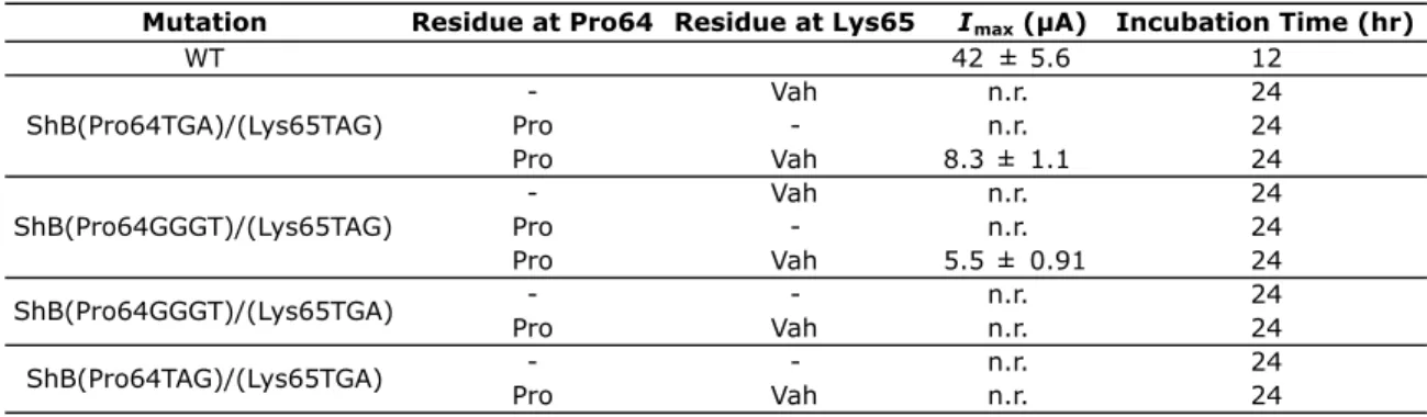
Several combinations of stop codons and the four-base codon were used in initial screens of Pro64 (a site that Npg was incorporated at) and the i+1 residue Lys65 (Table 6.1). Pro and Vah (valine, α-hydroxy) were used as indicators of the maximal current values that could be expected for nonsense suppression experiments with each codon combination. Unacylated suppressor tRNA (represented in Table 6.1 as a dash (-)) was also used as a misacylation control. As shown in Table 6.1, only the Pro65TGA/Lys65TAG and Pro65GGGT/Lys65TAG combinations gave current in these experiments, and no current was seen in misacylation control experiments after a 24 hr of incubation period. To our knowledge, this is the first example of double nonsense suppression at sites that are adjacent in sequence.
Table 6.1. Current (Imax) obtained for control studies of Pro64 and Lys65 in ShB using different codon combinations. “n.r.” stands for no response. A dash (-) represents a misacylation control experiment. “WT” is the wild-type ShB protein.
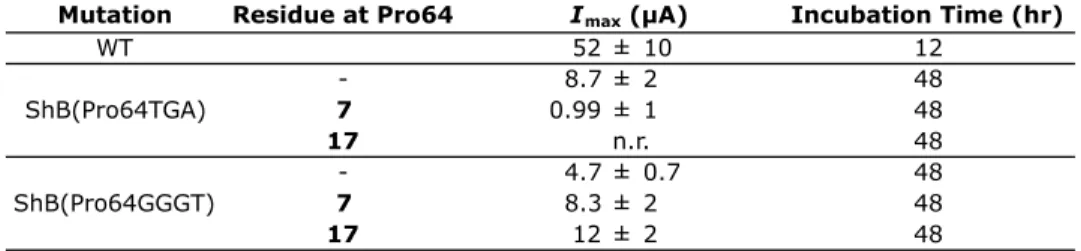
Given these results, we then incorporated 7 and 17 at Pro64 using the TAG stop codon or GGGT frameshift codon. After 48 hrs of incubation, current was seen for proteins injected with 7 and 17, but the Imax values were on the order of what was seen for misacylation control experiments (Table 6.2), which questioned the fidelity of these
Mutation Residue at Pro64 Residue at Lys65 Incubation Time (hr)
WT 42 ± 5.6 12
- Vah n.r. 24
Pro - n.r. 24
Pro Vah 8.3 ± 1.1 24
- Vah n.r. 24
Pro - n.r. 24
Pro Vah 5.5 ± 0.91 24
- - n.r. 24
Pro Vah n.r. 24
- - n.r. 24
Pro Vah n.r. 24
Imax (µA)
ShB(Pro64TGA)/(Lys65TAG)
ShB(Pro64GGGT)/(Lys65TAG)
ShB(Pro64GGGT)/(Lys65TGA) ShB(Pro64TAG)/(Lys65TGA)
152
experiments. Irradiation of oocytes injected with 7 and 17 had no effect on the inactivation of the ShB currents, suggesting that these residues cannot cleave the peptide backbone or that 7 and 17 did not actually incorporate into the protein and the currents observed are the result of misacylation of the suppressor tRNA (incorporation of a natural amino acid at the Pro64 site).
Table 6.2. Current (Imax) obtained for studies with 7 and 17 at Pro64 in ShB. “n.r” stands for no response. A dash (-) represents a misacylation control experiment. Note that no current was seen after 24 hours for 7 or 17. “WT” is the wild-type ShB protein.
We then repeated these experiments with 17 at Pro64 and Vah at Lys65 (to give a better leaving group for the cleavage reaction). Unfortunately, no current was seen for these experiments for reasons that we do not presently understand. Future work could revisit these studies by trying different residues in the ShB protein or a different α- hydroxy acid at the i+1 residue.
During the course of our studies, Schultz and co-workers reported a photochemical cleavage strategy that is nearly identical to Figure 6.4 and is based on the amino acid derivative of 7 (2-nitrophenylalanine).8 They showed that 2- nitrophenylalanine could promote backbone cleavage of model peptides and also of a model protein, T4 lysozyme, expressed in E. coli, albeit with only 30% photochemical cleavage efficiency of the latter system. These findings are certainly encouraging (if not
Mutation Residue at Pro64 Incubation Time (hr)
WT 52 ± 10 12
- 8.7 ± 2 48
7 0.99 ± 1 48
17 n.r. 48
- 4.7 ± 0.7 48
7 8.3 ± 2 48
17 12 ± 2 48
Imax (µA)
ShB(Pro64TGA) ShB(Pro64GGGT)
153
overlapping) and so it would be interesting to revisit the studies of 7 to determine the origin of our photolysis problems.
6.4 DISCUSSION
In summary, we first described novel chemistry in which a photochemically-liberated selenide undergoes an intramolecular SN2 reaction, cleaving an ester that results from the incorporation of the α-hydroxy acids 1 or 2 into a peptide. These studies suggest that for chemical biology applications, aryl selenide 2 is the preferred substrate. Two side reactions have been discovered that must be considered in possible chemical biology applications: dimerization and oxidation. For studies involving peptides prepared by SPPS, adding DTT and controlling the concentration might be appropriate. For in vivo nonsense suppression expression experiments, where protein concentrations are typically low, dimerization may be less likely. In addition, the reducing conditions inside cells likely will discourage dimerization and oxidation. Unfortunately, our initial efforts to see backbone cleavage by 1 or 2 in vivo and in vitro have been unsuccessful, but additional studies should be conducted to determine whether other applications of this methodology are possible.
We also described two alternative strategies for photochemical protein cleavage that were inspired by chemistry we encountered during our synthesis of 2. These strategies are based on (1) intramolecular cyclization and backbone cleavage by a photochemically-liberated aniline nucleophile and (2) the photochemistry of (2- nitrophenyl)ethyl derivatives. These new unnatural amino acids were successfully synthesized, but photolysis studies in proteins expressed in Xenopus oocytes have been unsuccessful thus far.
154 6.5 EXPERIMENTAL SECTION
Chemical synthesis. All reactions were performed at ambient temperature and pressure unless otherwise noted. All reactions involving potentially air-sensitive compounds were conducted under an inert atmosphere using Schlenk techniques. Solvents were purified by passage through alumina. Unless otherwise noted, all chemicals and reagents were used as received without further purification. Compounds 3,25 4,26 and 730 were prepared according to published protocols. The syntheses of 1 and 12 were performed by Dr. Amy L. Eastwood23 and Dr. Niki M. Zacharias24 and are described in their respective theses and also in Eastwood et al.44Flash chromatography was performed with EMD silica gel 60 (particle size 0.040-0.063 mm). Thin-layer chromatography (TLC) was performed using EMD silica gel 60 F254 precoated plates (0.25 mm) and visualized by UV light, potassium permanganate, ceric ammonium molybdate, or ninhydrin. Nuclear magnetic resonance spectroscopy (NMR) was performed on either a Varian Mercury 300 or a Varian Inova 500 instrument and spectra resonances are assigned relative to Me4Si (δ 0.0) or CD3OD (δ 3.31 for 1H NMR and δ 49.1 for 13C NMR). Data for 1H NMR spectra are reported as follows: chemical shift (δ ppm), integration, multiplicity, and coupling constant (Hz). Data for 13C NMR spectra are reported as chemical shift (δ ppm). High- resolution mass spectrometry (HRMS) spectra were obtained from the Caltech Mass Spectrometry Lab. Electrospray ionization mass spectrometry (ESI-MS) used to analyze the proteolysis reactions was performed using an LCQ Classic ion trap (ThermoFinnigan) in direct infusion mode. A uranium glass absorption sleeve was prepared by the Caltech Glassblowing Shop.
155
Synthesis of di-tert-butyl-protected nitrophenylalanine derivative (8) and tert-butyl- protected nitrophenylalanine derivative (where the secondary alcohol is not protected) (8’). α-Hydroxy acid 7 (0.200 g, 0.947 mmol, 1 eq) was
placed in a 2-neck round-bottom flask under Ar (g) and dissolved in THF (3 mL) and cyclohexane (3.5 mL). A solution of tert-butyl- 2,2,2-trichloroacetimidate (0.678 mL, 3.79 mmol, 4 eq) in cyclohexane (3.5 mL) was added simultaneously with boron
trifluoride diethyletherate (0.041 mL, 0.33 mmol, 0.35 eq). The reaction stirred for 45 minutes until it was quenched with saturated NaHCO3 (aq). The organics were extracted with Et2O (3×), washed with brine, dried over MgSO4, and concentrated. The resulting white sludge was suspended in hexanes, filtered, and then purified by flash column chromatography (15% EtOAc in hexanes) to afford two fractions: compound 8 (0.0550 g, 0.170 mmol, 18% yield) and compound 8’ (0.101 g, 0.379 mmol, 40% yield) as clear oils. Rf of 8 = 0.71 (30% EtOAc in hexanes); 1H NMR of 8 (300 MHz, CDCl3, 298 K) δ 7.91 (1H, d, J = 8.0 Hz), 7.50 (1H, m), 7.37 (2H, m), 4.14 (1H, dd, J = 8.9, 4.9 Hz), 3.29 (1H, dd, J = 13.2, 4.9 Hz), 3.13 (1H, dd, J = 13.2, 9.1 Hz) 1.39 (9H, s), 0.95 (9H, s); 13C NMR of 8 (75 MHz, CDCl3, 298 K) δ 172.9, 149.9, 134.5, 132.9, 132.6, 127.9, 124.7, 81.2, 75.2, 71.8, 37.7, 28.0, 27.6; HRMS (FAB) of 8 m/z calc’d for C17H26NO5 [M+H]:
324.1811, found 324.1820. Rf of 8’ = 0.44 (30% EtOAc in hexanes); 1H NMR of 8’ (300 MHz, CDCl3, 298 K) δ 7.90 (1H, d, J = 8.1 Hz), 7.51 (1H, m), 7.40 (2H, m), 4.34 (1H, dd, J = 8.1, 4.1 Hz), 3.48 (1H, dd, J = 13.8, 4.3 Hz), 3.13 (1H, dd, J = 13.9, 8.3 Hz), 1.42 (9H, s); 13C NMR of 8’ (75 MHz, CDCl3, 298 K) δ 173.5, 150.1, 133.4, 133.0, 132.4, 128.1, 125.0, 83.3, 70.7, 37.5, 28.1; HRMS (FAB) of 8’ m/z calc’d for C13H18NO5
O2N
HO O
O 8'
156
[M+H]: 268.1185, found 268.1193. An enantiomeric excess of 94% was obtained for 8’
by analytical chiral HPLC analysis using a Chiralcel OD-H column (4.6 mm × 25 cm) from Daicel Chemical Industries, Ltd. with 2% isopropyl alcohol in hexanes.
Synthesis of aniline (9). Di-tert-butyl-protected nitrophenylalanine derivative 8 (0.606 g, 1.87 mmol, 1 eq) was placed in a 2-neck round-bottom flask under Ar (g) and dissolved in THF (30 mL) and MeOH (15 mL) at 0 °C. To this was added 10 wt % palladium on activated carbon (0.050 g) and sodium borohydride (0.141 g, 3.74 mmol, 2 eq). The reaction was followed by TLC using ninhydrin staining. The solution was stirred for 40 minutes and was then quenched with water and filtered through a pad of Celite™. The filtrate was extracted with Et2O (3×), washed with brine, dried over MgSO4, and concentrated to afford a pale yellow oil. The resulting liquid was purified by flash column chromatography (15% EtOAc in hexanes) to afford aniline 9 as a pale yellow oil (0.493 g, 1.68 mmol, 90% yield). Rf = 0.59 (30% EtOAc in hexanes); 1H NMR (300 MHz, CDCl3, 298 K) δ 7.27 (1H, d, J = 8.9 Hz), 7.17 (1H, t, J = 7.4 Hz), 7.04 (1H, d, J = 7.3 Hz), 6.83 (1H, t, J = 7.3 Hz), 3.99 (1H, dd, J = 7.8, 2.3 Hz), 2.96 (1H, dd, J = 13.8, 10.4 Hz), 2.68 (1H, dd, J = 13.8, 9.1 Hz), 1.46 (9H, s), 0.98 (9H, s); 13C NMR (75 MHz, CDCl3, 298 K) δ 173.5, 149.9, 130.3, 127.8, 123.8, 121.0, 114.2, 81.6, 75.6, 74.8, 35.5, 28.0, 27.4; HRMS (ESI) m/z calc’d for C17H27NO3 [M+H]: 294.2069, found 294.2058.
Synthesis of selenocyanate (10). Aniline 9 (0.248 g, 0.845 mmol, 1.0 eq) was placed in a round-bottom flask, dissolved by sonication in AcOH (17 mL), and cooled to 0 °C. To this solution was quickly added 3 M sodium nitrite (0.34 mL, 1.02 mmol, 1.21 eq) via
157
staining. The pH of the solution was then increased to ~6 by the addition of 10 wt % NaOH. To this was added potassium selenocyanate (0.366 mL, 2.54 mmol, 4.2 eq) in H2O (36 mL) and the solution stirred for an additional 30 min. The organics were extracted with Et2O (3×), washed with brine, filtered through a pad of Celite™, dried over MgSO4, and concentrated. The resulting orange solid was purified by flash column chromatography (20% EtOAc in hexanes) to afford selenocyanate 10 as a yellow, non- crystalline solid (0.162 g, 0.423 mmol, 50% yield). Rf = 0.83 (40% EtOAc in hexanes);
1H NMR (300 MHz, CDCl3, 298 K) δ 7.82 (1H, d, J = 7.6 Hz), 7.26 (3H, m), 3.82 (1H, dd, J = 8.7, 4.7 Hz), 3.06 (2H, m, J = 9.48, 4.53 Hz), 1.46 (9H, s), 0.92 (9H, s); 13C NMR (75 MHz, CDCl3, 298 K) δ 172.4, 139.2, 134.0, 131.1, 129.6, 128.7, 127.6, 105.2, 81.5, 76.2, 73.1, 40.3, 28.0, 27.4; HRMS (ESI) m/z calc’d for C18H25NO3Se [M+H]: 384.1078, found 384.1073.
Synthesis of tert-butyl-protected arylalkyl selenide (11). Selenocyanate 10 (0.161 g, 0.421 mmol, 1 eq) was placed in a 2-neck round-bottom flask under Ar (g) and dissolved in THF (7 mL) at 0 °C. To this was added a solution of sodium borohydride (0.019 g, 0.505 mmol, 1.2 eq) in EtOH (1 mL) and the solution stirred for 1 hr. A solution of 2- nitrobenzylbromide (0.118 g, 0.547 mmol, 1.3 eq) in THF (5.5 mL) was then added.
After this addition, the solution was allowed to warm to room temperature and was stirred for 3 hours until it was quenched with H2O. The organics were extracted with Et2O (3×), washed with brine, filtered through a pad of Celite™, dried over MgSO4, and concentrated. The resulting solid was purified by flash column chromatography (15%
EtOAc in hexanes) to afford tert-butyl protected arylalkyl selenide 11 as a yellow, non- crystalline solid (0.145 g, 0.295 g, 70% yield). Rf = 0.65 (30% EtOAc in hexanes); 1H
158
NMR (300 MHz, CDCl3, 298 K) δ 8.00 (1H, d, J = 7.2 Hz), 7.45 (1H, d, J = 7.1 Hz), 7.35 (2H, m), 7.21 (2H, m), 7.09 (1H, m), 6.99 (1H, m), 4.33 (2H, s), 4.15 (1H, m), 3.16 (1H, dd, J = 9.90, 4.53 Hz), 2.96 (1H, dd, J = 8.24, 5.36 Hz), 1.43 (9H, s); 13C NMR (75 MHz, CDCl3, 298 K) δ 173.2, 141.2, 136.2, 135.5, 133.2, 132.1, 131.8, 130.9, 128.3, 128.0, 127.6, 125.6, 80.9, 74.9, 72.8, 40.9, 30.0, 28.0, 27.7, 22.4; HRMS (FAB) m/z calc’d for C24H31NO5Se [M+]: 493.1367, found 493.1374.
Synthesis of arylalkyl selenide α-hydroxy acid (2). tert-Butyl protected arylalkyl selenide 11 (0.533 g, 1.08 mmol) was placed in a 2-neck round-bottom flask under Ar (g) and dissolved in CH2Cl2 (8 mL). To this was added trifluoroacetic acid (4 mL) and the solution stirred for 16 hours. The reaction mixture was concentrated to afford a yellow- orange solid, which was triturated with ether. The resulting yellow solid was dissolved in EtOAc (3×), rinsed with 1 N HCl, rinsed with H2O, and then added to 5% NaHCO3. The organic layer (colorless) was discarded, and the pH of the aqueous layer was decreased to pH 2 by the addition of 6 N HCl. The organics were extracted into EtOAc (3×), rinsed with brine, dried over MgSO4, and concentrated to afford arylalkyl selenide α-hydroxy acid 2 as a yellow, non-crystalline solid (0.195 g, 0.512 mmol, 96% yield). [α]24 D =
−45.3° (c = 1, CHCl3); 1H NMR (300 MHz, CDCl3, 298 K) δ 7.99 (1H, d, J = 6.6 Hz), 7.47 (1H, d, J = 7.7 Hz), 7.37 (2H, m), 7.28 (2H, m), 7.14 (1H, m), 7.01 (1H, d, J = 6.9 Hz), 4.35 (3H, m), 3.30 (1H, dd, J = 13.7, 4.40 Hz), 3.03 (1H, dd, J = 13.2, 8.5 Hz); 13C NMR (75 MHz, CDCl3, 298 K) δ 178.3, 147.8, 139.9, 136.9, 135.2, 133.4, 132.2, 131.0, 130.7, 129.1, 128.2, 128.1, 125.7, 71.2, 40.4, 30.3; HRMS (FAB) m/z calc’d for C16H15NO5Se [M+H]: 382.0194, found 382.0191.
159
Synthesis of aryl selenide-glutamate hydroxy-peptide (13). Arylalkyl selenide α- hydroxy acid 2 (0.091 g, 0.239 mmol, 1 eq), di-tert-butyl-L-glutamate (0.106 g, 0.359 mmol, 1.5 eq), and benzotriazol-1-yl-oxytripyrrolidinophosphonium hexafluorophosphate (0.149 g, 0.287 mmol, 1.2 eq) were placed in a round-bottom flask under Ar (g) and dissolved in CH2Cl2 (1.8 mL). To this was added N-methylmorpholine (0.09 mL, 0.790 mmol, 3.3 eq) via syringe, and the reaction stirred for 24 hours. The reaction mixture was then diluted with EtOAc, and the resulting solution was washed with 1 M KHSO4 (2×), H2O, 5% NaHCO3 (2×), and brine. The organic layer was passed through a pad of Celite™, dried over MgSO4, and concentrated. The resulting solid was purified by flash column chromatography (30% EtOAc in hexanes) to afford hydroxy-peptide 13 as a yellow oil (0.134 g, 0.215 mmol, 90% yield). Rf = 0.49 (50% EtOAc in hexanes); 1H NMR (300 MHz, CDCl3, 298 K) δ 7.98 (1H, d, J = 7.4 Hz), 7.45 (1H, d, J = 7.4 Hz), 7.36 (2H, m), 7.26 (2H, m), 7.10 (1H, m), 6.99 (1H, m), 4.46 (1H, m), 4.25 (1H, m), 3.30 (1H, dd, J = 14.0, 3.6 Hz), 2.96 (2H, m), 2.14 (3H, m), 1.86 (1H, m), 1.45 (18H, s); 13C NMR (75 MHz, CDCl3, 298 K) δ 172.7, 172.2, 171.0, 148.0, 140.9, 136.9, 135.0, 133.4, 132.2, 131.1, 130.9, 129.2, 128.3, 128.0, 125.7, 82.6, 80.9, 72.9, 52.1, 41.1, 31.6, 30.3, 28.3, 28.2, 27.9; HRMS (FAB) m/z calc’d for C29H38N2O8Se [M+H]: 623.1871, found 623.1881.
Synthesis of tryptophan-aryl selenide-glutamate depsipeptide (14). Hydroxy-peptide 13 (0.140 g, 0.225 mmol, 1 eq), Nα-(tert-butoxycarbonyl)-L-tryptophan (0.137 g, 0.450 mmol, 2 eq), 4-(dimethylamino)pyridine (0.014 g, 0.113 mmol, 0.5 eq), and N,N’- dicyclohexylcarbodiimide (0.093g, 0.450 mmol, 2 eq) were placed in a round-bottom flask under Ar (g) and dissolved in CH2Cl2 (5 mL). After stirring for 48 hours, the
160
reaction mixture was diluted with EtOAc. The solution was washed with 1 M KHSO4
(2×), H2O, 5% NaHCO3 (2×), and brine. The organic layer was filtered through a pad of Celite™, dried over MgSO4, and concentrated. The resulting solid was purified by flash column chromatography (35% EtOAc in hexanes) to afford depsipeptide 14 as a pale yellow, non-crystalline solid (0.0817 g, 0.0900 mmol, 40% yield). Rf = 0.34 (50%
EtOAc in hexanes); 1H NMR (500 MHz, CDCl3, 298 K) δ 8.52 (1H, b), 7.98 (1H, m), 7.57 (1H, d, J = 7.9 Hz), 7.41 (1H, d, J = 7.7 Hz), 7.34 (3H, m), 7.18 (3H, m), 7.10 (2H, m), 7.02 (1H, s), 6.91 (1H, m), 5.25 (1H, m), 4.98 (1H, d, J = 7.5 Hz), 4.57 (1H, dd, J = 12.6, 5.3 Hz), 4.41 (1H, m), 4.24 (2H, s), 3.17 (2H, m), 2.20 (1H, m), 2.08 (2H, m), 1.79 (1H, m), 1.48 (9H, s), 1.45 (9H, s), 1.31 (9H, s). 13C NMR (125 MHz, CDCl3, 298 K) δ 172.4, 171.2, 169.0, 156.0, 148.0, 139.7, 136.7, 136.4, 135.1, 133.3, 132.1, 131.3, 131.0, 129.0, 128.2, 128.1, 128.0, 125.7, 123.9, 122.3, 122.3, 119.8, 118.9, 111.5, 109.6, 82.3, 80.9, 80.1, 75.1, 54.6, 52.4, 37.7, 34.2, 31.8, 30.3, 28.5, 28.4, 28.2, 27.6; HRMS (ESI) m/z calc’d for C45H56N4O11Se [M+H]: 909.3229, found 909.3238.
Synthesis of selenacyclopentane (15). Depsipeptide 12 (0.0722g, 0.088 mmol, 1 eq) was placed in a pyrex reaction vessel and dissolved in acetonitrile (125 mL). To this was added dithiothreitol (1.36 g, 8.8 mmol, 100 eq) and 20 mM phosphate buffer, pH 7.6 (125 mL). The resulting solution was stirred under N2 (g), and a 450 W medium-pressure mercury-vapor UV immersion lamp (ACE Glass), filtered with a pyrex glass absorption sleeve and equipped with a water cooling jacket, was assembled and attached to the reaction vessel. The progress of the reaction was followed by ESI-MS. After 1 hour of photolysis (at which point the temperature of the reaction had increased from 25 ºC to 32 ºC) the two major m/z ratios seen in the mass spectrum of the reaction were the
161
depsipeptide 12 ([M+Na+] = 844 m/z) and the nitrobenzyl-deprotected selenol ([M+Na+]
= 709 m/z). After 5 hours of photolysis, the m/z ratio attributed to 12 ([M+Na+] = 844 m/z) had diminished to a negligible level while the m/z ratios corresponding to the nitrobenzyl-deprotected selenol ([M+Na+] = 709 m/z), the nitrobenzyl-deprotected diselenide ([M+Na+] = 1391 m/z), the olefin-containing depsipeptide derived from the oxidative elimination of the selenium ([M+Na+] = 627 m/z), and the desired selenacyclopentane 15([M+Na+] = 444 m/z) persisted. At this time, the reaction was removed from the photoreactor, heated to 70 ºC, and monitored by ESI-MS. After 3 hours of heating at this temperature, both nitrobenzyl-deprotection products (m/z ratios 709 and 1391) were no longer detectable, but the m/z ratio attributed to the desired selenacyclopentane 15 ([M+Na+] = 444 m/z) and a m/z ratio corresponding to its dimer ([M+Na+] = 865 m/z) remained. The resulting mixture was extracted with EtOAc (2×), dried over MgSO4, and concentrated. The crude product was purified twice by flash column chromatography (both began with 11% EtOAc in hexanes then were changed to 33% EtOAc in hexanes after the DTT eluted, both were dry loaded in CH2Cl2) to afford selenacyclopentane 15 as a yellow oil (0.0107 g, 0.0255 mmol, 29% yield). Rf = 0.28 (33% EtOAC in hexanes); [α]24 D = −16.0° (c = 1, CHCl3); 1H NMR (500 MHz, CDCl3, 298 K) δ 7.08 (1H, d, J = 13.5 Hz), 4.44 (1H, dt, J = 13.5, 8 Hz), 4.08 (1H, m), 3.15 (1H, m), 2.97 (1H, m), 2.37 (1.5H, m), 2.26 (1.5H, m), 2.14 (4H, m), 1.92 (1H, m), 1.47 (9H, s), 1.44 (9H, s); 13C NMR (125 MHz, CDCl3, 298 K) δ 172.3, 172.1, 170.8, 82.3, 80.7, 52.845.4, 37.4, 32.4, 31.5, 28.1, 28.0, 27.5, 27.1. HRMS (TOF) m/z calc’d for C18H31NO5Se [M+H]: 422.1446, found 422.1465.
162
Synthesis of aryl selenacyclopentane (16). Depsipeptide 14 (0.0230 g, 0.0250 mmol, 1 eq) was placed in a pyrex reaction vessel and dissolved in acetonitrile (125 mL). To this was added dithiothreitol (0.0390 g, 0.250 mmol, 10 eq) and pH 8 H2O (125 mL). The resulting solution was stirred under N2 (g), and a 450 W medium-pressure mercury-vapor UV immersion lamp (ACE Glass), filtered with a uranium glass absorption sleeve and equipped with a water cooling jacket, was assembled and attached to the reaction vessel.
The progress of the reaction was followed by ESI-MS. After 1 hour of photolysis, three m/z ratios were seen in the mass spectrum of the reaction: the depsipeptide 14 ([M+Na+]
= 931 m/z), the nitrobenzyl deprotected aryl selenol ([M+Na+] = 796 m/z), and the desired aryl selenacyclopentane 16 ([M+Na+] = 492 m/z). No m/z ratio corresponding to the nitrobenzyl deprotected diselenide ([M+Na+] = 1567 m/z) was observed. After 2 hours, both m/z ratios attributed to 14 ([M+Na+] = 931 m/z) and the nitrobenzyl-deprotected aryl selenol ([M+Na+] = 796 m/z) had diminished to a negligible level while a m/z ratio corresponding to the desired aryl selenacyclopentane 16 ([M+Na+] = 492 m/z) persisted.
At this time, the reaction was removed from the photoreactor. The acetonitrile was then removed in vacuo, and the resulting aqueous solution was extracted with Et2O (3×), washed with brine, dried over MgSO4, and concentrated. The resulting yellow oil was purified by flash column chromatography (15% EtOAC in hexanes) to afford aryl selenacyclopentane 16 as a pale yellow oil (0.00843 g, 0.0180 mmol, 72% yield). Rf = 0.59 (30% EtOAC in hexanes); [α]24 D = −80.5° (c = 1, CHCl3); 1H NMR (300 MHz, CDCl3, 298 K) δ 7.30 (1H, m), 7.20 (1H, m), 7.12 (2H, m), 7.29 (2H, m), 6.92 (1H, d, J = 7.41 Hz), 4.50 (1H, dd, J = 8.2, 5.2 Hz), 4.43 (1H, dd, J = 7.8, 4.7 Hz), 2.25 (2H, m), 2.15 (1H, m), 1.91 (1H, m), 1.44 (9H, s), 1.41 (9H, s); 13C NMR (75 MHz, CDCl3, 298
163
K) δ 172.4, 171.4, 170.7, 141.7, 135.3, 128.0, 125.9, 125.7, 125.5, 82.6, 81.0, 53.1, 45.9, 41.7, 31.6, 29.9, 28.3, 28.1; HRMS (ESI) m/z calc’d for C22H31NO5Se [M+H]: 470.1446, found 470.1469.
Synthesis of the cyanomethyl ester of 2. Compound 2 (0.12 g, 0.032 mmol, 1 eq) was added to a 20 mL scintillation vial. To this was added chloroacetonitrile (0.10 mL, 1.6 mmol, 50 eq) and triethylamine (0.013 mL, 0.095 mmol, 3 eq). The resulting mixture stirred for 6 hours. Then 10 mL of H2O was added and the organics were extracted with CH2Cl2 (3×), washed with brine, dried over Na2SO4, concentrated, and then purified by flash column chromatography (50% EtOAc in hexanes) to afford the cyanomethyl ester of 2 as a yellow oil (0.010 g, 0.024 mmol, 76% yield). Rf = 0.40 (50% EtOAc in hexanes); 1H NMR (300 MHz, CDCl3, 298 K) δ 8.01 (1H, d, J = 9.5 Hz), 7.52 (1H, d, J
= 7.6 Hz), 7.34 (3H, m), 7.22 (1H, m), 7.17 (1H, m), 6.97 (1H, m), 4.77 (2H, b), 4.34 (3H, b), 3.16 (1H, dd, J = 13.7, 4.5 Hz), 2.94 (1H, dd, J = 13.8, 8.5 Hz), 2.64 (1H, d, J = 5.6 Hz) ; 13C NMR (75 MHz, CDCl3, 298 K) δ 172.7, 139.6, 137.2, 135.1, 133.3, 132.0, 132.0, 130.9, 130.6, 129.2, 128.3, 128.3, 125.7, 114.0, 71.1, 49.1, 40.6, 30.5; HRMS (FAB+) m/z calc’d for C18H17N2O5Se [M+H]: 421.0303, found 421.0303. A similar protocol was used to synthesize the cyanomethyl ester of 1 and is described in the theses of Dr. Amy L. Eastwood23 and Dr. Niki M. Zacharias.24
Synthesis of 4PO-protected nitrophenylalanine 18. L-2-nitrophenylalanine (CSPS Pharmaceuticals) (1.5 g, 7.1 mmol, 1 eq) was added to a 20 mL round-bottom flask under Ar (g) and suspended in 20 mL of THF and 10 mL of H2O. The resulting suspension was placed in an ice bath. To this was added triethylamine (2.1 mL, 15 mmol), 2.1 eq). Pent- 4-enoic anhydride (1.5 mL, 8.5 mmol, 1.2 eq) was then added dropwise. After 1 hour of
164
stirring, an addition 0.5 mL of triethylamine (3.6 mmol) and 0.5 mL of pent-4-enoic anhydride (2.8 mmol) was added. The solution was stirred for 1 more hour and then 50 mL of 0.2 M NaHSO4 was added. The pH of the resulting solution was increased to pH 9 by addition of 2M NaOH. The organics were extracted with Et2O and discarded. The pH of the aqueous layer was then decreased to pH 2 via addition of 6 M HCl. The organics were extracted with EtOAc (3×), washed with 0.2 M NaHSO4, washed with brine, dried over Na2SO4, filtered, and concentrated. The resulting yellow liquid was purified by flash column chromatography (0.9% formic acid and 32% hexanes in EtOAc) to afford 4PO- protected nitrophenylalanine 18 as a yellow oil (1.8 g, 6.2 mmol, 87% yield). Rf = 0.46 (0.9% formic acid and 32 % hexanes in EtOAc); 1H NMR (300 MHz, CDCl3, 298 K) δ 9.92 (1H, b), 7.95 (1H, d, J = 8.2 Hz), 7.59 (1H, m), 7.46 (2H, m), 5.69 (1H, m), 4.98 (3H, m), 3.62 (1H, dd, J = 20, 6.8 Hz), 3.34 (1H, dd, J = 24, 9.4 Hz) 2.19 (2H, m); 13C NMR (75 MHz, CDCl3, 298 K) δ 174.1, 173.7, 149.7, 136.4, 133.3, 132.8, 131.5, 128.3, 124.9, 115.8, 53.0, 35.3, 34.3, 29.2.
Synthesis of 4PO- t-butyl-protected nitrophenylalanine 19. Compound 18 (0.33 g, 1.1 mmol, 1 eq) was placed in a 2-neck round-bottom flask under Ar (g) and dissolved in THF (5 mL) and cyclohexane (5 mL). A solution of tert-butyl-2,2,2-trichloroacetimidate (0.82 mL, 4.6 mmol, 4 eq) in cyclohexane (5 mL) was added simultaneously with boron trifluoride diethyletherate (0.049 mL, 0.40 mmol, 0.35 eq). The reaction stirred for 1 hour until it was quenched with 20 mL of saturated NaHCO3 (aq). The organics were extracted with Et2O (3×), washed with brine, dried over Na2SO4, and concentrated. The resulting white sludge was suspended in hexanes, filtered, and then purified by flash column chromatography (30% EtOAc in hexanes) to afford 4PO- t-butyl-protected


![New 6-(3-Indolyl)benzo[b]carbazoles](data:image/gif;base64,R0lGODlhAQABAIAAAP///wAAACH5BAEAAAAALAAAAAABAAEAAAICRAEAOw==)