C/EBPBETA3 (LIP) INDUCES CELL DEATH IN BREAST CANCER CELLS
By
Maria M. Abreu
Dissertation
Submitted to the Faculty of the Graduate School of Vanderbilt University
in partial fulfillment of the requirements for the degree of
DOCTOR IN PHILOSOPHY
in Cancer Biology
May, 2012 Nashville, Tennessee
Approved:
Professor Vito Quaranta
Professor Barbara Fingleton
Professor Andries Zijlstra
Professor Linda Sealy
Para mi madre- Lourdes Abreu, gracias por todo tu apoyo.
To my mentor- Linda Sealy, thank you for believing in me.
To all the women that continue the battle and those that have lost their battle to breast cancer.
ACKNOWLEDGEMENTS
I would like to begin by thanking my mentor, role model, and friend Linda Sealy. Words cannot express how grateful I am to have the opportunity to learn from such a brilliant scientist, teacher, and woman. This work could not have been accomplished without her creativity, guidance, support, and relentless enthusiasm for science. It would be impossible to list the many things Linda has taught me regarding life and science. Still, one of the main things I have learned from Linda is the importance of “thinking outside of the box”. I sincerely appreciate all the time Linda has spent mentoring and preparing me for a successful career in science. I am also indebted to Roger Chalkley for his guidance and support throughout my graduate school years. I would like to thank my lab mates and friends Allison Atwood, Linda Bundy, Rachel Jerrell, and Alisha Russell. I would especially like to thank Allison, my lab partner, for her continuous support with science and life matters as well as Rachel, my work wife, for all her mathematic and technical skills. You four have helped me at different stages throughout this process, thank you for all the wonderful memories.
I would like to express my gratitude to my committee members Andries Zijlstra (chair), Vito Quaranta, Barbara Fingleton, and Mike Thomson - for their help with reagents, suggestions, and questions that have strengthened my project. Thank you as well to Cathy Alford of the VA flow cytometry core for her technical support, as well as the Vanderbilt University cores. I would like to thank the staff of the Department of Cancer Biology, especially Tracy Tveit. Thank you to the staff members of the BRET/IGP office and Vanderbilt University.
I was honored and very grateful for the various sources of funding I have been awarded throughout my graduate studies. The Virus, Nucleic Acid, and Cancer (VNAC) training grant at Vanderbilt University funded my first two years.
The NIH Ruth L. Kirschstein National Research Service Award GM089106 has funded the remaining years of my graduate studies. We are very grateful to the Vanderbilt Ingram Cancer Center (VICC) for core scholarship funds and the Vanderbilt Institute for Clinical and Translational Research award that also funded some of this study.
I am very fortunate to have a long list of friends that have made this an amazing experience. Some have been with me from the start of IGP (Mohammed Aiyegbo, Mike Lindquist, Veronica Placencio, Thomas Tomasiak), while others have been placed in my path along the way (Freddie Pruitt, Lisa McCorvey, Maggie Manning). I would like to thank my entire Nashville family for all their support, encouragement, patience, and willingness to listen to all of my life and science problems. Thank you for being there through thick and thin. I would also like to thank my long-time college friend Christina Stujenske, without her I would not have applied to Vanderbilt University. My sincerest thanks to my best friend, Trenis Palmer, for all his support, patience, and love throughout this entire graduate school process. I will always cherish the many memories we have shared in Nashville.
Finally, I would like to thank my family- Lourdes, Jorge, Frank, and Mario for all their unconditional love and support, as well as my extended family.
Muchas gracias a mi familia tan hermosa. My mother is an incredible and
intelligent woman, who has worked hard to make sure that I succeed in life. She has instilled in me some of the most important things required to succeed as a scientist: hard-work, optimism, and perseverance.
TABLE OF CONTENTS
DEDICATION……….…ii
ACKNOWLEDGEMENTS………...iii
LIST OF FIGURES………..ix
LIST OF TABLES……….xi
LIST OF ABBREVIATIONS………...xii
CHAPTER I. INTRODUCTION……….….1
Breast Cancer overview……….…….1
Different types of programmed cell death………...4
Apoptosis………...4
Autophagic cell death………..7
Necrosis………...11
Entosis and Cell engulfment……….………...13
CCAAT/enhancer-binding protein family……….….…...16
C/EBPbeta………...17
Isoform specific C/EBPbeta expression………...20
Role of C/EBPbeta in the mammary gland……….……...24
C/EBPbeta in breast cancer……….……29
Purpose of this Study……….….. 31
Significance………... 32
II. C/EBPBETA3 (LIP) AND AUTOPHAGY ……….... 34
Introduction……….…34
Materials and Methods……….….36
Adenoviral Constructs and Cell lines……….……. 36
Cell growth and proliferation assays………37
Colony formation assays………...38
Cell Cycle Analysis……….38
Electron Microscopy………...39
Whole cell lysates, cell fractionation, and immunoblot analysis……..39
Indirect immunostaining and image acquisition………..41
Quantification of Acidic Vesicles by Acridine Orange using Flow Cytometry………..42
Statistical analysis………43
Results………43
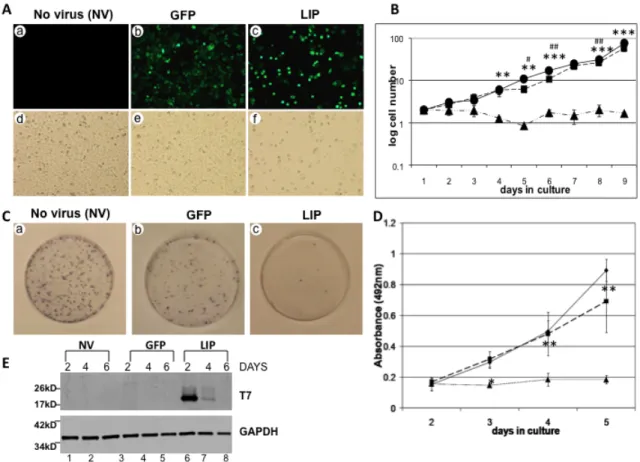
LIP expression attenuates proliferation of the MDA-MB-468 breast cancer cell line……….43
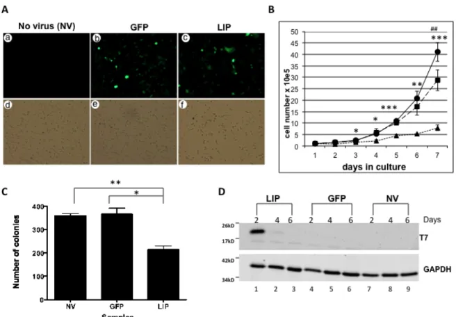
LIP expression attenuates proliferation of the MDA-MB-231 breast cancer cell line……….47
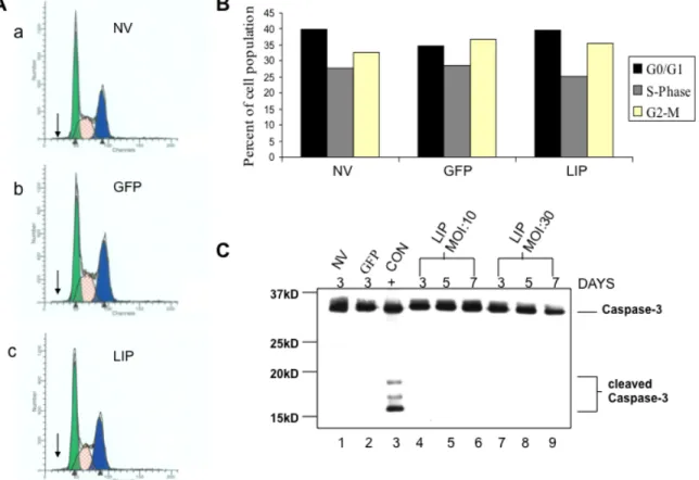
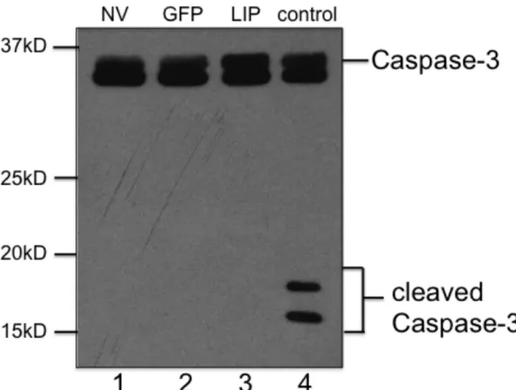
LIP does not block cell cycle progression or induces apoptosis………49
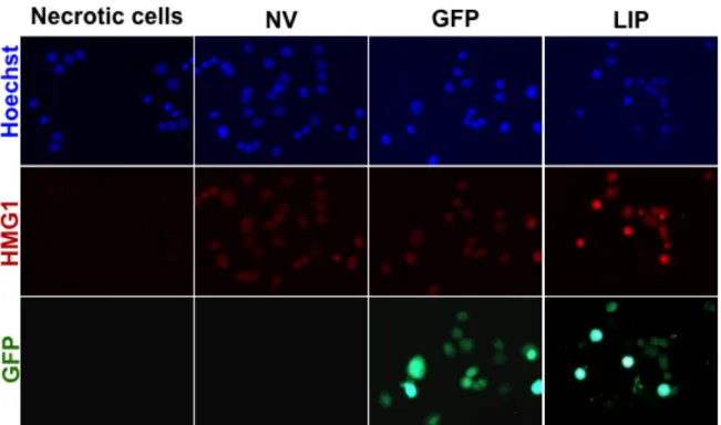
LIP does not induce necrosis……….52
Ultrastructure of autophagic vesicles (AV) formed during LIP-induced autophagy……….54
LIP overexpression leads to increase acidic vesicles……….57
LIP-induced activation of LC3……….57
LIP expression leads to diabetes- and obesity related (DOR) protein translocation………..61
Discussion………..61
III. C/EBPBETA3 (LIP) AND ENGULFMENT………..66
Introduction………...66
Materials and Methods……….…..68
Cell culture and adenoviral constructs……….…...68
Cell cycle analysis………..68
Cell internalization assays………....69
Electron Microscopy………...70
Quantitative cell engulfment assay using flow cytometry……….70
Phosphatidylserine exposure………... 71
Transfections………..………..71
Indirect immunostaining and image acquisition………..72
Time-lapse microscopy………...73
Confocal microscopy………...73
Immunoblot analysis of mouse mammary glands………..74
Statistical analysis………74
Results………74
Cell disintegration following exogenous expression of LIP………74
DNA content of LIP-expressing MDA-MB-468 cells………75
LIP-expressing MDA-MB-468 cells engulf neighboring cells………….77
Ultrastructure analysis of LIP-mediated cell engulfment………81
Quantitation of LIP-mediated cell engulfment………..83
LIP-mediated cell engulfment requires Rho……….85
LIP does not appear to stimulate the “eat me” signal……….85
LIP-mediated cell engulfment is not dependent on adenoviral infection……….87
LIP may play a physiological role during involution of the mammary gland……….90
Discussion……….92
IV. C/EBPBETA3 (LIP) AND TRANSCRIPTIONAL REGULATION……….97
Introduction………...97
Materials and Methods……….…..98
Cell culture and adenoviral constructs……….…...98
Genomic profiling………99
Real time PCR………...99
Whole cell lysates and immunoblot analysis………100
Quantitative cell engulfment assay using flow cytometry…………...102
Results………...102
Genomic profiling of LIP-expressing MDA-MB-231 cells………102
Genomic profiling of LIP-expressing MDA-MB-468 cells………104
HSPA1A mRNA increase is transient………106
HSP70 cellular protein levels do not correlate with HSPA1A mRNA increase……….109
LIP expression leads to increases in exosome secretion………109
LIP-derived exosomes are involved in cell engulfment………113
Discussion……….116
V. SUMMARY AND DISCUSSION……….119
Autophagy and cancer………121
C/EBPbeta3 (LIP) and cancer………...123
C/EBPbeta3 (LIP) and autophagy: explaining the paradox?...123
C/EBPbeta3 (LIP) and cell engulfment………126
Engulfment can be pro- or anti- tumorigenic………...128
C/EBPbeta3 (LIP) and transcriptional regulation………132
C/EBPbeta3 (LIP) and exosome secretion………..133
LIP expression may lead to many cell fates……….136
A possible physiological role for LIP-mediated cell engulfment ………..138
REFERENCES………....141
LIST OF FIGURES
Figure 1. Model of autophagosome formation……….…10
Figure 2. Schematic representation of C/EBPbeta……….…19 Figure 3. Mouse mammary gland development. ………...26 Figure 4. Lobuloalveolar development is compromised in
the C/EBPbeta null mice upon stimulation of pregnancy
with estrogen and progesterone. ………...28 Figure 5. C/EBPbeta isoform expression in mammary samples. ……….. 30 Figure 6. LIP expression attenuates proliferation
of the MDA-MB-468 breast cancer cell line. ………...44 Figure 7. LIP expression attenuates proliferation
of the MDA-MB-231 breast cancer cell line. ………..46 Figure 8. LIP expression attenuates proliferation of
MCF-7 breast cancer cell line………48
Figure 9. LIP does not block cell cycle progression
or induce apoptosis. ………...50 Figure 10. LIP expression does not induce caspase-3 activation. …………...51 Figure 11. LIP does not induce necrosis……….….53 Figure 12. Ultrastructure of autophagic vacuoles (AV)
formed during LIP-induced autophagy. ……….……..55 Figure 13. LIP overexpression leads to increase of
autophagic vacuoles (AV). ………... 56 Figure 14. LIP-induced activation of LC3. ……….………. 58 Figure 15. LIP-induced DOR nuclear to cytoplasmic translocation. …………...60
Figure 16. Cell disintegration following exogenous expression of LIP. ………..76 Figure 17. DNA content of LIP-expressing MDA-MB-468 cells………...78 Figure 18. LIP-expressing MDA-MB-468 cells engulf neighboring cells……....79 Figure 19. Ultrastructure analysis of LIP-mediated cell engulfment
in MDA-MB-468 cells……….….82 Figure 20. Quantification of LIP-mediated cell engulfment………...84 Figure 21. LIP does not appear to stimulate PtdSer exposure
in MDA-MB-468 cells. ………...86 Figure 22. LIP-mediated cell engulfment is not
dependent on adenoviral infection. ………..88 Figure 23. LIP engulfment of neighboring cells via confocal microscopy. …....89 Figure 24. LIP expression in mouse mammary glands. ………...91 Figure 25. Schematic of experimental design. ……….101 Figure 26. LIP induces HSPA1A expression in MDA-MB-231 cells. …………107 Figure 27. LIP induces HSPA1A expression in MDA-MB-468 cells. …………108 Figure 28. HSP70 protein levels in MDA-MB-231 cells. ……….110 Figure 29. LIP expression leads to increased exosome secretion. …………..112 Figure 30. LIP-derived exosomes are involved in cell engulfment. ………..…115 Figure 31. LIP may have dual roles in tumor development and progression. .125 Figure 32. LIP may have dual roles in tumor progression. ……….131 Figure 33. LIP-mediated cell engulfment. ……….134 Figure 34. The many cell fates of LIP-expressing cells………..137
LIST OF TABLES
Table. 1 LIP regulation of genes in MDA-MB-231 cells………103 Table 2. LIP regulation of genes in MDA-MB-468 cells………105
LIST OF ABBREVIATIONS
ACS-American Cancer Society Ad- adenoviral
AGP/EBP- alpha 1-acid glycoprotein enhancer-binding protein ATCC- American Type Culture Collection
ATP-adenosine triphosphate ATG- autophagy-related genes AV-autophagic acidic vesicles BECN1- Beclin 1
BME- 2-mercaptoethanol
BRCA1- breast cancer susceptibility gene 1 BRCA2- breast cancer susceptibility gene 2 BSA- bovine serum albumin
C-terminal- carboxyl-terminal CDK- cyclin-dependent kinase
C/EBPbeta- CCAAT/enhancer-binding protein beta COX-2- cyclooxygenase-2
CRT- calreticulin
CUGBP1- CUG triplet repeat binding protein DAPK- death-associated protein kinase-1 DMEM- Dulbecco’s Modified Eagles’s medium DOR- diabetes- and obesity-regulated
DRAM- damage-regulated autophagy modulator E2F1- E2 factor 1
EDTA- ethylenediaminetetraacetic acid EGF- epidermal growth factor
EGFR- epidermal growth factor receptor eIF- eukaryotic translation initiation factor EM- electron microscope
EMT- epithelial-to-mesenchymal transition ER-endoplasmic reticulum
FACS-fluorescence-activated cell sorting FBS- fetal bovine serum
FOXO- forkhead box O
GFP-green fluorescent protein
HER2/Erb2- human epidermal growth factor receptor 2 HIV- human immunodeficiency virus
HMGB1- high mobility group box 1 HR- homologous recombination HRP- horse radish peroxidase
IGF-IR- insulin-like growth factor I receptor IL- interleukin
IL6/DBP- IL-6 DNA binding protein JNK- c-Jun N-terminal kinase kD- kilodalton
LAP- liver-enriched activator protein LIP- liver-enriched inhibitory protein
LC3- microtuble-associated protein light chain 3 mTOR- mammalian target of rapamycin
MEC- mammary epithelial cells
MMTV/c-neu- mouse mammary tumor virus/ c-neu MOI- multiplicity of infection
mRNA- messenger RNA
MTS- 3-(4,5-dimethylthiazol-2-yl)-5-(3-carboxymethoxyphenyl)-2-(4 sulfophenyl)-2H-tetrazolium
NFDM- nonfat dried milk
NF-IL6- nuclear factor-interleukin 6 NF-κB- nuclear factor kappa B N-terminal- amino-terminal NK- natural killer cells Nec-1- necrostatin-1 NV- no virus
OsO4- osmium tetroxide
PBS- phosphate-buffered saline PCD- programmed cell death
PAGE- polyacrylamide gel electrophoresis PE- phosphatidylethanolamine
PEPCK- gluconeogenic phosphoenolpyruvate carboxykinase
PI3K- class Iphosphatidylinositol 3-kinase PKB- protein kinase B
PLAC1- placenta specific 1 PMS- phenazine methosulfate PtdSer- phosphatidylserine
PTEN- phosphatase and tensin homolog
RANKL- receptor activator of nuclear factor kappa B ligand RIP-1- receptor-interacting protein kinase-1
ROCK- Rho-associated protein kinase ROS- reactive oxygen species
SD- standard deviation SDS- sodium dodecyl sulfate SL- stem-loop
SUMO- sumoylation
TBS-T- Tris Buffered Saline + Tween-20 TEM- transmission electron microscopy THR-Thyroid hormone receptor
TLR- Toll-like receptors
TNFR1- tumor necrosis factor receptor 1 TOR- target of rapamycin
TRAIL-R- TNF-related apoptosis-inducing ligand receptor TSC- tuberous sclerosis
uORF- upstream open reading frame
UTP- uridine triphosphate v/v- volume/volume
WAP- whey acidic promoter WHO- World Health Organization w/v- weight/volume
CHAPTER I
Introduction
Breast cancer overview
Cancer remains a leading cause of death worldwide. According to the latest reports from the World Health Organization (WHO) in 2008, cancer accounted for 7.6 million deaths worldwide. About 70% of all cancer deaths occurred in low- and middle- income countries. Deaths from cancer worldwide are projected to continue to rise to over 13 million in 2030 (Siegel et al., 2011).
In the United States, the American Cancer Society (ACS) estimates approximately 1.7 million new cancer cases to be diagnosed in 2012. Invasive breast cancers diagnosed in women will account for about 226,870 of these new cases (Siegel et al., 2011). Breast cancer is still the most common cancer among American women, with the exception of skin cancers (Siegel et al., 2011).
In fact breast cancer remains the second leading cause of cancer-related death in women. The ACS estimates about 39,510 women will tragically lose their battle against breast cancer in 2012. Women diagnosed with aggressive and/or more advanced forms of breast cancer have a meager 26% five-year survival rate. While the battle against cancer continues, the ACS reports that earlier detection through screening and increased awareness, as well as advances in improved treatment options have significantly improved patient outcomes and survival. Death rates from breast cancer have been declining since about 1990, with larger decreases in women younger than 50. This decline is likely due to
increases in the prevalence of mammography screening and also decreased use of menopausal hormones following the publication of the Women’s Health Initiative randomized trial results (Ravdin et al., 2007 and Coombs et al., 2010).
Currently, there are more than 2.6 million breast cancer survivors in the United States (Siegel et al., 2011). Despite significant advances in diagnosing and treating breast cancer, several major unresolved clinical and scientific problems remain. Hence, the search for more effective preventative strategies and improved treatments for cancer patients continue to drive those involved in cancer research.
While the exact etiology of breast cancer is unknown, there are a number of important risk factors that have been identified. The use of hormone therapy following menopause, obesity, and alcohol consumption have all been shown to increase risk in women (Coombs et al., 2010; La Vecchia et al., 2011; Pelucchi et al., 2011). Other factors that increase risk include a long menstrual history, use of oral contraceptives, never having children, age at first full-term pregnancy, exposure to radiation and breast density (Madigan et al., 1995; McCormack and dos Santos, 2006). Although most women with breast cancer do not have a family history of the disease, it is one of the strongest determinants of risk, suggesting hereditary factors such as germline mutations in breast cancer susceptibility gene 1 (BRCA1) and breast cancer susceptibility gene 2 (BRCA2) increase risk (Miki et al., 1994; Roy et al., 2011). BRCA1 has been implicated in controlling the cell cycle by its ability to interact with various cyclins and cyclin- dependent kinases (CDKs) (Yoshida and Miki, 2004). Also, BRCA1 is known to
induce the activity/expression of the negative cell cycle regulators, CDK inhibitor p21 and the p53 tumor suppressor gene. Both of these breast cancer susceptibility genes also play a key role during homologous recombination (HR), a vital process employed during repair of DNA double strand breaks and stalled DNA replication (Moynahan et al., 1999). Therefore the loss of BRCA1 or BRCA2 may lead to impairment of HR providing an enhanced opportunity for a cell to become transformed. Notably, inherited mutations in breast cancer susceptibility genes account for approximately 5%-10% of all female and male breast cancer cases (Brody and Biesecker, 1998; Ellisen and Haber, 1998).
Like many other types of cancer, breast cancer is not a single disease but it is highly heterogeneous at both the molecular and clinical level. The natural course of breast tumorigenesis involves a multistep process through defined pathological and clinical stages, starting with ductal hyperproliferation, with subsequent evolution into in situ and invasive carcinomas, and finally into metastatic disease (Sakorafas and Tsiotou, 2000). These steps reflect genetic alterations that propel the progressive transformation of normal mammary epithelial cells (MEC) into highly malignant derivatives. During tumorigenesis there are a variety of biological capabilities acquired by tumor cells, recognized as the hallmarks of cancer (Hanahan and Weinberg, 2000). Included in these hallmarks of cancer is the ability of tumor cells to resist cell death. Over several decades, it has become evident that cell death serves as a natural barrier to tumorigenesis.
Different types of programmed cell death
Programmed cell death (PCD) plays a vital role during animal development and tissue homeostasis (Fuchs and Steller, 2011). Abnormal regulation of this process is associated with a wide variety of human diseases, including neurodegeneration, immunological and developmental disorders, as well as cancer (Thompson, 1995; Fuchs and Steller, 2011). Studies of normal and cancerous cells led to the definition of three main forms of cell death. It is generally understood that apoptotic (type I) cell death requires two cells: the dying cell and the phagocyte that digests the dead cell with the help of the phagocyte lysosome (Savill and Fadok, 2000). Autophagic (type II) cell death depends on the dying cells' own lysosomes and a self-cannibalization process known as autophagy (Klionsky and Emr, 2000). Nonlysosomal (type III) cell death, also known as necrosis, is associated with membrane leakage and inflammation without any role for the lysosome (Proskuryakov et al., 2003). It should also be noted that a single stimulus often triggers several distinct death programs concurrently. Normally, only the fastest and most effective death pathway is evident, but one cell may also display characteristics of several death programs simultaneously (Hirsch et al., 1997; Zeiss, 2003; Elmore, 2007).
Apoptosis
Kerr and his colleagues coined the expression “apoptosis” when describing specific morphological features of dying cells. Their ultrastructure studies revealed rounding-up of the dying cell (shrinking) while organelles and
plasma membrane retained their integrity (Kerr et al., 1972). In addition, they proposed this process reflects the operation of an active, intracellular death program that can be activated or inhibited by a variety of physiological or pathological environmental stimuli. It wasn’t until two decades later that our understanding of apoptosis progressed when Horvitz and colleagues performed screens to identify genes that are required for programmed cell death in the worm Caenorhabditis elegans (Horvitz, 1999). These studies provided many important advances in our understanding of apoptosis.
The apoptosis cascade can be initiated via two major pathways, involving either activation of death receptors in response to ligand binding (extrinsic or death receptor pathway) or the release of proapoptotic proteins, such as cytochrome c, from mitochondria to cytosol (intrinsic or mitochondrial pathway) (Elmore, 2007). However, evidence now suggests that both pathways are linked and that molecules in one pathway can influence the other (Igney and Krammer, 2002). The central players in both pathways are the caspases (the cysteine dependent, aspartate specific family of proteases), which function as the main executors of apoptotic cell death (Cohen, 1997). Regulated at the post- translational level, caspases are synthesized and exist within the cell as inactive pro-caspases or zymogens. Under stimulation of pro-apoptotic signals, pro- caspases are cleaved by proteases to become active caspases (Yuan et al., 1993; Miura et al., 1993; Cohen, 1997).
Fragmentation of internucleosomal DNA (karyorrhexis) is a feature of apoptosis (Elmore, 2007). Lamins are intra-nuclear proteins that maintain the
shape of the nucleus and mediate interactions between the nuclear membrane and chromatin. Degradation of lamins by caspases results in chromatin condensation (pyknosis) and nuclear fragmentation (Elmore, 2007). Retraction of pseudopods, blebbing of the plasma membrane along with formation of apoptotic bodies that contain nuclear or cytoplasmic material are also changes that occur before plasma membrane integrity is lost and the cell dies through apoptosis (Elmore, 2007).
Various techniques are commonly used to detect these apoptotic features such as caspase activation assays, DNA laddering assays, as well as measuring cellular DNA content by flow cytometry (Muppidi et al., 2004). When measuring cellular DNA content cells are permeabilized. During this procedure fragmented DNA multimers leak out of the cell. The result is a population of cells with reduced DNA content. If the cells are then stained with a DNA intercalating dye (e.g., propidium iodide) then a DNA profile representing cells in G1, S-phase, and G2M will be observed with apoptotic cells being represented by a sub G0/G1
population (Muppidi et al., 2004).
In addition to these biochemical features, changes in several cell surface molecules are also observed. The presence of cell surface molecules ensures that apoptotic cells are immediately recognized and phagocytized by neighboring cells in tissues, resulting in many cells being removed from tissues relatively quickly (Bratton et al., 1997). The clearance of such dying cells is mediated either by phagocytes that are professional engulfers: macrophages and immature dendritic cells (Aderem and Underhill, 1999; Sauter et al., 2000; Fadok et al.,
1992; 2001) or neighboring cells: fibroblasts and epithelial cells (Monks et al., 2008). Elimination of dying cells through apoptosis prevents an inflammatory response (Savill and Fadok, 2000; Kurosaka et al., 2003).
The multi-step process involved in the clearance of dying cells is a complex one. One of the first steps is the “find-me” signals (such as low levels of nucleotides adenosine triphosphate (ATP) and uridine triphosphate (UTP)) released by apoptotic cells (Lauber et al., 2004). The find-me signal helps attract motile phagocytes to the proximity of the cell undergoing apoptosis (Lauber et al., 2004). Although find-me signals can guide phagocytes to the proximity of apoptotic cells within a tissue, the specific recognition of the dying cell among the neighboring live cells depends on eat-me signals exposed by the apoptotic cells (Lauber et al., 2004; Gardai et al., 2006). To date, multiple eat-me signals have been identified (Gardai et al., 2006). Among these is exposure of phosphatidylserine (PtdSer) on the outer leaflet of the plasma membrane. This is the most universally seen alteration on the surface of apoptotic cells (Fadok et al., 1992, 2001). In fact, PtdSer exposure is the best studied and the most accepted definition for calling a cell apoptotic.
Autophagic cell death
Apoptotic cell death continues to be the most studied and best characterized cell death mechanism at both genetic and biochemical levels.
However, there is evidence that other PCD mechanisms exist. Autophagic (Type-II) PCD occurs in the absence of chromatin condensation but is
accompanied by massive autophagic vacuolization of the cytoplasm (Gozuacik and Kimchi, 2004). The existence of autophagic cell death continues to be a topic of debate and more recent evidence indicates that cell death is not executed by autophagy, but that autophagic cell death simply describes cell death with autophagy (Levine and Yuan, 2005).
The catabolic process of autophagy is an evolutionarily conserved cellular process and responsible for self-cannibalization through a lysosomal degradation pathway (Levine and Klionsky, 2004). It is a multi-step process that is characterized by the formation of double-membrane vesicles called autophagosomes. These autophagosomes engulf bulk cytoplasm, which leads to the degradation of long-lived or damaged proteins and the turnover of various cytoplasmic organelles such as mitochondria, endoplasmic reticulum (ER), and Golgi (Levine and Klionsky, 2004; Meijer and Codogno, 2004). Eventually, the outer membrane of autophagosomes fuse with lysosomes generating autolysosomes, where acidic lysosomal hydrolases degrade the cytoplasm- derived contents of the autophagosome together with its inner membrane (Levine and Klionsky, 2004). This process takes place in cells at a basal level where it plays a pivotal role in housekeeping or disposal of assorted cytoplasmic components comprising of damaged organelles and of toxic, aggregation-prone proteins. The individual components are then used as renewable resources to provide components and energy for cell survival (Meijer and Codogno, 2004;
Wang and Klionsky, 2003). In addition, autophagy is essential for maintaining cell survival following a variety of extracellular and intracellular stimuli including
ER stress, oxidative stress, nutrient-starvation, and growth factor deprivation (Levine and Klionsky, 2004). Amino acids, fatty acids, and nucleic acids generated by this process can be used for protein synthesis, or can be oxidized by the mitochondrial electron transport chain to produce ATP for cell survival under harsh conditions. Yet, this mechanism of cell autonomous survival is inevitably self-limited and the ultimate consequence is autophagy-mediated cell death if the stress imposed on the cell is sustained (Gozuacik and Kimchi, 2004).
Autophagy was first detected by electron microscopy (Klionsky, 2007).
Degradation of cytoplasmic areas sequestered by the phagophore, which matures into the prelysosomal autophagosome remains the hallmark of autophagy (Klionsky, 2007). The functional significance of this process was unknown until the 1990s when autophagy-related genes (ATG) in the yeast Saccharomyces cerevisiae were discovered in response to starvation. These genes are highly conserved among eukaryotes (Nakatogawa et al., 2009). To date, over 30 ATG genes have been identified in yeast and at least 11 (ATG1, 3, 4, 5, 6, 7, 8, 9, 10, 12 and 16) have orthologs in mammals (Krick et al., 2011).
ATG6 is also known as Beclin 1 (BECN1) and ATG8 is referred to as microtuble- associated protein light chain 3 (LC3) in mammals (Klionsky, 2007).
LC3 is the first mammalian protein known to specifically associate with autophagosomes (Kabeya et al., 2000). LC3 is synthesized as proLC3, which is cleaved by Atg4B to form LC3-I, with the carboxyl terminal glycine exposed (Figure 1). LC3-I binds the elongating preautophagosomal membrane after activation by Atg7. LC3-I is then transferred to Atg3, and finally conjugated to
Figure 1. Model of autophagosome formation. Atg5-Atg12 conjugate and Atg16L localize to the isolation membrane (phagophore) during the elongation process. ProLC3 is processed for recruitment to the membrane in an Atg5- dependent manner. Atg12-Atg5 and Atg16L dissociate from the membrane upon completion of autophagosome formation, while LC3-II remains on the autophagosome membrane. Modified from Gozuacik and Kimchi, 2004 and Yoshimori and Noda, 2008.
phosphatidylethanolamine (PE) (Kabeya et al., 2000; Geng and Klionsky, 2008).
The LC3-PE conjugate is referred to as LC3-II (Kabeya et al., 2000). The proteolytic cleavage converts LC3 from an 18kD (LC3-I) to a 16kD (LC3-II) protein. The conversion of soluble or cytosolic form of LC3 (LC3-I) to the autophagosome-associated form (LC3-II) is a well-accepted autophagosomal marker (Klionsky et al., 2007). LC3-II on the cytoplasmic surface of autophagosomes is delipidated by Atg4B to recycle LC3-I for further autophagosome formation.
To date, ultrastructural analysis of autophagosome formation using electron microscopy together with the detection of processed LC3 by western blot or fluorescence studies have been the conventional methods for autophagy detection (Klionsky et al., 2007). Mitochondrial dilation, extensive intracellular membrane remodeling, and the generation of other autophagic acidic vesicles (AV) are also characteristics of autophagy. There is a long list of guidelines for monitoring and interpreting the results of autophagy assays (Klionsky et al., 2008). As with any experimental methods, each has its strengths and limitations.
Necrosis
Necrotic (type III) cell death or necrosis is morphologically distinguishable by a gain in cell volume (oncosis), swelling of organelles, plasma membrane rupture and subsequent loss of intracellular contents (Elmore, 2007). Necrosis is caused by factors external to the cell or tissue, such as infection, toxins, or trauma. One major difference between type I-II cell death and type III necrotic
cell death is that necrosis can lead to local inflammation, presumably through the liberation of factors from dead cells that alert the innate immune system (Kanduc et al., 2002). As necrotic cells swell, they are internalized by a macropinocytotic mechanism, with the implication that phagocytes take up only parts of the cell.
As research in this area has progressed, sequences of biochemical events that define necrosis have been described. Production of reactive oxygen species (ROS) by mitochondria and swelling of mitochondria, ATP depletion, failure of calcium homeostasis, perinuclear clustering of organelles, activation of a few non-caspase proteases, specifically cathepsins and calpains, lysosomal rupture, and ultimately plasma membrane rupture occur in necrotic cells (Golstein and Kroemer, 2007).
While necrosis has been traditionally seen as a passive and uncontrolled form of cell death, proteins that finely regulate this process have been identified recently (Barros et al., 2001). Necrosis is activated by death domain receptors [e.g. tumor necrosis factor receptor 1 (TNFR1), Fas/CD95, and TNF-related apoptosis-inducing ligand receptor (TRAIL-R)] and Toll-like receptors (e.g. TLR3 and TLR4), in particular in the presence of caspase inhibitors (Festjens et al., 2006). This necrotic cell death seemingly depends on the serine/threonine kinase receptor-interacting protein kinase-1 (RIP-1); this has been demonstrated by its knockout/knockdown and chemical inhibition by a small molecule inhibitor necrostatin-1 (Nec-1) (Christofferson and Yuan, 2010). This form of cell death is termed ‘‘necroptosis” (Christofferson and Yuan, 2010; Yuan and Kroemer, 2010).
This area is relatively new and further studies are required to fully comprehend
the similarities and differences involved between necrosis and necroptosis.
Besides the conventional three types of PCD, other pathways have been discovered. These pathways are called "nonapoptotic programmed cell death" or
"caspase-independent programmed cell death". These alternative routes to death are as efficient as apoptosis and can occur concomitantly with apoptosis or function as an alternative cell death mechanism when apoptosis is impaired.
Increasing evidence indicates that nonapoptotic cell death mechanisms mediate such important developmental processes as interdigital cell death and hollowing of the mammary ducts during puberty (Chautan et al., 1999; Debnath et al., 2005; Degterev et al., 2005; Mailleux et al., 2007). One specific mechanism of cell death that has recently received much attention is the cell internalization process termed entosis (Overholtzer et al., 2007).
Entosis and Cell engulfment
Entosis is described as a nonapoptotic cell death mechanism that occurs in matrix-detached cells, where viable target cells invade into viable host cells, forming cell-in-cell structures (Overholtzer et al., 2007). While apoptosis can also result in the internalization of one cell inside of another, the mechanisms responsible for entosis are substantially distinct. The fate of entotic cells varies.
Internalized cells can be released; but the most common fate of internalized cells is cell death (Overholtzer et al., 2007). Entotic cells are targeted to lysosomal compartments where they are degraded. In trying to decipher a mechanism for entotic cell death, Florey et al. show a role for autophagy in entosis. During
entosis, the entotic vacuole membrane encircling internalized cells recruits the autophagy protein LC3 (Florey et al., 2011). This was shown to be independent of autophagosome formation, yet dependent on ATG5, ATG7, and VPS34, which are all part of autophagy machinery (Florey et al., 2011). LC3-targeted entotic vacuoles recruit lysosomes, resulting in the degradation of internalized cells, which are killed by their hosts. Autophagy protein inhibitors can significantly increase the level of transformed growth of cells undergoing high rates of entosis.
These data suggest that entosis may suppress transformed growth by inducing cell death, using proteins important in the autophagy process (Florey et al., 2011).
Interestingly, entosis is not the first description of a process that leads to cell-in-cell structures. Reports of cell-in-cell structures date back to the mid 1800’s (Overholtzer and Brugge, 2008). Many terms have been used in the literature to describe cell-in-cell structures including entosis, emperipolesis, cytophagocytosis, and cannibalism (xeno-cannibalism). Humble et al. were the first to introduce the term emperipolesis in the 1950’s to refer to a heterogeneous cell-in-cell phenomenon in which viable lymphocytes move into malignant cells (Humble et al., 1956). During cell cannibalism, the cannibalistic (host) cell comes in contact with the target cell. The next step is the gradual engulfment of the cell cytoplasm of the target cell. The nucleus of the target cell appears unaltered; yet the nucleus of the host cell is pressed to one side, changing into a crescent or semilunar shape (Humble et al., 1956; Sharma and Dey, 2011). Finally the target cell completely dies off and its nucleus disintegrates. There is some
evidence implying the lack of nutrients as the cause of death of target cells.
It has been proposed that emperipolesis denotes the process of cells entering, moving within, as well as exiting the cell, whereas cytophagocytosis, cannibalism, and entosis describe the specific mechanism of cell-in-cell formation (Overholtzer and Brugge, 2008). While there are some overlapping similarities among the various mechanisms, entosis is a mechanism whereby target cells invade the host cell (Overholtzer et al., 2007). Conversely, in cell cannibalism a host cell actively engulfs the target cell. The ability of cannibal tumor cells to engulf other tumor cells resembles autophagic digestion of cellular organelles.
Cell cannibalism has been frequently detected in highly malignant or metastatic tumors and has been correlated with poor prognosis (Sharma and Dey, 2011). This could possibly be due to the tumor cell’s ability to ingest immune cells such as lymphocytes and neutrophils for immune evasion (Overholtzer and Brugge, 2008; Sharma and Dey, 2011). In contrast, natural killer (NK) cell internalization has been shown to precede target tumor cell death and NK cell self-destruction, suggesting that this cell-in-cell pathway is a mechanism to kill tumor cells (Xia et al., 2008). This potential tumor suppressive function is similar to that observed in soft agar assays during entosis (Overholtzer et al., 2007). Nevertheless, the significance of cell-in-cell structures and the underlying mechanism(s) of their formation remain unknown.
Information on factors that stimulate or regulate cell engulfment is almost nonexistent. One such protein that may play a role in stimulating engulfment of
cells is the transcription factor CCAAT/enhancer-binding protein beta (C/EBPbeta).
CCAAT/enhancer-binding protein family
C/EBPbeta is one of the members of the CCAAT/enhancer-binding protein family. These DNA binding transcription factors belong to the basic leucine zipper superfamily of transcription factors (Tsukada et al., 2011). The following six genes encode the C/EBP family including: C/EBPalpha, C/EBPbeta, C/EBPgamma, C/EBPdelta, C/EBPepsilon, and C/EBPzeta (Lekstrom-Himes and Xanthopoulos, 1998; Ramji and Foka, 2002). Members of this superfamily are characterized by a leucine zipper motif, which allows them to homodimerize or heterodimerize with other members of the family (Tsukada et al, 2011). Upon dimerization, the C/EBPs exploit their basic domain to bind to the specific consensus sequence: TTnnG(C/T)AAT in the promoters of countless genes (Tsukada et al., 2011). The carboxyl (C)-terminal region contains both the DNA binding in addition to the dimerization motifs. It is this C-terminal region that is highly conserved within the family. The members of the C/EBPs are divergent in their amino (N)-terminal region containing regulatory and transactivation domains that interact with transcriptional coactivators, corepressors, and the basal transcription machinery (Tsukada et al., 2011). Although these family members can bind the same target sequences in gene promoters, it is the interaction with the numerous binding partners that successfully determines specificity. Overall, C/EBPs have been shown to play important roles in growth control and the induction of differentiation (Tsukada et al., 2011).
The C/EBPs are a fascinating and complex family of transcription factors.
C/EBPgamma, C/EBPdelta, and C/EBPzeta exist as single polypeptides, whereas multiple polypeptides are generated through alternative splicing and regulated translation initiation from the genes encoding C/EBPalpha, C/EBPbeta, and C/EBPepsilon (Ramji and Foka, 2002). C/EBPalpha is the founding member of the C/EBP family and was first identified as a liver-enriched DNA binding protein (Johnson and McKnight, 1989). Genetic deletion studies of individual C/EBPs in mice revealed information regarding expression patterns as well as their ability to cooperate in essential roles in the development and function of various tissue types (Lekstrom-Himes and Xanthopoulos, 1998; Ramji and Foka, 2002). Some of the members (e.g. C/EBPPbeta, C/EBPgamma, and C/EBPzeta) are ubiquitously expressed; most of the C/EBPs are expressed and involved in tissue-specific gene expression in the liver, lung, intestine, bone, adipose tissue, immune cells, ovary and breast (Ramji and Foka, 2002).
C/EBPbeta
In the early 1990’s C/EBPbeta was independently identified by separate groups of investigators working on different model systems. Shiazu Akira and colleagues identified the factor then cloned the protein from a human glioblastoma cell line and referred to it as nuclear factor-interleukin 6 (NF-IL6).
This was due to the fact that it is a nuclear factor and it can activate the interleukin-6 promoter (Akira et al., 1990). In regards to the rat and mouse proteins, Patrick Descombes and Ueli Schibler identified the protein from rat liver
extracts and named the larger, activator isoforms liver-enriched activator protein (LAP) and the smaller, inhibitory isoform liver-enriched inhibitory protein (LIP) (Descombes et al., 1990). AGP/EBP, IL6-DBP, CRP2, and NF-M (chicken protein) are terms scarcely used in the literature since it was later realized that these proteins displayed homology to the recently identified CCAAT/enhancer binging protein (now referred to as C/EBPalpha) cloned in the laboratory of Steve McKnight (Landschulz et al., 1988; Ramji and Foka, 2002).
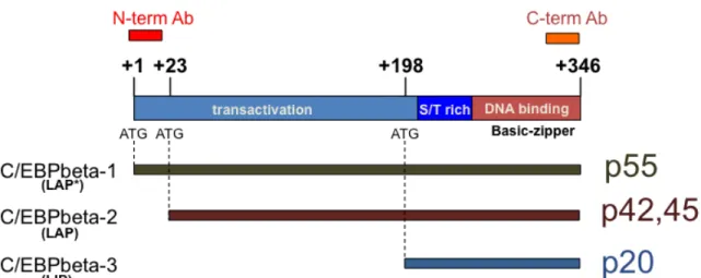
C/EBPbeta is transcribed from an intronless gene that gives rise to three protein isoforms from a single messenger RNA (mRNA) (Figure 2). This is due to alternative translation initiation at three in-frame methionine initiator codons or regulated proteolysis (Descombes et al., 1991; Ramji and Foka, 2002). In humans, full-length C/EBPbeta1 is 346 amino acids long (Akira et al., 1990) (297 in rat and mouse (Cao et al., 1991; Descombes et al., 1990) and has an apparent molecular weight of 55 kilodalton (kD). C/EBPbeta2 begins at the second in- frame methionine located 23 amino acid downstream from the first. C/EBPbeta2 appears as a doublet on immunoblots with an apparent molecular weight of 45kD and 42kD. The smallest isoform, C/EBPbeta3, begins at the final in-frame methionine at position 198 in the human protein and has an apparent molecular weight of 20kD (Descombes and Schibler, 1991). The structure of C/EBPbeta is such that the transactivation domain resides in the N-terminal region and the protein dimerization (leucine zipper) and DNA binding domains (basic region) reside in the C-terminal end. Unlike the first two transcription activator isoforms,
Figure 2. Schematic representation of C/EBPbeta. Three proteins isoforms are generated from a single mRNA by alternative translation. The positions of the three ATG start codons and the relative sizes of each isoform are shown.
The positions of epitopes recognized by two antibodies are also depicted.
C/EBPbeta1 and C/EBPbeta2 (termed LAP* and LAP in rodents), the third isoform, C/EBPbeta3 (termed LIP in rodents) lacks the entire N-terminal activation domain, while retaining the DNA binding/protein dimerization domain (Descombes and Schibler, 1991). Therefore, this protein acts as a transcriptional repressor since it can occupy the C/EBPbeta consensus DNA elements within promoters of target genes.
Isoform specific C/EBPbeta expression
Several mechanisms by which a cell regulates the production of a specific C/EBPbeta isoform have been proposed. One of the earliest explanations is that the three isoforms arise by a “leaky” ribosome scanning mechanism (Descombes and Schibler, 1991). Since then, there has been more complex mechanisms proposed; some that specifically lead to increases in C/EBPbeta3 (hereafter referred to as LIP) protein expression.
Mammalian target of rapamycin (mTOR) and additional signal transduction pathways that regulate the function of the translation initiation factors such as eukaryotic translation initiation factor (eIF2) alpha and eIF-4E have been shown to determine the ratio of C/EBP isoforms (Raught et al., 1996) Calkhoven and colleagues showed that high eIF2alpha activity leads to increased LIP production. Alternative translation initiation involves a highly conserved small upstream open reading frame (uORF) at the 5’ region of C/EBPbeta mRNA (Lincoln et al., 1998; Calkhoven et al., 2000; Wethmar et al., 2010). Studies of molecular mechanisms that control the initiation of translation
on the uORF-specific AUG codon suggested that certain RNA-binding proteins might regulate this process by interacting with the uORF region of C/EBPbeta mRNA. Two RNA-binding proteins, CUG triplet repeat binding protein (CUGBP1) and calreticulin (CRT), which specifically bind to the uORF were identified to be important players in regulating LIP translation (Timchenko et al., 1999; 2002;
2006). Although CUGBP1 and CRT interact with the same sequence of the 5′
region of C/EBPbeta mRNA, the consequences of these interactions are different. CRT binds to the 5′ region of C/EBPbeta mRNA and stabilizes a stem- loop (SL) structure, leading to the inhibition of translation of C/EBPbeta. On the contrary, the interaction of CUGBP1 with the 5′ region of C/EBPbeta mRNA increases translation of C/EBPbeta (Timchenko et al., 1999; 2002; 2006). It is not surprising that cells have highly regulated mechanisms to control translation initiation of the C/EBPbeta isoforms, since each have been linked to specific biological processes.
The second proposed mechanism to generate LIP is through specific proteolysis of the larger C/EBPbeta isoforms. In vivo studies have shown specific cleavage of the larger isoforms results in an increase in LIP protein levels (Welm et al., 2000; Baer and Johnson, 2000). This was shown to occur in the liver and depend on C/EBPalpha; it is likely that this occurs in other tissues as well (Welm et al., 2000). It is of great importance to note that in vitro cleavage of the larger C/EBPbeta isoforms can also occur (Baer and Johnson, 2000). LIP can be generated through artifactual proteolysis depending on the method used during sample preparation. Hence, there are discrepancies in the literature
regarding the production of LIP in cells.
Most of the research on C/EBPbeta has focused on the C/EBPbeta2 (LAP) and C/EBPbeta3 (LIP) isoforms. Until recently, many investigators failed to acknowledge the existence of C/EBPbeta1 (LAP*), due to its low expression levels in cultured cells. Historically there was this notion that C/EBPbeta1 and C/EBPbeta2 were functionally redundant, since they only differ by a 23 N- terminal amino acid truncation and both are transcriptional activators. However, mounting evidence by our lab and others demonstrate that the three C/EBPbeta isoforms have very different and unique roles in cells. C/EBPbeta1 plays important functions in differentiation and senescence (Kowenz-Leutz and Leutz, 1999; Eaton, et al., 2001; Eaton and Sealy, 2003; Atwood and Sealy, 2010), while C/EBPbeta2 participates in more proliferative roles in cells (Bundy and Sealy, 2003). This is evident in the ability of C/EBPbeta1, not C/EBPbeta2, to synergistically collaborate with c-Myb in co-expression assays to turn on differentiation genes in myeloid cells. Unlike C/EBPbeta2, C/EBPbeta1 was able to activate differentiation genes by interacting with and recruiting the SWI/SNF chromatin-remodeling complex (Kowenz-Leutz and Leutz, 1999). This recruitment was dependent on the N-terminal amino acids present in C/EBPbeta1, but not C/EBPbeta2. In addition, the N-terminal amino acids unique to C/EBPbeta1 are necessary for efficient sumoylation (SUMO) (Eaton and Sealy, 2003). Briefly, sumoylation is a post-translational modification that has been shown to regulate the function of various proteins (Geiss-Friedlander and Melchior, 2007). Specifically, sumoylation of transcription factors leads to
transcriptional repression by altering binding partners. C/EBPbeta1 is the only transactivator isoform that is sumoylated in COS cells even though both C/EBPbeta1 and C/EBPbeta2 contain a SUMO consensus sequence around lysine 173 (Eaton and Sealy, 2003). More recently, our lab found that C/EBPbeta1 more efficiently induces senescence than C/EBPbeta2 (Atwood and Sealy, 2010). This is likely due to the ability of C/EBPbeta1 to induce interleukin- 6 (IL-6) expression (Atwood and Sealy, 2010). Interestingly, expression of sumo- C/EBPbeta1 fusion protein does not induce IL-6 in comparison to expression of wild-type C/EBPbeta1, indicating that sumoylation of C/EBPbeta1 does lead to transcriptional repression (Atwood and Sealy, 2011).
Differences in protein expression have also been observed in mammary epithelial cells. C/EBPbeta1 is found in normal mammary epithelial cells from reduction mammoplasties, whereas C/EBPbeta2 is undetectable (Eaton et al., 2001). Interestingly, in human breast tumors, where cancer cells are actively proliferating, there is a significant gain of C/EBPbeta2 protein levels (Eaton et al., 2001). In fact, C/EBPbeta 2 but not C/EBPbeta1, was observed to transactivate the promoters of cyclin D1 (Eaton et al., 2001) and placenta specific 1 (PLAC1) genes (Koslowski, et al., 2009), which promote proliferation and are frequently upregulated in breast tumors. Moreover, overexpression of C/EBPbeta2 in the MCF10A mammary epithelial cell line leads to cell transformation (Bundy and Sealy, 2003). C/EBPbeta2-expressing MCF10A cells form foci, gain anchorage independence, express markers associated with having undergone an epithelial-to-mesenchymal transition (EMT), and acquire an
invasive phenotype (Bundy and Sealy, 2003). The role of LIP in cells continues to be a topic of debate and will be discussed further in greater detail in the next chapters.
Role of C/EBPbeta in the mammary gland
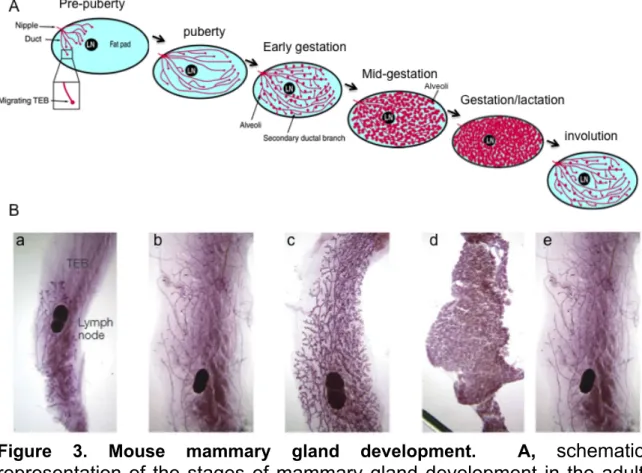
Genetic deletion of C/EBPbeta in mice revealed the importance of this transcription factor in a number of physiological processes. The C/EBPbeta null mice exhibit a number of dramatic phenotypes including impaired liver regeneration, increased sensitivity to infections, sterility, and defects in mammary gland development (Greenbaum et al., 1998; Sterneck et al., 1997; Robinson et al., 1998; Seagroves et al., 1998). The mammary gland is a dynamic organ, which undergoes growth and extraordinary remodeling in response to hormonal signals at puberty, pregnancy and continues through weaning of the offspring (Figure 3A). During embryonic development, a rudimentary epithelial ductal tree is formed which remains mostly unchanged until puberty. During puberty, estrogen stimulates the expansion and development of the ductal tree and lobuloalveolar structures at the ends of the ducts. During gestation, estrogen and progesterone stimulate additional proliferation and further development of the ducts and lobuloalveoli. Following parturition, epithelial cells differentiate into secretory epithelial cells for milk production and secretion. After lactation, the mammary epithelial ductal tree regresses to a quiescent state through the process of involution (Sternlicht et al., 2006). The involution process encompasses several changes including the reabsorption of residual milk, loss of
the epithelium by programmed cell death, clearance of these dying cells, and regrowth of the interstitial adipose cells. This regressed state has mistakenly been thought to be the same as the gland in the nulliparous state, before the first pregnancy and lactation. However, D’Cruz et al. have described a host of gene- expression changes of the mammary gland following pregnancy and lactation, (D’Cruz et al., 2002) and distinct genetic signatures have been reported for the parous and nulliparous human breast (Balogh et al., 2006). During the involution process, many secretory and epithelial cells undergo massive cell death; this has been shown to involve several forms of cell death including apoptosis, autophagy, and cell engulfment (Zarzynska and Motyl, 2008; Monks et al., 2008).
This process of epithelial cell growth, differentiation, and involution of the mammary gland occurs with each pregnancy.
In wild type mice extensive mammary gland development occurs around three weeks of age during puberty (Figure 3B). This fascinating process of developing normal mammary glands is well orchestrated and arises from an epithelial cell bud proximal to the nipple. At puberty, the epithelial cells divide and migrate to penetrate the mammary fat pad, forming the ductal tree.
Secondary and tertiary branching ducts stem from the primary ducts. These secondary and tertiary branching ducts also penetrate the fat pad. One way to measure the development of the mammary gland in mice is to monitor the position of the epithelial tree in relation to the distal lymph node in the inguinal mammary gland.
Figure 3. Mouse mammary gland development. A, schematic representation of the stages of mammary gland development in the adult mouse, from pre-puberty to gestation, lactation and involution. B, whole mount images of the different stages a, puberty; b, mature virgin; c, pregnancy; d, lactation; e, post-involution. LN, lymph node; TEB, terminal end bud. Modified from Watson and Khaled, 2008; Hennighausen and Robinson, 2005.
In 1998, studies were published describing the mammary gland deficiencies of two independently created C/EBPbeta knockout mice. Studies by Jeff Rosen and colleagues as well as Peter Johnson and colleagues reveal that C/EBPbeta is essential for proliferation of lobuloalveolar secretory units and ductal morphogenesis (Robinson et al., 1998; Seagroves et al., 1998).
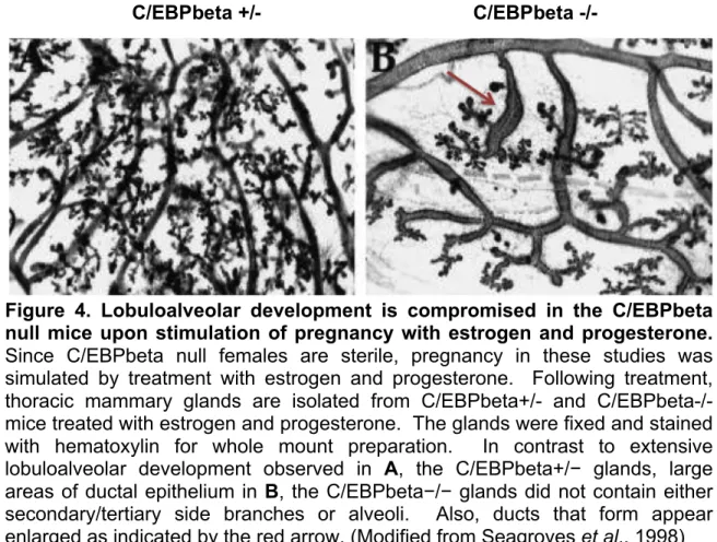
C/EBPbeta null mice have obvious defects in epithelial cell penetration into the mammary fat pad. In contrast to extensive lobuloalveolar development observed in C/EBPbeta heterozygous mammary glands, large areas of ductal epithelium of the C/EBPbeta null glands do not contain secondary/tertiary side branches or alveoli (Figure 4). Ducts that do form appear enlarged and produce distended ends. Both beta-casein and whey acidic protein expression are inhibited or absent in C/EBPbeta null mice (Grimm and Rosen, 2003). These data provide evidence for the essential role of C/EBPbeta in mammary epithelial proliferation in response to hormonal stimulation at puberty or gestation. Furthermore, the studies depict the importance of C/EBPbeta in functional differentiation of secretory epithelium in the mammary gland. Taken together, C/EBPbeta is a crucial component in both proliferation and differentiation in mammary epithelial cells. The mechanism by which C/EBPbeta is directing these antagonistic processes is likely due to the distinct functional properties of the three C/EBPbeta isoforms.
Figure 4. Lobuloalveolar development is compromised in the C/EBPbeta null mice upon stimulation of pregnancy with estrogen and progesterone.
Since C/EBPbeta null females are sterile, pregnancy in these studies was simulated by treatment with estrogen and progesterone. Following treatment, thoracic mammary glands are isolated from C/EBPbeta+/- and C/EBPbeta-/- mice treated with estrogen and progesterone. The glands were fixed and stained with hematoxylin for whole mount preparation. In contrast to extensive lobuloalveolar development observed in A, the C/EBPbeta+/− glands, large areas of ductal epithelium in B, the C/EBPbeta−/− glands did not contain either secondary/tertiary side branches or alveoli. Also, ducts that form appear enlarged as indicated by the red arrow. (Modified from Seagroves et al., 1998)
C/EBPbeta +/- C/EBPbeta -/-
C/EBPbeta in Breast cancer
With the establishment that C/EBPbeta is an important player in
proliferation during mammary gland development, many investigators focused their attention on the role of this protein in cancer. Increased C/EBPbeta expression has been detected in ovarian tumors, colorectal tumors, and breast tumors (Sundfeldt et al., 1999; Rask et al., 2000; Staiger et al., 2009; Grimm and Rosen, 2003; Bundy and Sealy, 2003). Increases in C/EBPbeta mRNA have been detected in 18 of 18 MMTV/c-neu mammary tumors, approximately 2-5 fold over levels expressed during lactation or involution (Dearth et al., 2001).
However, this does not provide information on which isoform is being upregulated. Some studies have described elevated levels of LIP in both murine mammary hyperplasia and in human breast cancers (Zahnow et al., 1997;
Zahnow et al., 2001). It has been argued that the transcriptional inhibitor LIP isoform is predominantly expressed during proliferative cellular responses and is associated with aggressive tumors (Zahnow et al., 1997). It has also been reported that expression of LIP under the control of the whey acidic promoter (WAP) in the mouse mammary gland results in the formation of hyperplastic tissue, and more infrequently, carcinomas (Zahnow et al., 2001). However, because the LIP transgene was not epitope-tagged in these mice it is not possible to ascertain transgene expression from any endogenous LIP expression. Moreover, the level of LIP expression (transgene or endogenous) in the mammary tumors was not actually examined. Zahnow et al. have stated that LIP is overexpressed in 23% of infiltrating ductal carcinomas specimens. Yet,
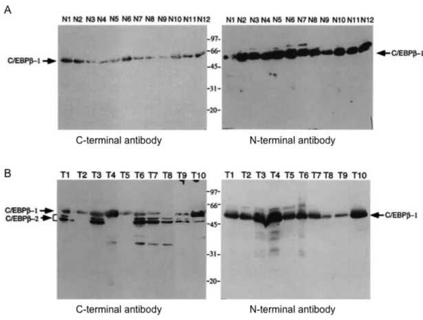
Figure 5. C/EBPbeta isoform expression in mammary samples. Whole cell extracts were prepared from A, normal breast tissue from reductive mammoplasty or B, breast tumors obtained from the Tissue Procurement Core at Vanderbilt University. Immunoblot analysis was performed using either the C- terminal or N-terminal C/EBPbeta antibody as indicated. Modified from Eaton, et al., 2001.
our own study on primary breast tumor samples found that high grade, invasive mammary carcinomas showed significant C/EBPbeta2 expression, but no LIP protein was detected in any of the samples (Figure 5) (Eaton et al., 2001). LIP is known to be easily generated by artifactual proteolysis of the larger isoforms, which could explain this discrepancy (Baer and Johnson, 2000).
We wanted to address this issue by overexpressing each C/EBPbeta isoform individually in a normal MEC line model. Overexpression of C/EBPbeta2 (but not C/EBPbeta1 or LIP) results in a variety of cancer phenotypes. In previous studies by our lab, we asked whether LIP could confer epidermal growth factor (EGF)-independent growth of MCF10A cells (Bundy et al., 2005). In contrast to C/EBPbeta2, a high level of LIP expression is incompatible with continued proliferation. In fact, our data reveals a remarkable increase in cell death of LIP-expressing cells, even in the presence of EGF (Bundy et al., 2005).
Purpose of this Study
In recent years, huge strides have been made in pursuing transcription factors as anticancer targets (Karamouzis et al., 2002; Frank, 2009).
Therapeutics directed at C/EBPbeta are promising, not only for cancer, but for many other diseases. C/EBPbeta is known to play key roles in diverse pathological conditions such as human immunodeficiency virus (HIV), diabetes, as well as various inflammatory diseases. Nevertheless, it is essential to find ways to target a specific isoform, since the three are functionally distinct. Recent findings in our laboratory corroborate the importance of C/EBPbeta, specifically
C/EBPbeta1, in oncogene-induced senescence (Atwood and Sealy, 2010). Also, we found C/EBPbeta1 to be negatively regulated during transformation. We previously demonstrated a role for C/EBPbeta2 in promoting tumor cell proliferation and transformation (Bundy and Sealy, 2003). On the other hand, overexpression of C/EBPbeta1 or LIP does not promote tumor cell proliferation.
Yet, the exact role of LIP in cancer is unclear. The main objective of this work is to examine the role of LIP in breast cancer in order to gain a better understanding of whether LIP is a tumor-promoter, tumor-suppressor, or both.
These studies demonstrate high levels of LIP stimulate cell death in breast cancer cell lines. Moreover, my work shows that LIP leads to a nonapoptotic cell death process in breast cancer cell lines involving cell engulfment and autophagy. The central hypothesis to be tested is that as a transcriptional repressor, LIP stimulates cell death in breast cancer cells by altering the cellular transcriptional program.
Significance
The apoptotic cell death pathway has been one of the major emphases in anticancer therapeutics. However, a critical step in tumor formation and progression is the ability of tumor cells to evade apoptosis, resulting in malignant cells that will not die (Hanahan and Weinberg, 2000). Apoptosis is complex mechanism and encompasses many pathways. Errors can transpire at any point along these pathways, leading to not only malignant transformation of the affected cells, but many times to tumor cell metastasis and resistance to
anticancer therapies.
Several alternative cell death pathways have been recently described. The aims of this work focus on characterizing the induction of autophagy as well as describing the ability to engulf neighboring cells which ultimately leads to cell death upon LIP expression in breast cancer cell lines. It is clear that knowledge of mechanisms and identification of new agents capable of inducing alternative cell death pathways has vast potential to improve cancer therapeutics.
CHAPTER II
C/EBPbeta3 (LIP) and autophagy Introduction
Autophagy is a process involving the bulk degradation of cellular components in the cytoplasm via the lysosomal degradation pathway (Levine and Klionsky, 2004). Autophagy manifests a protective role in stressful conditions such as nutrient or growth factor depletion; however, extensive degradation of regulatory molecules or organelles essential for survival can lead to the demise of the cell, or autophagy-mediated cell death (Gozuacik and Kimchi, 2004). The role of autophagy in cancer is complex with roles in both tumor suppression and tumor promotion proposed (Hippert et al., 2006).
Activation of autophagy may lead to different outcomes depending on the cell genetic background as well as the duration and strength of the stress- inducing stimulus. It has been suggested that in early stages of tumor formation, a defective autophagic system leads to the accumulation of damaged proteins and organelles. This increase in genotoxic substances may lead to both failure to constrain cell growth and mutations. Yet, at later stages of tumorigenesis, autophagy may be a means by which tumor cells survive under oxygen and nutrient limiting conditions, providing extra time for adaptation via the induction of angiogenesis and/or motility and invasion (Hippert et al., 2006). The mechanisms by which autophagy may be regulated to provide complete cellular destruction or a survival advantage remain unknown.