HIV
Thesis by Kenneth K. Yu
In Partial Fulfillment of the Requirements for the Degree of
Doctor of Philosophy
CALIFORNIA INSTITUTE OF TECHNOLOGY Pasadena, California
2012
(Defended 13 December 2011)
2012 Kenneth Yu All Rights Reserved
ACKNOWLEDGEMENTS
Some people say that science is an intensely personal enterprise, bringing to mind the image of a scientist working alone long into the night, in a solo quest to make Nature yield its secrets. There is some truth to that image, but it’s far from the whole story. For me, I would have to say that science is an inherently social experience. It’s hard to engage in it without becoming connected to people, and having one’s self enriched by their presence. And thus it’s a tremendous honor for me to write this acknowledgement, to express, perhaps inadequately, my deepest gratitude to the people without whom this work would not have been possible.
I want to thank my parents for their unconditional love, and for always supporting me to pursue my dreams, even when sometimes they appear seemingly incoherent. Dad, thank you for being my inspiration—you are the first physician I got to know in my life, and you are the one who taught me that caring for and helping those around us is what gives meaning to our lives. And thank you for showing me the meaning of Hebrews 11:1.
Mom, thank you for putting up with being separated from Dad across an ocean, so that we kids could pursue the American dream. And thank you for standing by me and encouraging me even when you did not understand the reasons for my choices. To you both I dedicate this thesis.
I want to thank my sister and brother, Karen and Kevin, for always believing in me, even at moments when I doubted myself. And thank you Kevin for introducing me to “Still Alive” and writing “Prepping Cells”.
I want to thank my soon-to-be wife, Jessica, for your prayers and understanding when I disappeared into the lab. Your love makes every moment of life full of joy.
I want to thank my first-grade science teacher, Dr. Yu-wen Zhuang. You first spotted in me the curiosity to ask questions about Nature, and nutured it by teaching me to do experiments to answer them, in the process leaving me more full of wonder than when I started. You inspired me to be a scientist.
I want to thank my high-school friend Morgan Gough, who took time off from work and came to take care of me when I nearly went blind in one eye in the second year of grad school.
And to the many wonderful and talented people that are the Baltimore Lab, thank you for sharing with me your excitement about science and showing me how to do it. Lili Yang, thank you for your critical input and discussions. Your creativity and discipline will always be an inspiration to me. Dinesh Rao, thank you for being a great bay-mate, friend, and role model. And I will always treasure the times we spent pondering questions about biology, or for that matter, the mysteries of life. Jonathan Tsai, thank you for being my first student. I enjoyed doing science with you as much as learning from you about the unique species known as the Caltech Undergrads. Kiefer Aguilar, thank you for being a student, teacher, and friend; science is best when shared with good company.
Alex Balazs, thank you for your tips on how to do experiments, thoughtful discussions, and the pHAGE vectors. Your rationality is an inspiration. Ryan O’Connell, thank you for your help with the HIS mice. Dev Majumdar, thank you for keeping me on my toes!
Your curiosity is impeccable. And thanks for sharing with me your optimism and
encouraging me to do what needed to be done when I most needed it. Eun-Mi Hur, thank you for collaborating with me and teaching me how to do experiments. Rachel Galimidi and Pri Gnanapragasam, thank you for your help with critical experiments and being great collaborators! Tom Su, thanks for all the good times hanging out in and out of the lab. Congrats to you and Connie on Parker! Michael Bethune, thank you for your warmth, encouragement, guidance, and help, and the many discussions we’ve had. We
should do more trips to the Shakespeare Theatre! Alex Sigal, thanks for enlightening me with your sense of humor. Evgenij Raskatov, great business plan; let’s have fun with some more mice!
And to all the other members of the Baltimore family, Arnav Mehta, Jimmy Zhao, Geoffrey Lovely, Aadel Chaudhuri, Param Ramakrishnan, Chee-Kwee Ea, Shengli Hao, Mark Boldin, Konstantin Taganov, Alex So, Jocelyn Kim, Yvette Garcia, Vanessa Jonsson, Yang Yu, Claret Liu, Stella Ouyang, Christin Hong, Joyce Chen, Eric Santiestevan, Joanne Laurence, and Julie Kelly, thank you for making my experience here a memorable and enjoyable one. The lab wouldn’t be the same without you.
I want to thank the members of my committee for your support and insights. Dr. Ellen Rothenberg, thank you for sharing with me your love and passion for science. Your immunology class was a wonderful window into the process of science and discovery.
Your excitement about science is contagious! Thank you for all the thoughtful
discussions we’ve had. Dr. Sarkis Mazmanian, thank you for your encouragement and discernment about what paths one should explore when doing science. Dr. Pamela
Bjorkman, thank you for your insight and support and making me feel as much a member of the Bjorkman lab as the Baltimore lab.
Last but not least, I want to thank my advisor Dr. David Baltimore, who is the ultimate source of my inspiration. You have assembled an environment for learning, doing, and experiencing science that is unparalleled. It could only be the embodiment and
expression of who you are as a scientist and mentor. You took a chance on an untested graduate student who had no clue what a transcription factor was when he first walked into your office in Parsons-Gates, and you taught him the spirit of experimental science by example. I recall walking back from a Tuesday afternoon general biology seminar with you one day. We had lingered to talk with the speaker. By the time we started back
for the lab, it was a little past 5 pm, the normal time they’d lock the doors to the
buildings. As we neared Braun’s east entrance, I started fumbling for my keys. Like a true experimentalist, you reached for the door handle and pulled the unlocked door open.
With a smile on your face, you said to me, “Do the experiment!” To a theoretical physicist by training, that changed my world. Dear David, I have done the experiments, and the results are exciting. Thank you for opening the door.
ABSTRACT
An effective vaccine against the human immunodeficiency virus (HIV)-1 has so far been elusive. Anti-viral vaccines against other viruses work by stimulating the production of neutralizing antibodies that block infection. To be useful, an anti-HIV vaccine preparation needs to elicit potent neutralizing antibody response with sufficient breadth to cover the diversity of HIV variants. Despite sustained research efforts, such an immunogen has been difficult to develop. We could overcome this difficulty by using gene therapy to directly instruct the body to produce anti-HIV broadly neutralizing antibodies (bNAbs). In this thesis, I describe a technology I developed termed the
“Molecular Rheostat” for directing the simultaneous expression of anti-HIV surface and secreted immunoglobulins using mutant 2A “self-cleaving” peptides. I describe the application of this system to the programming of hematopoeitic stem cells to generate anti-HIV B cells as a strategy to “vaccinate” against HIV infection. I then pivot to consider alternatives to B-cell programming to produce antibodies against HIV. I investigate the modification of non-lymphoid hematopoietic cells to produce antibodies using retroviral vectors and describe the use of lentiviral vectors to program muscle to produce anti-HIV broadly neutralizing antibodies. In addition to presenting a novel tool for controlling the simultaneous expression of full-length and truncated proteins, the work described here furnishes a foundation for future development into potential gene- therapeutic prophylaxis against HIV.
TABLE OF CONTENTS
Acknowledgements ... iii
Abstract ... iv
Table of Contents ...v
Chapter 1: Introduction ...1
AIDS at 30 ...1
Toward an AIDS Vaccine: The Broadly Neutralizing Antibodies ...5
Engineering Immunity Against HIV ...7
Overview of Thesis ...10
References ...12
Chapter 2: Use of Mutated “Self-Cleaving” 2A Peptides as “Molecular Rheostats” to Direct Simultaneous Formation of Membrane and Secreted Immunoglobulins ...14
Abstract ...14
Introduction ...15
Materials and Methods ...17
Results ...22
Discussion ...30
References ...44
Chapter 3: In Vivo Characterization of the Molecular Rheostat Immunoglobulins ...46
Introduction ...46
Materials and Methods ...47
Results and Discussion ...49
References ...63
Chapter 4: The Use of Non-Lymphoid Hematopoietic Cells for Antibody Production ...64
Introduction ...64
Materials and Methods ...65
Results and Discussion ...66
References ...77
Chapter 5: Lentiviral-Vector-Mediated-Broadly-Neutralizing Antibody Production from Muscle ...78
Introduction ...78
Materials and Methods ...79
Results and Discussion ...80
References ...92
Chapter 5: Looking Ahead ...98
Summary and Future Directions ...98
Concluding Remarks ...101
References ...102
C
HAPTER1: I
NTRODUCTIONAIDS at 30
The year 2011 marks the 30th anniversary of the first formal report of the disease that came to be known as AIDS (Acquired Immune-Deficiency Syndrome) caused by the human immunodeficiency virus (HIV). According to the most recent statistics available through UNAIDS, it is estimated that 34 million people globally were living with HIV/AIDS at the end of 2010, with 2.6 million new infections, and 1.8 million deaths directly attributed to AIDS (UNAIDS 2011). While the number of new infections has stabilized, the burden of the disease continues to grow globally.
In the summer of 2007, I went on a medical and humanitarian mission with the aid group, Project Africa Global, to Swaziland. Swaziland is a small sovereign kingdom inside South Africa with a population of a little over a million and the size of the State of New Jersey. We were the guests of the king and one of the princes, and we travelled in relative comfort and our lodging was pleasant. It was there that I witnessed with my own eyes the tragic consequences of the HIV/AIDS pandemic both on the individual level and on a society as a whole. That year, over 30 percent of Swazis were living with HIV/AIDS (and by some estimates, more than 40 percent). I remember the drives from our residence to the clinics in the countryside. Despite the seasonably warm weather and a clear blue sky, there was a palpable sense of doom and depression as we travelled on the rural roads. Fields lay fallow and unused, not because it wasn’t the right season, but because
there was not enough man power to cultivate them. As we drove through the cities, at the height of the day, the streets seemed strangely quiet and abandoned. HIV/AIDS had cut down entire generations from Swaziland’s population pyramid. The largest groups of people were those under the age of 18 and those older than 40. We arrived at a make- shift countryside clinic, and I beheld a sight that I would never be able to forget. As I worked with patients with various ailments (there were exactly 41 doctors in a country of one million, so a medical student was as close to a doctor as some of these people would ever see), a mother and her son came in to see us. Lovingly she carried her son in her arms. She came in for “severe ear infections”, along with cough, blood tinged sputum, and fever at night that rattled the bones—the latter three being the classic signs of TB infection, and in the setting of this patient population, the ominous harbinger of full blown AIDS. But as I examined her, the thing that caught my eye, and the attention of everyone in the room, was a red, florid, alien-looking, fungal growth that had colonized much of her right ear and had also put down roots on the left side of her face. We tried our best to clean the wounds and gave her anti-fungal medications, but with extremely limited access to consistent anti-retroviral therapy and nutritional support, we realized that she did not have long. Her son suffered from diarrhea and a slew of other ailments that suggested to us that he too was infected. As the day in the clinic wound down, I began to reflect on the patients I saw. I thought of the people who were dying with HIV/AIDS and those who they would leave behind; then I thought of the young boy who
would lose his mother, and whose own life was threatened by the disease. I never wished more fervently for a vaccine.
Here in the United States, the CDC estimates that 1.2 million people are living with HIV infection and over half a million people have died since the epidemic began (National Center for HIV/AIDS 2011). The annualized medical costs per HIV infection in the U.S. was estimated to be approximately $24,000 per person per year (Farnham, Holtgrave et al. 2010). These numbers do not begin to capture the magnitude of the human suffering caused by HIV/AIDS; they say nothing of the fear and stigma associated with the disease.
The identification of the causative agent of AIDS, the human immunodeficiency virus or HIV, was announced by two separate research groups led respectively by Luc Montagnier and Robert Gallo in 1983 (Barre-Sinoussi, Chermann et al. 1983; Gallo, Sarin et al. 1983), for which Montagnier was awarded the Nobel Prize in Physiology or Medicine in 2008. Gallo and Montagnier’s discovery brought great advances in the understanding of the molecular biology of the virus and the mechanisms of disease transmission and pathogenesis, and stimulated new developments in medicine at treatment, prevention, and control. The recognition that AIDS was transmissible by a virus led to early hopes for a quick vaccine. Those hopes were not entirely unreasonable, as our experiences with other viral diseases such as polio and smallpox suggested to us that it might be relatively easy to make a vaccine.
History turned out differently. Nearly 30 years after its initial identification, we still do not have a vaccine (yet). HIV is a virus so very different from those other viruses we had made effective vaccines for before that conventional vaccines did not work against it. For one thing, HIV belongs to the larger family of retroviruses that use the enzyme reverse transcriptase to make a copy of themselves that allows them to integrate into the host genome. Thus, once an infection is established, the virus effectively becomes a part of the host’s genetic make-up. The reverse transcriptase, discovered independently by Dr. David Baltimore and Dr. Howard Temin in 1970, and for which discovery they shared the Nobel Prize in 1975, is the target of the first class of antiviral drugs. Several other classes of drugs have come online since then, through our improved understanding of the molecular biology of the virus. This knowledge has led to the development of HAART (highly active antiretroviral therapy), inspired by the work by Dr. David Ho, in which multiple drugs that target different molecular aspects of the HIV life cycle are given in combination. HAART was shown to be capable of suppressing the virus and controlling the progression of the disease for periods up to decades. However, these drugs do not provide a cure, are expensive, and cause significant side effects. The best way to stop or slow the epidemic is an effective vaccine that can prevent infection in the first place, and the need for it is as acute as ever (Baltimore 2002; Letvin, Barouch et al. 2002).
Toward an AIDS Vaccine: The Broadly Neutralizing Antibodies
Vaccines are antigen preparations that elicit immune responses against pathogens.
The utility of vaccines is limited by the kinds of antibodies (Abs) that are made by the host immune system after vaccination. Most currently used anti-viral vaccines work by stimulating production of neutralizing antibodies (NAbs), which block viral infection (Zinkernagel, LaMarre et al. 2001; Burton 2002). HIV is an enveloped retrovirus that presents problems for antibody-based vaccine strategies. The virus rapidly mutates to change residues on its surface, sheds immunodominant decoy epitopes, masks
immunogenic sites on its surface with host-derived carbohydrates, and/or hides conserved regions in the interfaces of oligomeric proteins (Burton, Stanfield et al. 2005; Berkley and Koff 2007). While neutralizing antibodies against HIV do emerge in the natural course of infection, they occur too late, after an infection has already been established, and are thwarted by rapid genetic mutation of the virus (Richman, Wrin et al. 2003). For all of these reasons, it has been exceedingly difficult to design an immunogen that would elicit an anti-HIV antibody response of sufficient quality and breadth to be protective (Burton, Desrosiers et al. 2004; Flynn, Forthal et al. 2005; Pitisuttithum, Gilbert et al.
2006; Johnston and Fauci 2007; Fauci, Johnston et al. 2008).
Broadly neutralizing antibodies (bNAbs) against HIV do exist; they are an unusual class of antibodies that neutralize a broad range of HIV variants (Burton,
Stanfield et al. 2005). Produced by few individuals, bNAbs are rare, but some have been
immortalized as monoclonal Abs (Burton, Stanfield et al. 2005). Passive immunization with bNAbs has been shown to protect animals against simian-HIV (SHIV) challenge (Mascola 2002). Indeed, long-term-non-progressors (individuals who have remained free of disease without treatment more than 10 years after HIV infection) exhibit broadly cross-reactive bNAb responses (Pilgrim, Pantaleo et al. 1997). The existence of bNAbs suggests that it might be possible to prevent HIV infection and subsequent disease by producing bNAbs in individuals at risk for AIDS (Burton 2002; Ferrantelli, Rasmussen et al. 2002; Burton, Desrosiers et al. 2004). Broadly reactive human bNAbs shown to protect against HIV challenge in animal models include b12, 2G12, 2F5, 4E10, and more recently VRC01 (through work done in our lab), among some others. Some of these bNAbs exert their effects by preventing the trimeric HIV-1 envelope complex (GP120- GP41) from binding to the host receptor (CD4) or co-receptor (usually CCR5 or CXCR4); others inhibit fusion of the virus with a host target cell by binding to the envelope protein after virions have attached to a target cell (Xiao, Dong et al. 2002;
Burton, Desrosiers et al. 2004). The bNAbs 2G12 and 2F5 have been verified to be safe for use in humans in phase I clinical trials (Wolfe, Cavacini et al. 1996; Cavacini, Samore et al. 1998; Armbruster, Stiegler et al. 2002), and evidence for anti-viral activity was seen in HIV-infected patients treated with these bNAbs (Stiegler, Armbruster et al. 2002).
Engineering Immunity Against HIV
It occurred to Dr. Baltimore that if we could directly instruct the immune system to produce broadly neutralizing anti-HIV antibodies, we might be able to use them as
“vaccines” to prevent HIV infections. Gene therapy technology provides the means for us to genetically program immune cells. Using gene therapy to deliver broadly neutralizing antibodies, we would be able to provide them to people as a prophylaxis against HIV infections. We call this approach “Engineering Immunity”.
The most natural way to provide broadly neutralizing antibodies to people is by engineering B cells, as they are the immune cells that are responsible for the production of antibodies. They begin their life in the bone marrow as descendants of the more primitive common hematopoietic stem and progenitor cells. As these cells develop into B cells, they undergo sequential RAG1/2-mediated DNA rearrangement of the heavy and light chain immunoglobulin gene loci in a process called V(D)J rearrangement. This is a pseudo-random process in which different V, (D), and J segments are combined together with the addition of certain non-templated nucleotides to produce a great diversity of antigen binding regions. Cells that successfully complete this process and assemble a functional IgM B cell receptor (BCR) on their surface are able to leave the bone marrow to continue further development in the peripheral lymphoid compartments (Burrows and Cooper 1993; Chen and Alt 1993). It has been shown in transgenic animals that provision of a pre-rearranged IgM heavy chain and light chain transgene shuts down the
rearrangement of endogenous heavy and light chain genes (allelic exclusion), and guides the ordered development of functional B cells with specificity defined by the transgene (Spanopoulou, Roman et al. 1994; Young, Ardman et al. 1994).
The mature B cells patrol the body in the general and lymphatic circulations, using their BCRs as antigen sensors. When a cognate antigen engages the BCR, the B cell becomes activated and enters into germinal center reactions in the lymph node or spleen in a dance of mutual activation with T cells; this process leads to further
development into memory B cells or differentiation into antibody-producing plasma cells.
The memory B cells will provide a more rapid and higher quality antibody response in the future when the same antigens are encountered again. The plasma cells produce antibodies against the inciting antigens, which leads to their eventual clearance from the body (McHeyzer-Williams and McHeyzer-Williams 2005).
As B cells differentiate into plasma cells, they switch from producing the membrane-bound BCR to making a soluble, secreted antibody. The switch is
accomplished on the level of RNA processing by alternative splicing of the 3’ end of the heavy-chain primary RNA transcript (Peterson, Gimmi et al. 1991; Peterson 2007). This replaces the hydrophobic amino acids that form the membrane anchor with a hydrophilic tail that enables the secretion of the BCR as free antibody. The antibody retains the same specificity and isotype as the BCR.
Some of the cells in the germinal center reactions also go through a process called isotype switching, in which the heavy chain constant regions of the initial IgM BCR are replaced with that of another isotype, which encodes different effector functions. This involves a DNA rearrangement mediated by the enzyme activation-induced cytidine deaminase (AID) (Muramatsu, Kinoshita et al. 2000). While the IgM BCR is required for the normal development of B cells in the bone marrow, and the IgM antibody is generally the first antibody isotype produced against an antigen, alternate isotypes provide additional effector functions that enhance the ability of the antibody to clear certain types of pathogens or to function in different body compartments. For example, in addition to fixing complements on target cells, as IgM antibodies can, IgG antibodies also have the ability to direct the killing of antibody-bound infected cells by engaging Fcγ receptors on NK cells (termed ADCC, or antibody-dependent cell-mediated cytotoxicity).
IgA antibodies are produced by plasma cells in mucosal areas and are transported across the epithelial barriers of the lung, gut, and genital tracts by binding the polymeric
immunoglobulin receptors (pIgR) with their Fc portions. These antibodies are critical in the defense of mucosal surfaces from pathogens.
Our understanding of the humoral immune response as summarized above forms the framework for my efforts to engineer the immune system. This framework suggests to us that by delivering a cleverly designed, synthetic immunoglobulin gene to the
hematopoietic stem and progenitor cells using gene therapy, we would be able to direct
the development of B cells that would produce broadly neutralizing antibodies against HIV with pre-programmed specificity and effector functions. Specifically, the synthetic immunoglobulin gene should 1) encode a mechanism that directs the production of both an IgM-like membrane-bound BCR and a secreted immunoglobulin isotype that has the desired effector properties, such as those of an IgG antibody, and 2) it should bind and neutralize HIV with the specificity and affinity of an anti-HIV broadly neutralizing antibody. Those were the two objectives that I set out to accomplish in my work when I joined Dr. Baltimore’s team in 2006, and this work will be described in detail below. As the project proceeds, I also explored a few other alternatives to this original approach by looking at cell types other than B cells as targets for engineering to produce anti-HIV antibodies, and the results obtained are summarized in separate chapters of this thesis.
Overview of Thesis
Chapter 1 gives a short, personal introduction to the HIV/AIDS epidemic and briefly reviews some significant scientific advances that have been made in the fight against HIV/AIDS. It then gives a succinct review of the broadly neutralizing antibodies against HIV and the biology of B cells that form the background of the Engineering Immunity project.
Chapter 2 of this thesis describes a novel approach I have developed to genetically program hematopoietic cells to become anti-HIV B cells that we call a
“Molecular Rheostat” for antibody genes. The focus will be on the in vitro development
and characterization of this technology. I will show that the Molecular Rheostats provides a useful tool for manipulating B cell specificity and gives us the ability to program them to produce a bNAb against HIV.
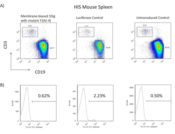
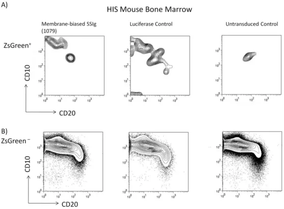
Chapter 3 describes my attempt to use the Molecular Rheostats to program B cells in vivo. It summarizes what we have learned from testing the system in both the human- immune-system (HIS) mouse model and the murine bone marrow adoptive transfer model. We describe certain limitations of the lentiviral vector system we developed and suggest what we might do to overcome the limitations
In Chapters 4 and 5 I pivot to look at alternative approaches to make broadly neutralizing antibodies in vivo. Chapter 4 describes the use of retroviral vectors to program non-lymphoid hematopoietic cells to produce antibody long-term. Chapter 5 describes my effort to study the feasibility of using lentiviral vectors to program muscle.
In Chapter 6 I give a short summary of this work, speculate on directions for future investigations, and offer some concluding remarks.
References
Armbruster, C., G. M. Stiegler, et al. (2002). "A phase I trial with two human monoclonal antibodies (hMAb 2F5, 2G12) against HIV-1." AIDS 16(2): 227-233.
Baltimore, D. (2002). "Steering a course to an AIDS vaccine." Science 296(5577): 2297.
Barre-Sinoussi, F., J. C. Chermann, et al. (1983). "Isolation of a T-lymphotropic retrovirus from a patient at risk for acquired immune deficiency syndrome (AIDS)." Science 220(4599): 868-871.
Berkley, S. F. and W. C. Koff (2007). "Scientific and policy challenges to development of an AIDS vaccine." Lancet 370(9581): 94-101.
Burrows, P. D. and M. D. Cooper (1993). "B-cell development in man." Curr Opin Immunol 5(2): 201-206.
Burton, D. R. (2002). "Antibodies, viruses and vaccines." Nat Rev Immunol 2(9): 706- 713.
Burton, D. R., R. C. Desrosiers, et al. (2004). "HIV vaccine design and the neutralizing antibody problem." Nat Immunol 5(3): 233-236.
Burton, D. R., R. L. Stanfield, et al. (2005). "Antibody vs. HIV in a clash of evolutionary titans." Proc Natl Acad Sci U S A 102(42): 14943-14948.
Cavacini, L. A., M. H. Samore, et al. (1998). "Phase I study of a human monoclonal antibody directed against the CD4-binding site of HIV type 1 glycoprotein 120."
AIDS Res Hum Retroviruses 14(7): 545-550.
Chen, J. and F. W. Alt (1993). "Gene rearrangement and B-cell development." Curr Opin Immunol 5(2): 194-200.
Farnham, P. G., D. R. Holtgrave, et al. (2010). "Medical costs averted by HIV prevention efforts in the United States, 1991-2006." J Acquir Immune Defic Syndr 54(5):
565-567.
Fauci, A. S., M. I. Johnston, et al. (2008). "HIV vaccine research: the way forward."
Science 321(5888): 530-532.
Ferrantelli, F., R. A. Rasmussen, et al. (2002). "Do not underestimate the power of antibodies—lessons from adoptive transfer of antibodies against HIV." Vaccine 20 Suppl 4: A61-65.
Flynn, N. M., D. N. Forthal, et al. (2005). "Placebo-controlled phase 3 trial of a
recombinant glycoprotein 120 vaccine to prevent HIV-1 infection." J Infect Dis 191(5): 654-665.
Gallo, R. C., P. S. Sarin, et al. (1983). "Isolation of human T-cell leukemia virus in acquired immune deficiency syndrome (AIDS)." Science 220(4599): 865-867.
Johnston, M. I. and A. S. Fauci (2007). "An HIV vaccine--evolving concepts." N Engl J Med 356(20): 2073-2081.
Letvin, N. L., D. H. Barouch, et al. (2002). "Prospects for vaccine protection against HIV-1 infection and AIDS." Annu Rev Immunol 20: 73-99.
Mascola, J. R. (2002). "Passive transfer studies to elucidate the role of antibody-mediated protection against HIV-1." Vaccine 20(15): 1922-1925.
McHeyzer-Williams, L. J. and M. G. McHeyzer-Williams (2005). "Antigen-specific memory B cell development." Annu Rev Immunol 23: 487-513.
Muramatsu, M., K. Kinoshita, et al. (2000). "Class switch recombination and
hypermutation require activation-induced cytidine deaminase (AID), a potential RNA editing enzyme." Cell 102(5): 553-563.
National Center for HIV/AIDS, V. H., STD, and TB Prevention (CDC) (2011). "HIV in the United States."
Peterson, M. L. (2007). "Mechanisms controlling production of membrane and secreted immunoglobulin during B cell development." Immunol Res 37(1): 33-46.
Peterson, M. L., E. R. Gimmi, et al. (1991). "The developmentally regulated shift from membrane to secreted mu mRNA production is accompanied by an increase in cleavage-polyadenylation efficiency but no measurable change in splicing efficiency." Mol Cell Biol 11(4): 2324-2327.
Pilgrim, A. K., G. Pantaleo, et al. (1997). "Neutralizing antibody responses to human immunodeficiency virus type 1 in primary infection and long-term-
nonprogressive infection." J Infect Dis 176(4): 924-932.
Pitisuttithum, P., P. Gilbert, et al. (2006). "Randomized, double-blind, placebo-controlled efficacy trial of a bivalent recombinant glycoprotein 120 HIV-1 vaccine among injection drug users in Bangkok, Thailand." J Infect Dis 194(12): 1661-1671.
Richman, D. D., T. Wrin, et al. (2003). "Rapid evolution of the neutralizing antibody response to HIV type 1 infection." Proc Natl Acad Sci U S A 100(7): 4144-4149.
Spanopoulou, E., C. A. Roman, et al. (1994). "Functional immunoglobulin transgenes guide ordered B-cell differentiation in Rag-1-deficient mice." Genes Dev 8(9):
1030-1042.
Stiegler, G., C. Armbruster, et al. (2002). "Antiviral activity of the neutralizing antibodies 2F5 and 2G12 in asymptomatic HIV-1-infected humans: a phase I evaluation."
Aids 16(15): 2019-2025.
UNAIDS (2011). AIDS at 30: Nations at Crossroads.
Wolfe, E. J., L. A. Cavacini, et al. (1996). "Pharmacokinetics of F105, a human monoclonal antibody, in persons infected with human immunodeficiency virus type 1." Clin Pharmacol Ther 59(6): 662-667.
Xiao, Y., X. Dong, et al. (2002). "Neutralizing antibodies mechanism of neutralization and protective activity against HIV-1." Immunol Res 25(3): 193-200.
Young, F., B. Ardman, et al. (1994). "Influence of immunoglobulin heavy- and light- chain expression on B-cell differentiation." Genes Dev 8(9): 1043-1057.
Zinkernagel, R. M., A. LaMarre, et al. (2001). "Neutralizing antiviral antibody responses." Adv Immunol 79: 1-53.
CHAPTER 2:USE OF MUTATED “SELF-CLEAVING”2APEPTIDES AS “MOLECULAR
RHEOSTATS” TO DIRECT SIMULTANEOUS FORMAION OF MEMBRANE AND SECRETED
IMMUNOGLOBULINS
Abstract
In nature, B cells produce surface immunoglobulin and secreted antibody from the same immunoglobulin gene via alternative splicing of the pre-messenger RNA. Here we present a novel system for genetically programming B cells to direct the simultaneous formation of membrane-bound and secreted immunoglobulins that we term a “Molecular Rheostat” Immunoglobulin gene, based on the use of mutated “self-cleaving” 2A
peptides. The Molecular Rheostats are designed so that the ratio of secreted to membrane-bound immunoglobulins can be controlled. Lentiviral transgenesis of the Molecular Rheostat constructs into B cell lines enables the expression of functional b12- based BCRs that signal to the cells and mediate the secretion of b12 IgG broadly
neutralizing antibodies that can bind and neutralize HIV-1 pseudovirus. We show that these b12-based Molecular Rheostat constructs promote the maturation of EU12 B cells in an in vitro model of B lymphopoiesis. The Molecular Rheostat Immunoglobulins offer a novel tool for genetically manipulating B cell specificity with implications for B-cell based gene therapy.
Introduction
B cells are responsible for the production of antibodies in response to foreign antigens. The ability to manipulate the antigen specificity of B cells and that of the antibody produced by these cells could be useful for achieving immunization against deadly pathogens such as HIV. In this chapter, I describe a novel way of programming B cells by using mutated 2A peptides to direct the simultaneous formation of an IgM-like BCR and IgG antibody. The system is designed so that the ratio of surface-to-secreted immunoglobulins can be controlled by appropriate choice of mutations. We call this system a “Molecular Rheostat” for immunoglobulin gene expression.
B cells begin their life in the bone marrow as descendants of the more primitive common hematopoietic stem and progenitor cells. As these cells develop into B cells, they undergo sequential RAG1/2-mediated DNA rearrangement of the heavy and light chain immunoglobulin gene loci in a process called V(D)J rearrangment. Cells that successfully complete this process and assemble a functional B cell receptor (BCR) of the IgM isotype on their surface are able to leave the bone marrow to continue further
development in the peripheral lymphoid compartments (Burrows and Cooper 1993; Chen and Alt 1993). The generation of the IgM BCR is central to B cell devevelopment and function. It is both necessary for the normal development of B cells (Kitamura, Roes et al. 1991; Kitamura and Rajewsky 1992; Wagner, Williams et al. 1994), and sufficient for directing B cell development. In transgenic animals. the provision of a pre-rearranged
IgM heavy chain and light chain transgene shuts down the rearrangement of endogenous heavy and light chain genes (allelic exclusion), and guides the ordered development of functional B cells with specificity defined by the transgene (Spanopoulou, Roman et al.
1994; Young, Ardman et al. 1994). See Chapter 1, pp. 7-9, for more details on the process of B cell development and developmentally regulated switch from membrane to secreted Ig production.
2A peptides are “self-cleaving” peptides that are derived from animal viruses and multicellular parasites of mammals (de Felipe 2004; Szymczak and Vignali 2005). They are involved in the processing and expression of polyproteins. Mechanistically, these peptides do not really undergo a “self-cleaving” event in the sense of breaking a pre- existing peptide bond; rather the presence of the 2A element in the mRNA causes the translating ribosome to undergo an intra-ribosomal, translational termination-and-restart event during the synthesis of nascent polypeptide chains. The peptide bond between the first and second polypeptide deriving from the same mRNA is in fact not formed during translation. As a result, when these two polypeptides are liberated from the ribosome, they appear as two separate proteins (de Felipe, Hughes et al. 2003; Doronina, de Felipe et al. 2008; Doronina, Wu et al. 2008). Because the apparent effect is as if a single polypeptide had been cleaved by an enzyme post-translationally into two separate polypeptides, for consistency with their historic description, I will still refer to 2A peptides as “self-cleaving” peptides, even though in reality they mediate a ribosomal
stop-and-restart event. Several 2A peptides appear to have near 100% cleavage efficiency in their native contexts, but they can be made to cleave at lower efficiencies when they are mutated at key amino acid residues or introduced into non-native sequences (Ryan and Drew 1994; Donnelly, Hughes et al. 2001; Donnelly, Luke et al.
2001). By engineering the peptides with reduced efficiency of cleavage, we show that we can co-express the BCR and antibody molecule simultaneously. We will call the system a
“Molecular Rheostat” for immunoglobulin genes.
Materials and Methods
Constructs
The Molecular Rheostat constructs were created by cloning a transgene containing the EEK promoter, the b12 light and heavy chains, the 2A sequences, and and the 3’ region of the human IgM BCR gene corresponding to the last 41 amino acids into either a pHAGE2 or pHAGE6 vector system. The Igα and Igβ genes were cloned into a FUW vector.
Transfections
293T cells were grown to 50–75% confluence on 30 cm dishes and were transfected in 15 ml D10 media (DMEM plus 10% heat-inactivated fetal bovine serum, supplemented with 20 mM L-glutamine, 1000 IU/ml penicillin, and 1000 µg/ml streptomycin, filtered
through a 0.22 µm PES membrane bottle-top filter) for 24 h. The transfections used the
TransIT-293 reagent (Mirus Bio, Madison WI) or BioT (Bioland Scientific, Paramount CA) according to manufacturer's instructions using a total of 40 µ g DNA.
Lentiviral Vector Production
293T cells were transfected with lentiviral vectors. After 24 h of incubation, the supernatant was pipetted off the cells and filtered through a 0.22 µm PES membrane bottle-top filter into a collection bottle. 15 ml of fresh D10 media was then filtered through the bottle-top filter into the collection bottle to reduce virus waste from
supernatant that the filter absorbed. The collected supernatant was stored at 4⁰C, and 30 ml of fresh D10 media was added to the dish. This collection process into the same collection bottle was repeated 4 to 5 additional times at 12 h intervals. All of the
collected supernatant was centrifuged at 10000 rpm for 12–24 h at 4⁰C to pellet the virus, and the supernatant was poured off the pellet. The pellet was re-suspended in 500–
1000µL DMEM media (for 293T transductions) or RPMI media 1640 (for OCI-Ly7 or EU12 transductions) and incubated on ice at 4⁰C for 12 h.
Lentiviral Transductions
0.5–1 × 10⁶ 293T, OCI-Ly7, or EU12 cells were suspended in 1 mL of D10 media for 293T transductions or C10 media (RPMI 1640 plus 10% heat-inactivated fetal bovine serum, supplemented with 25 µM β-mercaptoethanol, 1000 IU/ml penicillin, and 1000 µg/ml streptomycin, filtered through a 0.22 µm PES membrane bottle-top filter) for OCI-
Ly7 or EU12 transductions in 12 well plates, and 400–600µL of virus re-suspensions or dilutions thereof was added to each well. 10 mg/mL polybrene (Millipore, Billerica, MA) was added so that the final polybrene concentration was 10 µg/mL in each well.
The transductions were incubated for 24 h before the cells were passaged.
Cell Line
The 293T-Igα/β cell line was created from a vector carrying the Igα and Igβ genes using the transfection, lentiviral production, and lentiviral transduction procedures above.
Tissue Culture
293T and 293T Ig-αβ cells were grown in D10 media. The cells were passaged 1:5 every other days. OCI-Ly7 and EU12 cells were grown in C10 media. The cells were passaged 1:5–1:10 every other day to maintain a density between 105–106 cells/ml.
Flow Cytometry
For flow cytometric analysis, cells were first washed in PBS with 2% FBS, and then stained with combinations of the following antibodies: anti-human-IgG-APC (BD Pharmingen, San Diego, CA), anti-human-IgG-PE (BD Pharmingen), anti-human-IgM- PE/Cy5 (BD Pharmingen), anti-CD10-PE (Biolegend, San Diego, CA). The cells were then analyzed on a BD FACSCalibur flow cytometer.
Cell Sorting
Cells were prepared as in flow cytometric analysis and were sorted with the assistance of Sylvia Chavira at the University of Southern California’s Clinical Pathology Laboratory using a MoFlo FACS cell sorter.
Calcium Flux Assay
Calcium flux measurements were made essentially using the protocol described by Bondada, et al. [29], with the following modifications: cells were washed, pelleted, and resuspended in dye loading buffer (HBSS with Ca2+ and Mg2+ plus 4% 100mM
probenecid, 2% 1 M HEPES buffer, and 1% heat-inactivated fetal bovine serum) and were incubated with 4 µg/mL Fluo-3 AM and 1 µg/mL FuraRed AM dyes in the presence of 0.02% (w/v) pluronic F-127 for 30 m. The cells were again washed, pelleted, and resuspended in dye loading buffer and were kept at room temperature until they were analyzed on a BD FACSCalibur flow cytometer equipped with a circulating 37⁰C water bath on the sample port. During analysis, cells were stimulated with goat F(ab’)2 anti- human IgG γ Fc-specific antibodies (Invitrogen, Carlsbad, CA) or with goat F(ab’)2 anti- human IgM µ Fc-specific antibodies (Southern Biotech, Birmingham, AL) and a
ratiometric measurement between the Fluo-3 AM and FuraRed AM dye channels was made for 512 s. On some samples, ionomycin controls were performed to calibrate the dynamic signaling range.
ELISA
Supernatants from cultured cells were analyzed using Human IgG ELISA Quantitation Set (Bethyl Laboratories, Montgomery, TX) according to manufacturer’s instructions.
Biacore Binding Assay
Biacore binding assays were performed as previously described by Klein et al. (2009), with the following modifications: All experiments were done in-house. b12 antibody supernatants were produced from transfection of 293T cells.
In Vitro Neutralization Assay
In vitro neutralization assays were performed as previously described by West et al. [30], with the following modifications: All experiments were done in-house. Pseudoviruses were produced by co-transfecting HEK293T cells with an Env SF162 expression plasmid and a replication-defective backbone plasmid, PSG3minusEnv. Each mutant Fc and unmodified fragment version of b12 samples was tested in duplicates.
Results
IgM Molecular Rheostat Immunoglobulin Genes Mediate Co-Expression of IgM-Like BCR and Secreted IgM Antibody
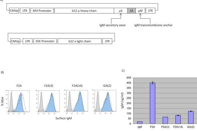
As a pilot experiment to test whether the mutated 2A peptides can mediate co- expression of surface and secreted immunoglobulins, we constructed the first-generation Molecular Rheostat Immunoglobulin genes by joining the secreted version of the b12 IgM heavy chain to the transmembrane domain of the IgM BCR via a mutated 2A peptide. The transmembrane domain is defined as the M1 and M2 exons from the human IgM locus and comprises the last 41 amino acids of the membrane bound IgM BCR (Figure 2.1A). We call these “IgM Molecular Rheostats”. We chose the wild type F2A and two mutant peptides as well as another F2A-like element derived from a silk-worm virus, based on previous work by Donnelly et al. (Donnelly, Hughes et al. 2001), in which they observed reduced cleavage efficiencies when certain mutations are introduced. The four mutants we chose are designated F2A, F2A(3), F2A(14), and I2A(2). See Table 2.1 for the nomenclature and the amino acid sequence for each of the 2A elements.
We cloned these IgM Molecular Rheostat genes into a lentiviral vector plasmid (FMHW) that doubles as a mammalian expression vector under the control of a CMV promoter. We co-transfected this vector together with a separate vector carrying the b12
light chain (FEEK-b12L) and a mammalian expression vector carrying the human Igα and Igβ genes (phIgαβ) into 293T cells (Figure 2.1A). We analyzed the cells and their supernatants by FACS and human IgM ELISA 48 hours later. All transfected cells showed surface expression of the IgM Molecular Rheostat BCR and secreted IgM into their supernatants (Figure 2.1B and 2.1C).
IgG Molecular Rheostat Mediates Expression of an IgG/M Chimeric BCR and Secreted IgG Antibody
We next attempted to adapt the Molecular Rheostat format to the production of an IgG antibody in an effort to mimic an isotype-switched secretory IgG while preserving the signaling properties of an IgM, which is required for normal B cell development.
Furthermore, we wished to explore whether we could manipulate the ratio of surface- bound to secreted immunoglobulins by making appropriate mutations in the 2A elements.
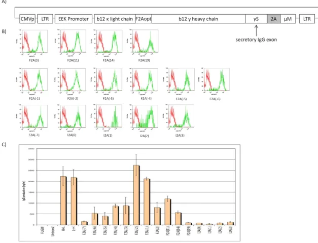
To test these ideas, we constructed a library of chimeric IgG Molecular Rheostat
immunoglobulins, in which a complete secretory b12 IgG is joined to the transmembrane anchor of the IgM BCR via different 2A peptides (Figure 2.2). The library includes all 2A peptides listed in Table 2.1.
To reduce the number of vectors that need to be transfected and anticipating the need to use the vectors in the context of lentiviral transduction, where it would be advantageous to work with a single vector, we fused the b12 light chain with the
Molecular Rheostat transgene by joining the b12 light chain to the b12 heavy chain via a
different F2A element, F2Aopt. F2Aopt is codon-optimized for human expression and contains a furin cleavage site before the 2A element.
Additionally, to ensure consistency of Igα and Igβ expression across the cells used to test the Molecular Rheostat constructs and reduce the number of vectors that need to be transfected, we engineered 293T cells that express human Igα and Igβ by repeatedly co-infecting 293T cells with two lentiviral vectors, FUW-Igα and FUW- Igβ, which carry the Igα and Igβ transgenes, repectively, under the control of a ubiquitin C promoter. The resulting cells are denoted 293T-Igαβ cells.
We transfected the library of IgG Molecular Rheostat constructs into the 293T- Igαβ cells, and 48 hours later analyzed the cells and their supernatants for surface IgG by FACS and secreted IgG by ELISA, respectively. All transfected cells showed surface expression of the IgG Molecular Rheostat BCR and secreted IgG into the culture supernatant (Figure 2.2B and 2.2C). Significantly, while the surface expression of the Molecular Rheostat BCR appears comparable across all constructs, there is a range of levels of secreted IgG. This suggests that the different Molecular Rheostats could be used to produce a range of ratios of surface to secreted immunoglobulins.
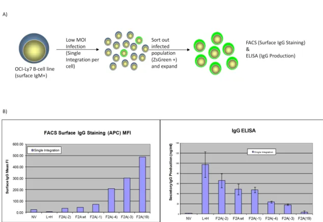
IgG Molecular Rheostat Mediates Expression of a Range of Ratios of Surface BCR to Secretory IgG in the Human B-Cell Line OCI-Ly7
To validate the results that the IgG/M Molecular Rheostat constructs can mediatea range of expression ratios of surface BCR to secreted antibodies in human B cells, we used lentiviral vectors to deliver the constructs into the OCI-Ly7 B cell line, which expresses an endogenous IgM BCR on its surface and therefore should possess the necessary machinery (such as Igα and Igβ co-receptors) for BCR surface expression. To provide an independent marker of lentiviral transduction than the expression of the Molecular Rheostat immunoglobulins, we constructed a lentiviral vector, pHAGE2-EEK- IRES-ZsGreen, which contains an Internal Ribosomal Entry Site (IRES) driving a
ZsGreen fluorescent protein gene. Based on the results in Figure 2.2B, we selected six of the IgG/M Molecular Rheostat genes and cloned them into the first position (before the IRES-ZsGreen) of the pHAGE2-EEK-IRES-ZsGreen vector. We then infected OCI-Ly7 cells with the IgG Molecular Rheostat vectors at low MOI (~ 0.1) to ensure that every cell that was infected had at most one copy of the transgene (Figure 2.3A). 48 hours after infection, we FACS-sorted out the ZsGreen positive cells and allowed these cells to expand for another 48 hours.
The cells and supernatants were analyzed by FACS and ELISA (Figure 2.3B, left and right panels, repectively). The different mutants produced a range of ratios of surface-to-secreted immunoglobulins. Significantly, there is an inverse relationship
between the amount of IgG Molecular Rheostat BCR expressed on the surface of the cells vs. the amount of IgG antibody that was detected in the supernatants, indicating that the mutant 2A elements were behaving like a “rheostat”, tuning the ratios of surface-to- secreted immunoglobulins. Also notably, the rank order of the ratios of surface-to- secreted immunoglobulin expression recapitulates what was observed from the
transfection into 293T-Igαβ cells (see Figure 2.2B and C). For example, from Figure 2.2B and C, F2A(-2) would be expected to make more secreted IgG than F2A(-4), and this was indeed the case when the constructs were expressed in the OCI-Ly7 B cell line.
Furthermore, F2A(-2) made less surface Molecular Rheostat BCR than F2A(-4), as would be expected if the F2A(-2) peptide mediated more efficient cleavage than the F2A(-4) peptide. The library of mutants thus gives us a Molecular Rheostat system that we can use to direct tunable ratios of expression of surface vs. secreted immunoglobulins.
IgG Molecular Rheostat Constructs Produce Functional b12 IgG/M Chimeric BCRs are Signaling Competent and Bind to HIV GP120
To test whether the IgG/M chimeric BCR produced by the IgG Molecular Rheostat genes is functional, we developed a ratiometric Fluo-3/FuraRed calcium flux assay in which anti-BCR crosslinking antibodies are used to examine whether the BCRs are able to signal in the OCI-Ly7 B cells. We chose two of the 2A peptides from the library, F2A, which cleaves with high efficiency, and I2A(2), which does not cleave well.
As the ZsGreen protein interferes with the Fluo-3 calcium-sensitive dye used in the assay, we cloned those two IgG Molecular Rheostat immunoglobulins into lentiviral vectors that do not have the IRES-ZsGreen marker gene. Lentiviral infections of OCI-Ly7 B cells with these vectors resulted in a variegated pattern of expression of the BCRs. The vector containing the I2A(2) element showed generally higher levels of surface BCR expression than F2A, as expected. While both populations responded to BCR stimulation using a control anti-IgM antibody (Southern Biotech, Birmingham, AB) and an anti-IgG antibody (Sigma, St Louis, MO), the responses were detectable but modest (data not shown). We believe the modest response was due to the effect of averaging the calcium signals over the large range of surface expressions. To ensure we have more homogenous populations for use in BCR stimulations, we FACS sorted out the top 10% of IgG positive cells from each of the populations (Figure 2.4B), and performed the calcium flux assays on the sorted cells. The cells responded robustly to anti-BCR stimulation (Figure 2.4A), with a dose-response correlating with the levels of surface IgG Molecular Rheostat BCR expression and the concentrations of anti-Ig used. The higher anti-IgG dose (100 ug/ml) gives a stronger calcium signal than the lower dose (20 ug/ml); the higher amount of surface Molecular Rheostat BCR also generates a stronger and more lasting response.
Additionally, to see whether the IgG/M chimeric BCR would bind to HIV antigens, we co-stained the sorted OCI-Ly7 cells with fluorescently labeled HIV gp120MN and anti-IgG interacting with the anti-GP120 epitope of b12 and the γ heavy
chain constant region of b12 IgG, respectively (Figure 2.4C). We found that the Molecular Rheostat BCRs on the cells bound to HIV GP120.
IgG Molecular Rheostat Constructs Produce b12 IgG Antibody that Neutralizes HIV Pseudovirus with Same Potency as Unmodified b12 IgG
To determine whether secreted b12 IgG from the Molecular Rheostat system can neutralize infectious virus, we performed an in vitro pseudovirus neutralization assay using an Env SF162 pseudotyped HIV-1 pseudovirus on the TMZ-b1 reporter cell line with supernatants from 293T cells transfected with several different IgG Molecular Rheostat constructs according to a protocol previously described by Klein et al. (Klein, Gnanapragasam et al. 2009). The neutralization curves demonstrated that secreted Molecular Rheostat b12 IgG antibodies neutralized the Env SF162 pseudovirus as potently as the control b12 IgG antibody (L+H), with IC50 values nearly identical to that of the control b12 IgG (Figure 2.5A). We also performed a surface-plasmon resonance GP120-binding assay. The antibodies tested bound GP120 as well as the control b12 IgG antibody, consistent with the neutralization assay results (Figure 2.5B).
Expression of IgG Molecular Rheostat Immunoglobulins Promote Maturation of EU12 Cells in an In Vitro Model of B Cell Development
The promotion of B cell development is one of the major functions performed by the IgM BCR. It thus also offers a stringent test of BCR function. To test whether the IgG
Molecular Rheostat Immunoglobulin BCR can direct B cell development, we adopted a model of human B cell development using the EU12 system (Zhang, Wang et al. 2003;
Zhang 2007). The EU12 cells are derived from a B cell leukemia patient, and the cells are CD19+ and exist in a spectrum of primitive (CD34+ and CD10-, or CD34+ and CD10+) to more mature (CD34- and CD10+, or CD34- and CD10-) states. These cells lack a
functional BCR, but rarely an IgM BCR is generated spontaneously and the cells proceed to acquire a more mature phenotype.
We isolated early-stage, CD 34+ EU12 cells by FACS sorting. These cells were then transduced with lentiviral vectors carrying IgG Molecular Rheostats that give rise to respectively low, intermediate, and high surface BCR expression. A luciferase-carrying vector was used as a control. The cells were allowed to expand, and 4 weeks after transduction the surface expression of IgG Molecular Rheostat BCR and maturation markers were analyzed by FACS (Figure 2.6). The EU12 cells transduced with Molecular Rheostat constructs tuned for different levels of surface BCR vs. secreted antibody
expression showed the expected levels of surface BCR expression (F2A was used for maximum secretion; F2A(11) for intermediate; F2A(19) for maximal surface). Using ZsGreen as a measure of the amount of gene expression from the entire cassette in each cell, the level of surface IgG Molecular Rheostat BCR expression correlates with the ZsGreen expression level for each of the three Molecular Rheostat constructs (Figure 2.6A).
Gating on the high-expressing cells, we analyzed CD34 and CD10 expression by FACS. We found that the cells that had been transduced with Molecular Rheostats tuned to higher BCR expression and less secreted antibody have larger populations of cells that down-regulated CD10 (Figure 2.6B). This provides further evidence that the IgG/M chimeric BCRs produced by the IgG Molecular Rheostat Immunoglobulins are functional BCRs and can promote maturation of B lineage cells.
Discussion
To provide a compact system for genetically manipulating the BCR and antibody specificity of B cells with a lentiviral vector, we created the Molecular Rheostat
Immunoglobulins to direct tunable simultaneous formation of the membrane-bound and secreted immunoglobulins by using mutant 2A “self-cleaving” peptides (Figure 2.7).
This system provides a synthetic approximation to the natural process of the mRNA alternative splicing-mediated switch to make membrane and secreted
immunoglobulins. By fusing an IgG to the membrane anchor of IgM through a mutant 2A peptide that functions as a Molecular Rheostat, we constructed both IgM and IgG/M chimeric versions of Molecular Rheostat immunoglobulins. We showed that such a design could produce both membrane bound and secreted immunoglobulins and
demonstrated that we could generate a library of mutant 2A elements to provide a range of tunable ratios of membrane-bound to secreted immunoglobulins by appropriate choice of mutations. We also showed that the surface chimeric IgG Molecular Rheostat BCRs signal to B cells and that these BCRs bind to HIV gp120 antigens. We showed that the secreted version of b12 IgG produced by the Molecular Rheostat constructs also bound GP120 and neutralized HIV-1 pseudovirus equally as well as unmodified b12 IgG. While the responsiveness of the BCRs was seen upon stimulation with anti-IgG antibodies that can cross-link the BCRs, it is possible that this responsiveness would be observed upon stimulation with any molecule(s) that can cross-link the BCRs, including multimeric
forms of gp120 and possibly HIV spike complexes. Finally, we provided evidence suggesting that the chimeric BCR produced by the Molecular Rheostat system can direct maturation of B cells using a cell line model of B cell maturation. In EU12 cells
transduced with vectors carrying the Molecular Rheostat Immunoglobulins, we observed increasing CD10-/CD 34- populations in the cells that received increasingly more surface- biased Molecular Rheostat constructs, suggesting that the chimeric IgG Molecular Rheostat BCRs are capable of directing the normal B cell maturation progression from CD10-/CD34+ to CD10+/CD34+ to CD10+/CD34- to CD10-/CD34-. We note, however, that the CD10-/CD34+ populations were also greater in cells that were treated with Molecular Rheostat immunoglobulins biased toward higher surface BCR expression. At first glance, this might be explained by the downregulation of CD10 alone as a result of the expression of chimeric BCR. However, that the ratio of the most mature CD10-, CD34- double negative population to the most primitive CD10-,CD34+ population also increases with the use of surface-baised Molecular Rheostat immunoglobulins suggests that the chimeric Molecular Rheostat BCR gives the more mature cells a proliferative advantage over the more primitive cells. This is consistent with the hypothesis that the Molecular Rheostat immunoglobulin genes promoted maturation of the cells.
While one might imagine encoding the entire heavy chain and light chain locus into a vector to program B cells, the heavy chain locus alone is ~ 1 Mb, too big to incorporate into a lentiviral vector with a coding capacity of ~ 10 kb. We had attempted
earlier to remove the introns of the heavy chain locus, except for the one required for the alternative splicing of the secreted and transmembrane exons. Our efforts to get those constructs to splice were not successful. We thus created the Molecular Rheostat system to mimic the natural system, incorporating the additional feature of expressing isotype- switched IgG antibodies while maintaining the signaling properties of an IgM
transmembrane domain. Our results suggest that the Molecular Rheostat system, which is small enough to be introduced into cells with a lentiviral vector, could be used to direct the in vivo maturation of anti-HIV B cells. A detailed in vivo characterization of the Molecular Rheostat system in animal models would be necessary to test this idea. We propose to use this system to transduce hematopoietic stem cells or B cells in transplant models as a prophylactic “vaccine” against HIV infections. Work is currently under way to study the use of these Molecular Rheostat Immunoglobulins in vivo as a vaccination strategy against HIV, but the system may be used to manipulate B cells to target other antigens.
Figure 2.4 Molecular Rheostat BCRs generate calcium signals in response to anti-BCR stimulations and bind to HIV gp120. A) Calcium response of cells to anti-BCR
stimulation. First column: response of endogenous IgM BCR to anti-IgM stimulation.
Second column: high dose (100 ug/ml) anti-IgG stimulation. Third column: low dose (20 ug/ml) anti-IgG stimulation. B) BCR expression post-sorting. Endogenous IgM
expression (vertical) vs. surface IgG staining from IgG Molecular Rheostat BCR (horizontal). Red: sorted cells expressing the Molecular Rheostat Immunoglobulins.
Green: uninfected control cells. C) Anti-IgG and gp120MN labeling of sorted cells. Red
and Blue: I2A(2) and F2A Molecular Rheostat Immunoglobulin vector transduced cells, repectively. Green: untransduced control cells.
2A
Mutant Mutation Type Amino Acid Sequence
F2A Wild-type QLLNFDLLKLAGDVESNPGP
F2A(-7) 7aa N-terminal deletion
LKLAGDVESNPGP F2A(-6) 6aa N-terminal
deletion
LLKLAGDVESNPGP F2A(-5) 5aa N-terminal
deletion
DLLKLAGDVESNPGP F2A(-4) 4aa N-terminal
deletion
FDLLKLAGDVESNPGP F2A(-3) 3aa N-terminal
deletion
NFDLLKLAGDVESNPGP F2A(-2) 2aa N-terminal
deletion
LNFDLLKLAGDVESNPGP F2A(-1) 1aa N-terminal
deletion
LLNFDLLKLAGDVESNPGP
F2A(3) Point mutation QLLNFDLLKLAGDVQSNPGP
F2A(11) Point mutation QLLNFDLLKLAGDVEINPGP F2A(14) Point mutation QLLNFDLLKLAGDVESEPGP F2A(19) Point mutation QLLNFDLLKLAGDVESNPAP
I2A(0) Wild-type TRAEIEDELIRRGIESNPGP
I2A(1) Point mutation TRAEIEDELIRAGIESNPGP I2A(2) Alternative codon TRAEIEDELIRRGIESNPGP I2A(3) Point mutation TRAEIEDELIRRGIESNPAP Table 2.1 Nomenclature and amino acid sequences of different 2A peptides.
Figure 2.1 A) Schematic representation of the IgM Molecular Rheostat
Immunoglobulin contructs. 2A: location of self-cleaving 2A elements. CMVp: CMV promoter. LTR: long terminal repeat. MH and EEK promoters: internal B cell specific promoters. b12 µ heavy chain: IgM heavy chain with variable region corresponding to that of the b12 broadly neutralizing antibody. B) Surface staining for human IgM. All 293T cells transfected with first-generation Molecular Rheostat contructs show surface staining for IgM. White: Membrane-bound IgM control. Blue: Molecular Rheostat
contructs. Gray: GFP control. C) IgM ELISA of supernatants of transfected cells. IgM:
membrane-bound IgM control.
Figure 2.2 A) Schematic representation of the IgG/M Molecular Rheostat constructs.
2A: location of mutant self-cleaving 2A elements. 2Aopt: optimized 2A element with a furin cleavage site at 5’ end. CMVp: CMV promoter. LTR: long terminal repeat. EEK:
internal B cell specific promoter. b12 γ heavy chain: IgG heavy chain with the variable region corresponding to that of the b12 broadly neutralizing antibody. B) Surface staining for human IgG. Green: Molecular Rheostat Contructs. Red: Secretory IgG (L+H)
control. C) IgG ELISA of supernatants of transfected cells. FUGW: GFP containing
vector control. L+H and H+L: secretory b12 IgG controls. H+L has the light chain in the first position and heavy chain in the second position; L+H is in the opposite order.
Figure 2.3 A) Experimental design for measuring the ratioed expression of surface-to- secreted immunoglobulins by IgG Molecular Rheostat constructs. B) Inverse relationship between surface expression of IgG Molecular Rheostat BCRs and secreted IgGs in the supernatants of sorted cells. NV: untransduced control. L+H: secretion only b12 control.
The Molecular Rheostat contructs are denoted by the mutant 2A elements they contain.
Figure 2.5 Secreted b12 IgG from the Molecular Rheostat constructs neutralized Env SF162 pseudovirus and bound to GP120 as well as control b12 IgG. A) In vitro
neutralization assay against Env SF162 pseudovirus. Green: L+H, the control b12 IgG.
Red, Magenta, and Orange: different b12 IgG Molecular Rheostat antibodies. Black: a different batch of b12 IgG purified included as positive control for the assay. B) Surface Plasmon Resonance GP120 binding assay.
Figure 2.6 Molecular Rheostat BCRs promote maturation of EU12 cells. A) CD 34+ EU12 cells (early B cells) transduced with IRES-driven ZsGreen expressing Molecular Rhestat constructs were analyzed by flow cytometry. Surface BCR levels correlate with ZsGreen intensity. Cells transduced with Molecular Rheostats tuned for higher surface expression showed more surface BCR expression with the same ZsGreen expression. B) Red arrows indicate the normal B cell development progression. Cells transduced with constructs that express higher surface IgG/M BCR levels show greater mature B cell populations.
Figure 2.7. A model of how the b12 IgG Molecular Rheostat immunoglobulin system directs tunable simultaneous formation of surface BCR and secreted IgG.
References
Burrows, P. D. and M. D. Cooper (1993). "B-cell development in man." Curr Opin Immunol 5(2): 201-206.
Chen, J. and F. W. Alt (1993). "Gene rearrangement and B-cell development." Curr Opin Immunol 5(2): 194-200.
de Felipe, P. (2004). "Skipping the co-expression problem: the new 2A "CHYSEL"
technology." Genet Vaccines Ther 2(1): 13.
de Felipe, P., L. E. Hughes, et al. (2003). "Co-translational, intraribosomal cleavage of polypeptides by the foot-and-mouth disease virus 2A peptide." J Biol Chem 278(13): 11441-11448.
Donnelly, M. L., L. E. Hughes, et al. (2001). "The 'cleavage' activities of foot-and-mouth disease virus 2A site-directed mutants and naturally occurring '2A-like'
sequences." J Gen Virol 82(Pt 5): 1027-1041.
Donnelly, M. L., G. Luke, et al. (2001). "Analysis of the aphthovirus 2A/2B polyprotein 'cleavage' mechanism indicates not a proteolytic reaction, but a novel translational effect: a putative ribosomal 'skip'." J Gen Virol 82(Pt 5): 1013-1025.
Doronina, V. A., P. de Felipe, et al. (2008). "Dissection of a co-translational nascent chain separation event." Biochem Soc Trans 36(Pt 4): 712-716.
Doronina, V. A., C. Wu, et al. (2008). "Site-specific release of nascent chains from ribosomes at a sense codon." Mol Cell Biol 28(13): 4227-4239.
Kitamura, D. and K. Rajewsky (1992). "Targeted disruption of mu chain membrane exon causes loss of heavy-chain allelic exclusion." Nature 356(6365): 154-156.
Kitamura, D., J. Roes, et al. (1991). "A B cell-deficient mouse by targeted disruption of the membrane exon of the immunoglobulin mu chain gene." Nature 350(6317):
423-426.
Klein, J. S., P. N. Gnanapragasam, et al. (2009). "Examination of the contributions of size and avidity to the neutralization mechanisms of the anti-HIV antibodies b12 and 4E10." Proc Natl Acad Sci U S A 106(18): 7385-7390.
Ryan, M. D. and J. Drew (1994). "Foot-and-mouth disease virus 2A oligopeptide mediated cleavage of an artificial polyprotein." EMBO J 13(4): 928-933.
Spanopoulou, E., C. A. Roman, et al. (1994). "Functional immunoglobulin transgenes guide ordered B-cell differentiation in Rag-1-deficient mice." Genes Dev 8(9):
1030-1042.
Szymczak, A. L. and D. A. Vignali (2005). "Development of 2A peptide-based strategies in the design of multicistronic vectors." Expert Opin Biol Ther 5(5): 627-638.
Wagner, S. D., G. T. Williams, et al. (1994). "Antibodies generated from human immunoglobulin miniloci in transgenic mice." Nucleic Acids Res 22(8): 1389- 1393.
Young, F., B. Ardman, et al. (1994). "Influence of immunoglobulin heavy- and light- chain expression on B-cell differentiation." Genes Dev 8(9): 1043-1057.
Zhang, Z. (2007). "VH replacement in mice and humans." Trends Immunol 28(3): 132- 137.
Zhang, Z., Y. H. Wang, et al. (2003). "Molecular mechanism of serial VH gene replacement." Ann N Y Acad Sci 987: 270-273.