From my labmates, I would like to thank Sydney Henriques for her assistance with in vivo studies and former lab members Dr. I also want to thank my stepfather, Brad Mattie, and my stepmother, Mona Glass, who also supported me and provided positive words when I
Low Response Rates in Current Immunotherapies
In this way, macrophages can be used to assist in the antitumor immune response and the generation of immunological memory. In addition, recent work has also demonstrated the ability of macrophages to induce innate immune memory, a function previously thought to be exclusive to the adaptive immune system. long-term therapies can be developed.

Techniques for Repolarizing TAMs to a Pro-Inflammatory Phenotype
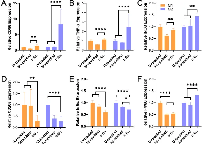
Alternatively to knockdown inhibitors of NF-κB, M1 polarization can also be induced by direct stimulation with inflammatory cytokines. Inflammatory cytokines, such as interferons (IFNs) and tumor necrosis factors (TNFs), promote M1 polarization and lead to more.
Injectable Biomaterials for Localized Drug Delivery
For our applications, we have developed a unique polymer specifically designed using siRNA loading and endosomal escape. In particular, injectable hydrogels for cancer therapy have gained popularity with scaffolds loaded with immune checkpoint blockades to sustain anti-PD-173 release, proton scavengers to modulate acidity in the TME,74 neoantigens to promote adaptive immune responses,75 among others reviewed by Leach, et al.
Polymeric Nanoparticles for Tailored Targeting
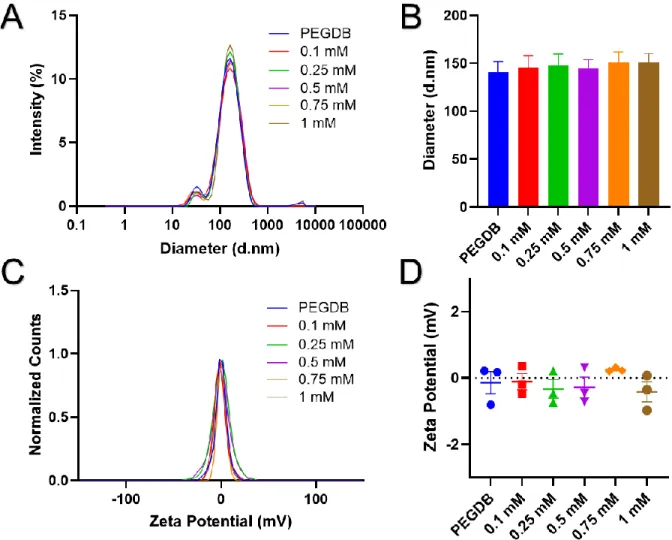
The endosomal escape mechanism of the PEGDB polymer has been previously described.79 The final component of the polymer NP system is the corona decoration for macrophage targeting. Ascites is the accumulation of fluid in the peritoneal cavity associated with the development of ovarian cancer.
Macroporous Cryogels as Localized Repolarization Depots
The concept of developing cancer immunotherapies using TAMs is not new in itself, but most TAM-based therapies have focused on preventing macrophage recruitment to the tumor or removing the TAM population in the TME .31–33 Other therapies have been developed more recently, and are summarized in a review by Duan and Luo.88 These therapies mainly consist of ablation of TAMs, inhibition of recruitment or prevention of tumor-promoting functions such as blocking angiogenesis and immunosuppression.88 However, a more promising and robust response is assumed if the TAMs are repolarized and employed as part of the anti-tumor immune response. The experiment performed in this aim will test the hypothesis that mannosylated NPs can provide targeted delivery to TAMs in the ascites and solid tumor and that delivery of IκBα siRNA to the TAMs will drive an inflammatory M1 response to suppress tumor development.
Introduction
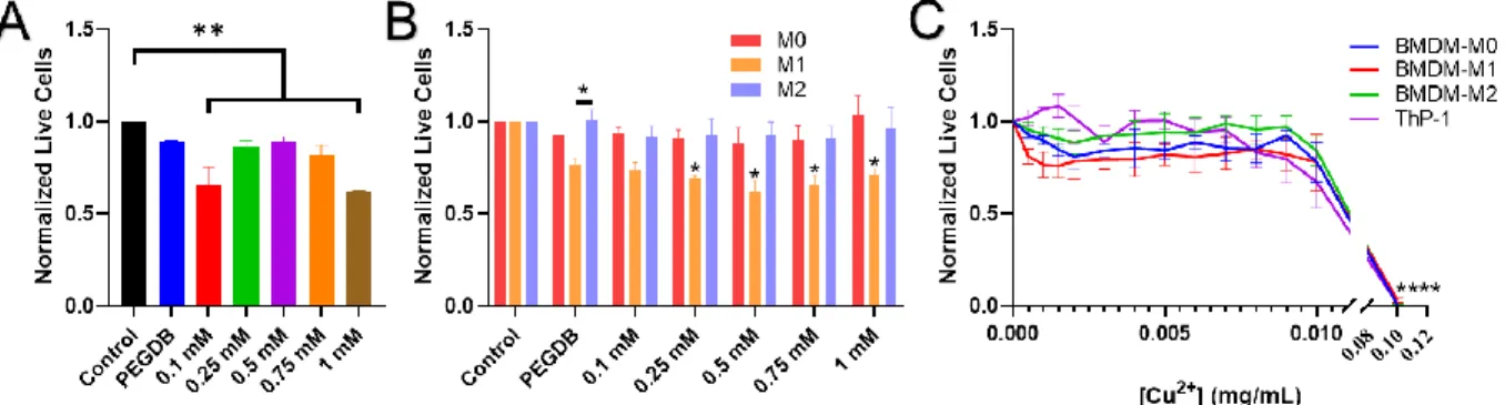
Cells were incubated for 24 hours before performing the CellTiter-Glo ® Luminescence Assay (Promega, Madison, WI). Cells were plated in 96-well plates and treated with 50 nM polyplexes loaded with Cy5-dsDNA.
Results and Discussion
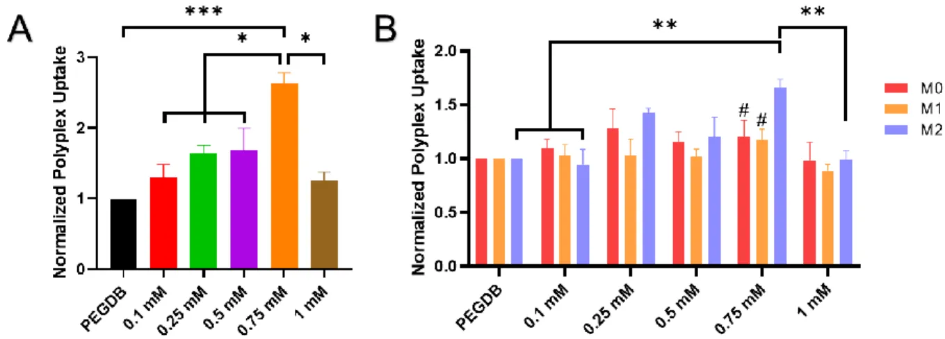
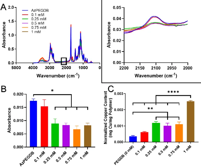
In ThP-1 human macrophages, MnPEGDB produced with 0.75 mM copper catalyst significantly increased uptake efficiency compared to all other polymer groups (Figure 2.4A). However, in the M2 polarized macrophages, the polymer prepared with 0.75 mM copper catalyst led to significantly increased uptake compared to the 0.1 and 1 mM groups, but not the 0.25 and 0.5 mM catalyst groups (Figure 2.4B).
Conclusions
After the nitrogen purge was removed, the solution was heated to 40°C in an oil bath and stirred for 24 hours. The final solution was added to equal volume of diethyl ether for extraction of excess reagents.
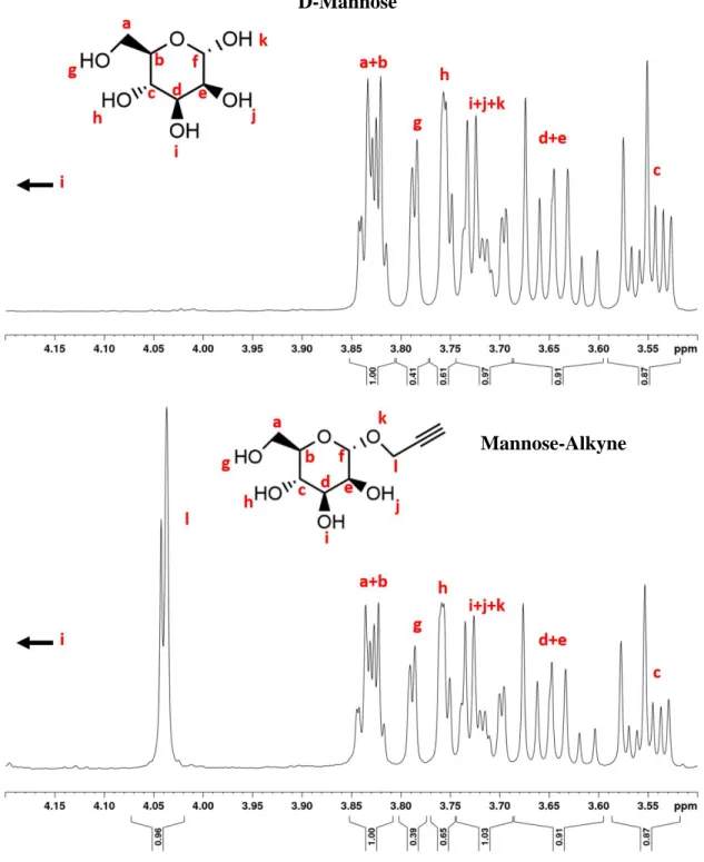
Mannose
Background
ID8 ovarian tumor cells and TBR5 genetically engineered ovarian tumor cells were used as previously described. 174–177 ID8 cells were from dr. ID8 cells were used in syngeneic mice in the C57Bl/6 background, while TBR5 cells were used in syngeneic mice in the FVB background. The resulting cells were resuspended in PBS with 1% BSA and snap frozen in liquid nitrogen for RNA.
After staining for 30 minutes, the cells were rinsed in PBS and resuspended in lysis buffer before lysis. For the TBR5 biodistribution study, flow cytometry was performed as previously described.183 The cells were incubated in an Fc block (BD Biosciences; 553142) for 10 minutes at RT, stained for surface markers for 15 minutes at RT, washed with a FACS buffer. containing PBS with 2% (v/v) FBS, and resuspended in the FACS buffer for flow analysis on a Miltenyi MACSQuant Analyzer 10 or 16.
Results
Macrophages displayed significantly high %MnNP+ levels compared to most other immune cells in tumor (C), (D) ascites, and (E) spleen. In contrast to the ID8 model, MnNP treatment in the TBR5 model significantly decreased ascites accumulation and decreased tumor burden (Figure 3.5E,F). A prolonged MnNP treatment in the TBR5 model was used to further evaluate the effects of the treatment on tumor progression and immune cell composition.
The percentage of tumor cells (gated CD45-/SSChi) in the ascites and tumor was significantly reduced in the IκBα-MnNP treatment compared to the PBS control (Figure 3.6D,E). Furthermore, the immune cells in the tumor and ascites were altered as a result of MnNP treatments.
Discussion
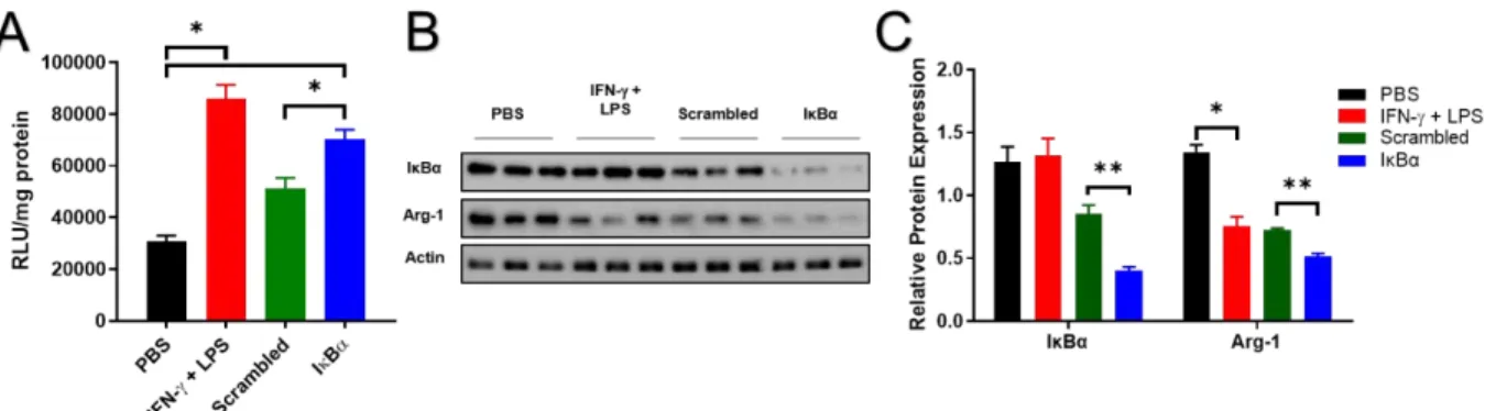
These results revealed that even 24 h after a single treatment, MnNPs preferentially associate with macrophages in ascites and tumor without targeting to the spleen. Encouraging results in late-stage treatment provided insight into the effects that IκBα-MnNPs have on immune cells in the TME. Western blot analysis of cells in ascites revealed that IκBα-MnNPs reduced IκBα protein levels, contributing to changes in immune cell composition.
A two-week treatment allowed MnNPs to successfully deliver TAMs in a solid tumor and change the phenotype of immune cells. These results immediately revealed significant therapeutic benefits of MnNP delivery, with ascites volume and tumor weight significantly reduced upon treatment.
Conclusions
Likewise, the positive therapeutic effects observed with early treatment of the aggressive TBR5 model, in terms of tumor progression and immune cell composition, provide context for future directions in combining MnNP treatments with T cell-targeted immunotherapies. Delivery of MnNPs to macrophages in the ascites led to a slight decrease in ascites build-up in the late-stage model, but a substantial decrease in the aggressive TBR5 model. The positive effect of mannosylated carriers on preventing ascites development in the aggressive TBR5 model indicates a potential for synergistic effects of IκBα siRNA with MnNPs.
While treatment of late-stage ID8 tumors did not significantly alter tumor progression, there were notable differences in the RNA expression of various M1- and M2-related markers, indicating beneficial immunostimulatory changes in the TME after MnNP treatment. In addition, we demonstrated that extended MnNP treatment in the TBR5 model significantly inhibited ascites accumulation and tumor development while altering the composition of immune cells in the TME.
Supplemental Figures
Supplementary Figure 3.S6: Polyplexes were formulated with scrambled siRNA or IκBα-siRNA and suspended in PBS (-/-, pH = 7.4). Supplementary Figure 3.S7: Gating strategy for flow cytometry of the ID8 ovarian tumor model with single 24-hour MnNP release. Supplementary Figure 3.S8: Gating strategy for flow cytometry of solid tumors in the TBR5 ovarian tumor model with biweekly MnNP release.
Supplementary Figure 3.S9: Passage strategy for flow cytometry of ascites cells in the TBR5 ovarian tumor model with biweekly delivery of MnNPs. Supplementary Figure 3.S10: Passage strategy for flow cytometry of spleens in the TBR5 ovarian tumor model with biweekly delivery of MnNPs.
Spleen
Introduction
Sodium alginate functionalization with aminoethyl methacrylate (AEMA) to form methacrylated alginate (MA alginate) and subsequent cross-linking of methacrylate groups were confirmed by H nuclear magnetic resonance (NMR) spectroscopy (Supplementary Figure 4.S1). However, IFN-γ treatment reduced Arginase-1 expression only 10-fold compared to a 74.8-fold decrease with IL-12 treatment (Supplementary Figure 4.S7). However, the BMDMs treated with ILC gels showed a significant increase in the receptor MHCII, up to the level of the M1 controls (Figure 4.5F).
Several markers showed significant upregulation in the ILC-treated BMDMs, including IL-1β, TNF-α, CXCL10, and CXCL9 (Figure 4.5G-H). Of these populations, there were no changes in the percentage of M2-like TAMs (CD206+/MHCII-) or M1-like TAMs (CD206-/MHCII+) in any of the treatments (Supplementary Figure 4.S13A,B).

Materials and methods
Collected cells were centrifuged (1000 g , 5 min) and blood cells were lysed by resuspending them in 2 ml of ACK lysis buffer (KD Medical, Columbia, MD) and incubating on ice for 2 min. Cells were cultured for four days in L929-conditioned medium to allow macrophage maturation. Cells were washed again before permeabilization with 0.5% Triton X100 in PBS (−/−) for 5 min at room temperature.
The cells were stained with Hoechst at 1μg/ml for 5 minutes at room temperature in the dark. Supplemental Figure 4.S11: PyMT tumor explants were cultured for 96 h in vitro and then evaluated with a CellTiter-Glo viability assay to ensure that viable cells were present after 4 days of culture.
Future Work and Potential Applications
In current applications, although the current MnNP formulation demonstrated high specificity for macrophage uptake in tumors and ascites after IP injections, other targeting moieties may allow tumor-specific targeting after IV injections. Furthermore, to increase NP accumulation at the tumor site after IV injection while avoiding macrophage uptake in other organs, this polymer system can be mixed with another polymer with an extended PEG chain (20 kDa) containing a matrix metalloproteinase (MMP)-cleavable peptide linker. . With the extended PEG polymer combined with MnPEGDB, the longer 20K PEG will “hide” the mannose (or other target moiety) from circulation.
In this way, the free chemokine would be released immediately, while the bound molecules would remain in the gel until the MMPs reach them and cleave the peptides. In the PyMT mouse model, this can be achieved by treating the primary tumor in the left flank #4 mammary fat pad, followed by the injection of tumor cells into the right flank #4 mammary fat pad after regression is seen in the first tumor.
Conclusions
Glass EB, Hoover AA, Bullock KK, Madden MZ, Reinfeld BI, Harris W, Parker D, Hufnagel DH, Crispens MA, Khabele D, Rathmell WK, Rathmell JC, Wilson AJ, Giorgio TD and Yull FE. Glass EB, Roy S, Manning AE, Hacker BC, Haycook CP, Dollinger BR, Rafat M, Kim YJ and Giorgio TD: Injectable alginate cryogels as repolarization depots for tumor-associated macrophages. Haycook CP, Balsamo JA, Glass EB, Williams CH, Hong CC, Major AS and Giorgio TD.
Hoover AA, Hufnagel DH, Harris W, Bullock K, Glass EB, Liu E, Barham W, Crispens MA, Khabele D, Giorgio TD, Wilson AJ, and Yull FE. Carr MH, Glass EB, Masjedi S and Giorgio TD: Analysis of the Effects of IFN-γ Induced Repolarization on the Viability of Bone Marrow-Derived Macrophages.
Tumor-associated macrophages correlate with the phenomenon of epithelial-mesenchymal transition and contribute to poor prognosis in triple-negative breast cancer patients. A novel liposomal clodronate depletes tumor-associated macrophages in primary and metastatic melanoma: antiangiogenic and antitumor effects. Evaluating the polarization of tumor-associated macrophages into M1 and M2 phenotypes in human cancer tissue: technical details and challenges in routine clinical practice.
A high M1/M2 ratio of tumor-associated macrophages is associated with prolonged survival in patients with ovarian cancer. Biocompatible mannosylated endosomal escape nanoparticles enhance the selective delivery of short nucleotide sequences to tumor-associated macrophages.