Notably, however, the CT reaction was significantly more efficient on the 3' side of the triplex. These data also highlight the importance of the p-stacked array as the critical pathway for CT.
Towards a Mechanistic Understanding of Long-Range Charge
Significant lesions at the distal site of the guanine doublet were observed in all cases with up to 10 intervening A, T, or alternating AT sequences. Thus, shifting the negative charge to the end more proximal to the rhodium intercalator dramatically reduced oxidative damage to the distal guanine doublet.
Spectroscopic Identification of Radical Intermediates in Long-Range CT
Remarkably, in series containing I at the injection site and no intervening guanines, radical product formation was also observed at doses ≥ 107 s-1. Even more intriguingly, the 600 nm signal is significantly larger for sequences containing I at the injection site, indicating a higher yield of radical formation.
Electrochemical Detection of Base Stacking Perturbations and
Perhaps DNA-binding proteins containing redox-active cofactors or structural elements such as flavins or Fe-S clusters benefit from DNA CT for communication in vivo. The demonstration of long-range oxidative damage in NCP and nuclei extends DNA CT as a viable mechanism for generating basic cellular lesions.
RERFERENCES
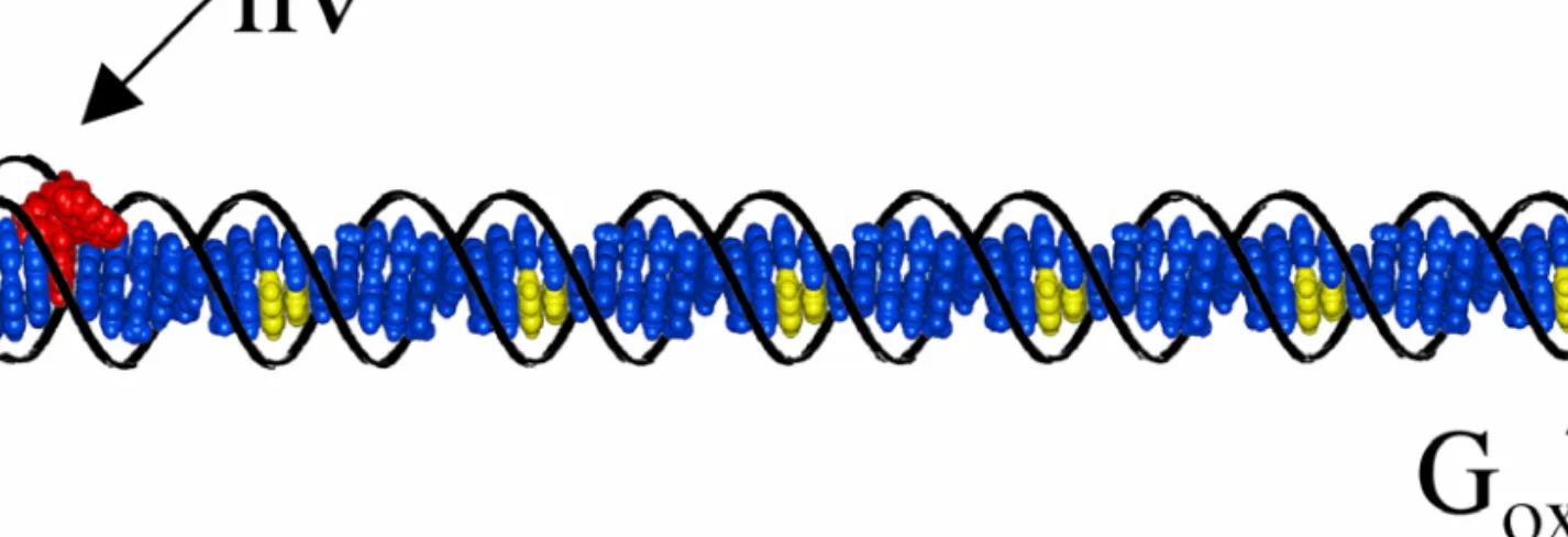
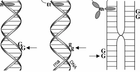
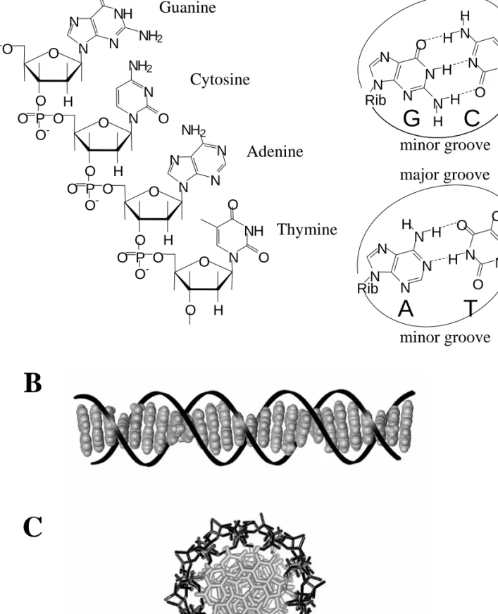
Schematic illustrations of some of the DNA structures studied for their ability to mediate CT. Upon photooxidant excitation, these DNA structures efficiently mediate long-range oxidative damage.
INTRODUCTION
Ruthenium Dppz complexes have been extensively studied because of their unique luminescence properties when bound to DNA (16, 17). The binding of ruthenium dpq and dpqC complexes to DNA has been structurally explored using NMR methods (18, 19).
METHODS
Electrochemistry. Ground state oxidation and reduction potentials for the ruthenium complexes were obtained on a Bioanalytical Systems (BAS) Model CV-50W
Emission intensity was determined by integration of the luminescence spectrum and standardized against [Ru(bpy)3]2+ as a calibration for. Luminescence titrations in an ISS-K2 fluorometer were performed to determine affinity constants for ruthenium.
Determination of Binding Constants to DNA. Luminescence titrations on an ISS-K2 fluorometer were performed to determine affinity constants for the ruthenium
Quantification was performed on a Beckman DU 7400 spectrophotometer using ε260 values estimated for single-stranded DNA (30). Ruthenium-conjugated oligonucleotides were characterized by mass spectrometry and quantified using the following extinction coefficients: [Ru(bpy')(dppz)(phen)]2+ modified oligonucleotides ε M-1cm-1; [Ru(bpy)2(bpy’)]2+ modified oligonucleotides ε M-.
Assay of Oxidative DNA Damage. For experiments conducted using noncovalently bound ruthenium, single strands containing the guanine doublet site were
For experiments performed using noncovalently bound ruthenium, it was the single strands that contained the double guanine site. After irradiation, samples were treated with 10% piperidine at 90 °C for 30 min, dried, and electrophoresed through a 20% denaturing polyacrylamide gel.
RESULTS
- Luminescence Characteristics in the Absence and Presence of DNA
- Binding Affinities Determined through Luminescence Titration and Support for an Intercalative Binding Mode. Spectroscopic titrations of the ruthenium
- Oxidative Damage by Noncovalently Bound Ruthenium Complexes
- Oxidative Damage by Covalently Bound Ruthenium Complexes. To probe the importance of intercalation to oxidative damage by long-range CT most
In the presence of the oxidative quencher [Ru(NH3)6]3+, all the DNA-bound ruthenium complexes investigated exhibit shorter excited state lifetimes indicative of dynamic quenching by the groove-bound ruthenium hexamine. Spectroscopic titrations of the ruthenium complexes with ct-DNA were performed over a range of metal concentrations.
DISCUSSION
Intercalative Binding by the Family of Ruthenium Complexes. The data shown here provide support for intercalative binding by the full family of ruthenium
The first consists of dppz and dppx complexes; these show the highest binding affinities, reflecting deep intercalation within the helix. Because of the hydrophobicity associated with dpq and dpqC, the groove-binding linkage seemed reasonable to consider as a.
Different Modes of Reactivity. Photoactivation of the ruthenium complexes bound to DNA leads to two distinct routes for oxidative damage, and these
Photoactivation of the ruthenium complexes bound to DNA leads to two distinct pathways of oxidative damage, and these. Instead, the reactivity depends on redox potentials, the ruthenium(III/II) couple and the guanines.
Direct Correlation Between Intercalation and DNA Charge Transport
We attribute this lack of reactivity at distal positions to the lack of coupling of the ruthenium oxidant into the base pair stack. By studying a family of ruthenium complexes containing the dppz ligand or derivatives, it was found that the ability of the complex to intercalate into the π-strand of DNA directly affects the extent of DNA CT and the resulting damage.
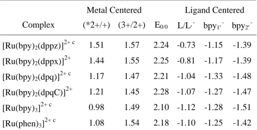
The E0/0 values are obtained from the luminescent triplet excited state and are therefore underestimates of the actual values. When present, the concentration of CT-DNA was 1 mM nucleotide, and the glycerol samples contained 60% glycerol by volume. Samples were irradiated at 450 nm and emission was monitored at 610 nm using a 495 nm cutoff filter.
5’-TGATCGGTGCGTCTGAGACT-3’
3’-ACTAGCCACGCAGACTCTGA-5’
INTRODUCTION
The molecular orbitals involved in this CT event are the HOMOs of the intervening bases of DNA (Figure 3.1). HPLC was used to monitor recovery of the thymine dimer over Å with intervening A – T base pairs; a very weak distance dependence on ET was observed and no dependence on. Therefore, the authors suggest that the rate of cleavage of the thymine dimer after reductive repair is comparable to that of ET by DNA.
METHODS
- Electrochemistry. Ground state oxidation and reduction potentials for the metal complexes were obtained on a Bioanalytical Systems (BAS) Model CV-50W
The ground state oxidation and reduction potentials for the metal complexes were obtained on a Bioanalytical Systems (BAS) Model CV-50W. Metal complexes were obtained on a Bioanalytical Systems (BAS) Model CV-50W electrochemical analyzer. E1/2 values were taken as the average of the voltage of maximum current for the forward and reverse electrochemical processes. The emission intensities were determined by integration of the luminescence spectrum and standardized against [Ru(bpy)3]2+ as a calibration for the instrument.
RESULTS
- Assay for Reductive DNA Damage
- Polyacrylamide Gel Electrophoresis. Products of oxidative DNA flash/quench experiments are readily visualized via denaturing polyacrylamide gel
- High Performance Liquid Chromatography. HPLC offers an
- Transient Absorption Spectroscopy. Transient absorption spectroscopy has been utilized to probe reductive flash/quench systems for transient species
Direct strand breaks and alkaline-labile lesions were visualized by denaturing PAGE ( 44 ) and attributed to hydrogen atom abstraction from the C1′ and C2′ position of the adjacent 5′-nucleotide. After piperidine treatment, damage is observed in FU, however, this damage is also present in dark controls, indicating that the damage is not a result of turn-on/quench chemistry, but rather site sensitivity to piperidine treatment. These changes were not observed after irradiation of the on/off samples at 442 nm for up to 1 h.
![Table 3.2. Fluorescence Quenching Data for *[Ru(phen) 2 (dppz)] 2+ by Electron Donating Molecules](https://thumb-ap.123doks.com/thumbv2/123dok/10408630.0/90.918.161.814.670.845/table-fluorescence-quenching-data-phen-electron-donating-molecules.webp)
DISCUSSION
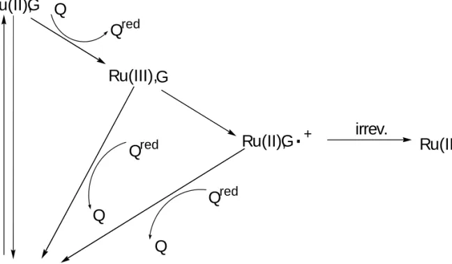
In this case, the ruthenium complex was covalently bound to the 5' end of the DNA duplex, and the back reaction with reduced quencher, [Ru(NH3)6]2+, was greatest with guanine at the charge injection site. In comparison, trapping of the guanine cation radical with H2O and/or O2 occurs at a rate of 104 s-1. Future work will include incorporation of the CPC nucleotide into DNA and monitoring for subsequent ring opening.
G -1.29V HOMO
C 1.1VLUMO
- INTRODUCTION
- METHODS
- Oligonucleotide Synthesis. The oligonucleotides were synthesized on an Applied Biosystems 394 DNA synthesizer using standard phosphoramidite chemistry
- RESULTS AND DISCUSSION
- Transient Absorption Spectroscopy on Methylindole Containing Assemblies. Figure 4.2 shows transient absorption data monitored at 600 nm after laser
- Oxidative DNA Damage Products Observed by Gel Electrophoresis
- REFERENCES
Below is the autoradiogram after irradiation of the oligonucleotide in the presence of Ru(bpy)2(dppz)2+. Below is the autoradiogram after irradiation of the oligonucleotide in the presence of Ru(phen)2(dppz)2+ and KI (lanes 5, 6, 9 and 10). For compound Ru-I-A-M, the data at 600 nm clearly show the rise of a positive signal corresponding to the formation of the methylene indole radical cation.
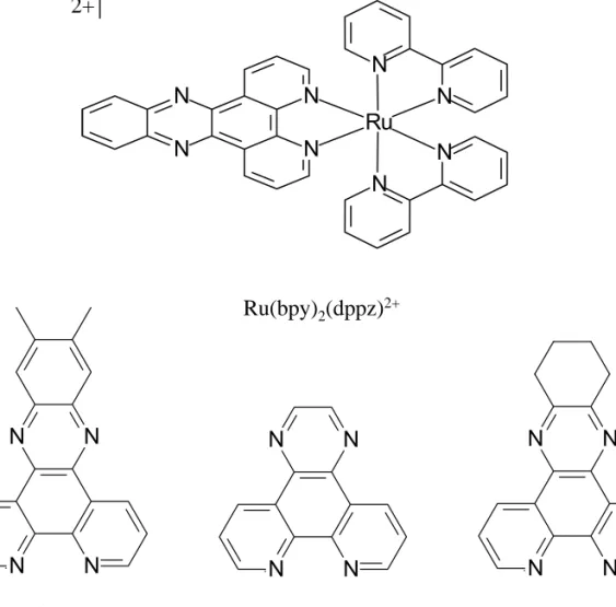
![Figure 3.3. Shown above is [Ru(bpy) 2 (dppz)] 2+ , the parent complex. Below is shown the derivatives of the dppz ligand utilized in these studies: dppx, dpq and dpqC where dppx = 7,8-dimethyldipyridophenazine, dpq = dipyridoquinoxaline and dpqC =](https://thumb-ap.123doks.com/thumbv2/123dok/10408630.0/110.918.185.754.132.691/figure-shown-complex-derivatives-utilized-studies-dimethyldipyridophenazine-dipyridoquinoxaline.webp)
TCA GAAAAAAGMGTCTA
TCA IAAAAAAGMGTCTA
TCA GAAAAAAGGGTCTA
TCA IAAAAAAGGGTCTA
TCA GAAAAAAGMGTCTA -AIT CTTTTTTCCCAGAT
TCA IAAAAAAGMGTCTA -AIT CTTTTTTCCCAGAT
TCA IAAAAAAGMGTCTA -AIT CTTTATTCCCAGAT
INTRODUCTION
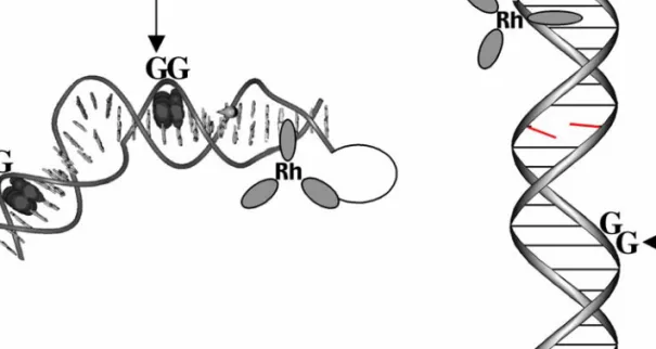
In this assembly, photoinduced oxidative damage of the 5'-G of 5'-GG-3' sites was observed; this damage pattern is considered the. Using rigid stilbene-modified hairpins, Lewis and colleagues observed a steep distance dependence of CT rates by monitoring the formation of the stilbene radical anion (13). Recent work by Kawai, Majima and colleagues investigated the yields of the charge-separated state in DNA hairpins.
METHODS
Purification of the ruthenium-modified DNA by reversed-phase HPLC afforded four isomers, which were characterized by UV-vis spectroscopy and MALDI-TOF mass spectrometry. DNA duplexes were formed by mixing equal concentrations of complementary strands in 50 mM NaCl, 15 mM sodium phosphate, pH 7 and heating to 90 °C, followed by slow cooling to 20 °C over 120 min. Emission intensity was obtained by integrating the area under the luminescence decay curve.
RESULTS
- Charge Transport Chemistry Is an Intraduplex Reaction. To initiate CT chemistry using dppz complexes of ruthenium the flash/quench technique is utilized (31)
- Oxidative Damage Products Observed by Gel Electrophoresis
For Ru-GGG-GMG and Ru-GMG-GGG, damage is also observed at the low-energy GGG site. For Ru-GMG, a large positive signal is observed at 600 nm, consistent with the formation of the methylindole radical. Also evident in Figure 5.7 is the difference in the decay rate of the positive signal at 600 nm for Ru-GMG, Ru-GGG-GMG and Ru-GMG-GGG.
DISCUSSION
- Singlet Oxygen Chemistry to Confirm an Intraduplex Charge Transport Reaction. The ruthenium photochemistry (31) is valuable not only in
- Competition between Two Oxidatively Sensitive Sites in DNA. The biochemical experiments confirm the competition between the methylindole and GGG
- Kinetic and Themodynamic Traps of Charge Transport Damage. When examining oxidative damage at two sites in a DNA duplex, it is important to consider the
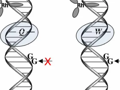
Clearly, it is not surprising that in Ru-GGG-GMG, the presence of the GGG site decreases the yield of the methylindole cation radical. Most interestingly, as CT decreases in the distal region of GGG as a result of base swelling, an increase in oxidative damage is observed in methylindole compared to Ru-GMG-GGG. In Ru-GMG, Ru-GGG-GMG, and Ru-GMG-GGG, the hole can probe the entire length of the duplex before returning at a cr rate.
PAGE after irradiation of Ru-GMG-GGG(left) and the duplex containing bulge Ru-GMG-blg-GGG(right) in the absence of quencher. The samples contained equimolar amounts of unlabeled duplexes containing the ruthenium oxidant and 32P-labeled duplex pa.
INTRODUCTION
Upon triplex targeting of a photooxidant to long DNA restriction fragments, the typical distance regime for long-range oxidative damage to DNA by CT was shown to be ∼60 base pairs ( 21 ). Oxidative damage to DNA from a distance through DNA CT chemistry thus provides a possible mechanism for the generation of cellular base lesions and now warrants consideration as a mechanism of DNA damage in the cell. Our studies of the distance regime of long-range oxidative damage indicate that funneling of all damage to telomeres is unlikely, and electrochemical studies of oxidative damage in quadruplexes have suggested that the structure is no more reactive to available oxidants than duplex DNA (25).
METHODS
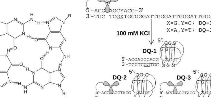
The quadruplex-forming single strand was 5'-32P-end-labeled using standard protocols and the conjugate was annealed against a was 5'-32P-end-labeled using standard protocols and the conjugate was annealed against ' a concentration of 4 µM. 32P-end-labeled using standard protocols and the conjugate was annealed at a concentration of 4 µM in 10 mM potassium phosphate, pH 7 with 100 mM KCl. The quadruplex-forming single strand was 5'-32P-end-labeled as above and the conjugates were annealed at a concentration of 4 were 5'-32P-end-labeled as above and the conjugates were annealed at a concentration of 4 µM.
RESULTS
- Characterization of DNA Duplex/Quadruplex Conjugates by Circular Dichroism. Antiparallel and parallel guanine quadruplexes possess characteristic and
- Melting Temperature Studies of the Duplex/Quadruplex Conjugates
- Charge Transport Chemistry in DNA Duplex/Quadruplex Conjugates
- Characterization of and Charge Transport Chemistry in a Guanine Quadruplex Containing Four Stacked Tetrads. The charge transport chemistry and
Interestingly, with irradiation at 313 nm, no damage is observed in the quadruplex portion of the conjugate. Upon irradiation of DQ-1 at 365 nm in the presence of rhodium photooxidant, damage is observed only in the quadruplex area. It is noteworthy that little damage is observed in the duplex region, which includes a guanine doublet site.
DISCUSSION
CT observation of the quadruplex conjugates designed here indicates that there is sufficient base-base overlap at the duplex/quadruplex junction. In the absence of a linker, however, sufficient interaction between the two regions provides a pathway for CT. Access to diffusible molecular oxygen may be limited in the quadruplex core, leading to reduced radical scavenging efficiency in the central tetrad.
In the presence of 100 mM KCl, the single-stranded overhang folds intramolecularly into an antiparallel quadruplex. Spectra were acquired at room temperature in 10 mM potassium phosphate, pH 7 with 100 mM KCl and at concentrations of 4 µM for the quadruplex-forming and duplex chains and 2.5 µM for DQ-1.