Matrix stiffness induces epithelial-to-mesenchymal transition via Piezo1-regulated calcium flux in prostate cancer cells”. PA-gel crosslinker application for PA-gel collagen coating was performed at the Vanderbilt Institute of Nanoscale Science and Engineering. This work was supported by the National Institutes of Health National Cancer Institute grant number CA203991.
4 Effects of pharmacological activation and inhibition on intracellular calcium concentration in PC3 cells at different stiffnesses……….
Introduction
Cancer cells can sense these forces via mechanosensitive ion channels, which transduce mechanical cues in the cells. Under healthy conditions, Piezo1 allows endothelial cells to sense and respond to shear forces in the bloodstream, helping to control vascular tone and the baroreceptor reflex (Fang et al., 2021). In addition, Piezo1 plays a crucial role in the digestive tract, urinary tract, joints, lungs and touch sensation.
In prostate cancer, Piezo1 is strongly upregulated compared to healthy prostate tissue (Han et al., 2019). Cells can modulate calcium flux into the intracellular space via membrane mechanosensitive ion channels such as Piezo1. Piezo1 is opened by stresses in the cell membrane as the matrix resists deformation when a cell pulls on it to migrate.
This study links how the increase in matrix stiffness due to disease progression activates Piezo1, increases calcium influx, and leads to EMT in PC3 and DU145 prostate cancer cell lines. Next, we report the effects of matrix stiffness and pharmacological inactivation and activation of Piezo1 on EMT-related changes in metastatic prostate cancer cell lines PC3 and DU145.
Materials & Methods
- Cell Culture and Reagents
- Polyacrylamide Gel Preparation
- Calcium Imaging
- Cell Shape and Actin Polymerization Assay
- Western Blotting
A 40 μL drop of the precursor solution was pipetted onto the activated side of the coverslip and sandwiched with another 22x22mm coverslip coated in an anti-adhesion solution for 1 hour. Cells were incubated in the dark at room temperature for 45 min and then washed with supplemented with each of the different treatments. The original image was then reopened, the ROIs were overlaid, and the mean fluorescence intensity was measured for each cell.
Faverage is the average of the mean fluorescence of all cells for each of the 5 images acquired. This was tested using the square tool in FIJI at 3 random locations and then averaged for each image. A total of 3 biological replicates were performed and the ΔF/F0 values for each of the 5 images for each were averaged and plotted in GraphPad Prism version 9 for Windows (GraphPad Software, San Diego, California USA).
A total of 3 biological replicates were performed for each of the treatments on 3 different substrates. 1 was used to calculate the ΔF/F0 value for each cell, where Faverage is the average fluorescence of each individual cell and F0 represents the average of 3 background measurements.
Results
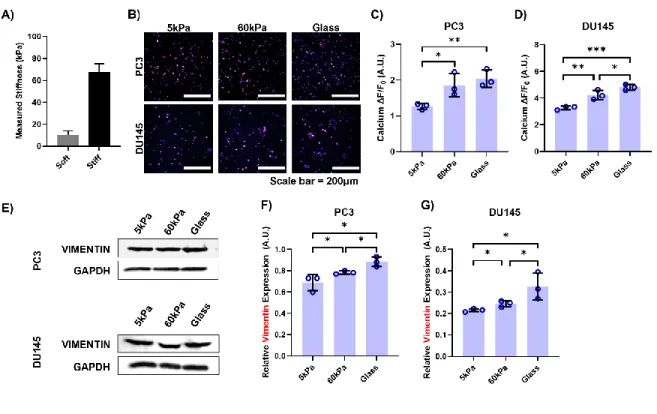
- Stiffer Substrates Lead to Increased Calcium Flux and Vimentin Expression
- EMT Associated Characteristics Change with Stiffness
- Calcium Flux Depends on Treatment and Stiffness
- Effect of Yoda1 and GsMTx-4 on Vimentin Expression
- Effect of Yoda1 and GsMTx-4 on EMT Morphology
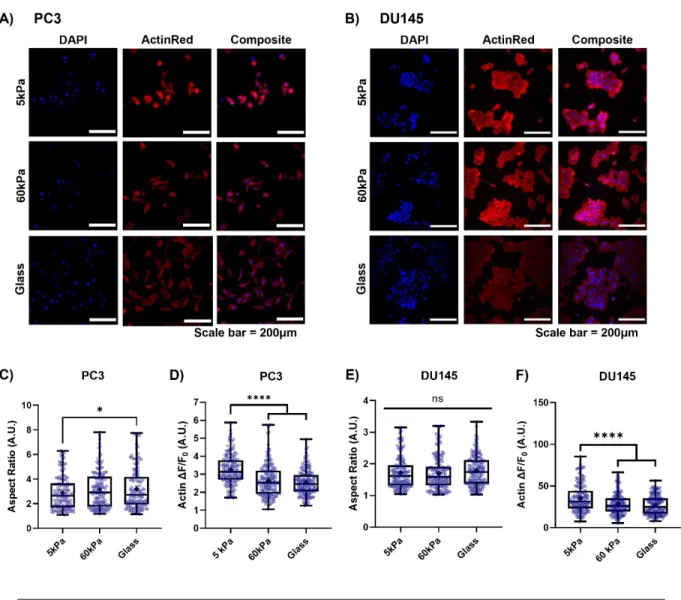
Notably, there was a significant difference in the ΔF/F0 measurement between DU145 cells grown on 60 kPa gels and those on glass coverslips. Consistently, DU145 cells plated on glass coverslips showed a 1.5-fold increase in vimentin expression compared to cells grown on softer PA gel. Micrographs of A) PC3 cells and B) DU145 cells grown on 3 different substrates and stained with ActinRed555 and DAPI.
Interestingly, F-actin fluorescence in the DU145 cells decreased by 33% from cells grown on the 5kPa PA gels to cells grown on both stiffer substrates (Fig. 3F). The PC3 cells grown on the 5 kPa (Fig. 4B) and 60 kPa (Fig. 4C) PA gels showed a significantly increased intracellular calcium concentration when treated with Yoda1 compared to the DMSO vehicle control. No difference was observed between the DMSO vehicle control and the Yoda1-treated cells on the glass control substrate (Fig. 4D).
When the DU145 cells were treated with GsMTx-4, the changes in calcium concentration were similar to those in the PC3 cells, especially on the stiffer substrates (Fig. 5E). Pharmacological inactivation of Piezo1 had no significant effect on AF/F0 in the cells grown on the softest substrate (Fig. 5F). There was no difference in vimentin expression between the vehicle control and Yoda1-treated cells on the control glass substrates.
There was no significant change in vimentin expression between the vehicle control and treatment groups in the cells grown on the 5kPa gels. Interestingly, the cells on the 60kPa gel had a 25% decrease in vimentin expression after GsMTx-4 treatment compared to the DPBS control. Regarding aspect ratio, Yoda1 had the greatest effect on the cells grown on the 5kPa gels (Fig. 7B).
Compared to the DMSO control, the number of cells on the 60 kPa gels and the glass control was reduced by 13%. Regarding F-actin expression, cells on 5kPa gels treated with vehicle control had no significant difference compared to those treated with GsMTx-4 (Figure 7F). A slight increase in the aspect ratio of cells grown on 5kPa gels was observed when treated with Yoda1 compared to the control, but this difference was not significant.
A significant increase in aspect ratio was seen in both groups of cells cultured on the 60 kPa gels and the glass controls with Yoda1 treatment. In the DU145 cells cultured on the 5kPa gels, F-actin ΔF/F0 was 46% lower in the treatment group compared to the vehicle control.
Discussion
This could be due to the fact that DU145 cells themselves have higher stiffness than the PC3 cells, potentially allowing them to resist deformation to a greater extent (Hope et al., 2021). When at rest, Piezo1 adopts the shape of an inverted dome, storing potential energy in this way (Coste et al., 2012). When the PC3 cells were plated on the stiffer substrates, the measured calcium fluorescence reached a plateau, suggesting that the calcium channel had likely reached a maximal open state due to the resistance of substrates to deformation (Fig. 2C).
Yoda1 acts as a molecular wedge that lowers the mechanical threshold for activation to increase the flow of calcium through the pore (Botello-Smith et al., 2019). Therefore, when cells were treated with Yoda1 on the soft substrates, calcium fluorescence increased significantly compared to the vehicle control (Figs. 4A, 4B, 4I, 5A, 5B and 5I). Conversely, GsMTx-4 ports Piezo1 to stabilize its closed state and in this way raises the energy barrier required to open the pore and decrease calcium flux into the cell (Bae et al., 2011).
Therefore, it was able to reduce the calcium flux into the cells at the stiffnesses of all three substrates, especially on the stiffer gels where Piezo1 showed more activity (Fig. 5E-H, 5E-H). In a study by Leggett et al., a modeling approach for EMT characterization based on vimentin expression and cytoplasm elongation was developed (Leggett et al., 2016). By treating PC3 and DU145 cells with Yoda1, increases in both vimentin expression (Fig. 6A, 6B, 6D and 6E) and aspect ratio (Fig. 7A, 7B, 8A and 8B) were observed compared to cells incubated with DMSO when grown on the softer substrates.
Similar results were observed in the groups that received no treatment and only stiffness increased (Figs. 2E-G, 2A-C and 3E). These determine motility and structural integrity while working together, and strongly influence each other (Jiu et al., 2017). Furthermore, the increases in vimentin expression with Yoda1 treatment compared to the vehicle control correlate with the decreases in F-actin fluorescence (Fig. 7C and 8C).
Pharmacological inhibition or knockdown of the TRPV4 calcium channel is known to prevent EMT morphology in breast cancer even at higher matrix stiffnesses (Sharma et al., 2019). These effects were observed in DU145 cells in vimentin expression (Fig. 6F), aspect ratio (Figs. 8D and 8E), and F-actin fluorescence (Fig. No significant changes in aspect ratio, F-actin fluorescence, or vimentin expression in these groups (Fig. 6C, 7D, 7E and 7F).
This finding could be due to the fact that alternative integrin signaling pathways are activated with the high substrate stiffness and bypass the pharmacological inhibition of Piezo1 ( Levental et al., 2009 ). Choi et al.'s study showed that the high shear forces in the circulation were associated with increased EMT in circulating tumor cells isolated from breast cancer patients (Choi et al., 2019).