MORPHOLOGICAL DIVERSITY AND EVOLUTION OF EGG AND CLUTCH STRUCTURE IN AMPHIBIANS
RONALDALTIG1,3AND ROYW. MCDIARMID2
1Department of Biological Sciences, Mississippi State University, Mississippi State, MS 39762-5759, USA
2USGS Patuxent Wildlife Research Center, National Museum of Natural History, Washington, DC 20560-0111, USA
ABSTRACT: The first part of this synthesis summarizes the morphology of the jelly layers surrounding an amphibian ovum. We propose a standard terminology and discuss the evolution of jelly layers. The second part reviews the morphological diversity and arrangement of deposited eggs—the ovipositional mode; we recognize 5 morphological classes including 14 modes. We discuss some of the oviductal, ovipositional, and postovipositional events that contribute to these morphologies. We have incorporated data from taxa from throughout the world but recognize that other types will be discovered that may modify understanding of these modes. Finally, we discuss the evolutionary context of the diversity of clutch structure and present a first estimate of its evolution.
Key words: Amphibia; Clutch; Eggs; Evolution; Jelly layers; Oviposition; Ovulation.
AMPHIBIANSsurely have the most varied and least known life histories of all terrestrial vertebrates. Typically, terrestrial adults return to aquatic sites where courtship, egg de- position, and fertilization occur. Eggs hatch into larvae that are mobile feeding forms.
After a period of growth and development, these larvae or tadpoles usually undergo some form of metamorphosis and move onto land where they grow into reproductive adults.
Variations on this pattern most often repre- sent evolutionary changes in developmental or life history traits (e.g., paedomorphy and pedotypy, egg placement, tadpole develop- ment) expressed during the ontogeny of an individual prior to its becoming an adult.
McDiarmid and Altig (1999) summarized the biology of tadpoles, and we know even more about the adult stage (e.g., Duellman and Trueb, 1986). Although the research efforts of embryologists and cell biologists have pro- vided extensive information on ovum pro- duction and early development, even if in- volving a relative few taxa, nearly every facet of the field biology of amphibian eggs is poorly documented. In this paper we focus on egg structure, particularly the jelly layers, and patterns of egg deposition. We also consider the characteristics of the physical and bi- ological environments in which the immobile eggs are laid that likely account for the
diversity of egg jellies and ovipositional modes.
Field identification of amphibian eggs is difficult at best, in part because authors of published descriptions of eggs have used inconsistent and inexact terminology and misinterpreted certain structures. For exam- ple, the vitelline membranesensu latoactually takes on three successive states with different biochemical and physical properties and functions: coelomic, vitelline, and fertilization membranes (Carotenuto, 2001; Gerton and Hedrick, 1986; Takamune et al., 1987), but most authors fail to distinguish among them.
In addition, different observational techniques can lead to different results, so that research- ers often disagree on traits as seemingly easy to observe as the number of jelly layers around an ovum.
While writing a key to the eggs of North American amphibians, we realized that egg identifications based on properly described ovipositional modes were likely to be more accurate than those based on the more labile and poorly documented features of individual eggs. Clutch morphology is easier to see, more readily definable, and usually less variable within species. By combining these definitions with careful field observations (e.g., Pombal and Haddad, 2005) made under well-docu- mented environmental conditions (e.g., Wright and Wright, 1924), a good observer should be able to identify the eggs of North American amphibian taxa at least to genus with reason-
3CORRESPONDENCE: e-mail, [email protected] E
1
able confidence. We suspect that the same could be done with other faunas as well.
Our initial goal in this review is to develop a generalized framework for observed di- versity of amphibian eggs and ovipositional modes that will contribute to a broad un- derstanding of the evolution of amphibian reproduction. We believe that the oviposition- al mode is a factor that can profitably augment our notions of breeding or reproductive modes (e.g., Crump, 1974; Duellman and Trueb, 1986; Haddad and Prado, 2005). In the process, we present a standardized terminol- ogy for egg jellies and clutch structures and discuss the evolution of these features. Al- though data acquired by cell biologists, de- velopmental geneticists, and particularly his- tochemists will enhance the eventual understanding of the biology of ova and egg jellies, we do not delve into this extensive literature.
Also, one should be constantly aware of the integration of the data discussed herein with the concept of breeding mode that is being constantly revised and expanded (e.g., Crump, 1974; Duellman and Trueb, 1986). What we present is a subset of the larger concept, although this integration is left for a future synthesis when egg biology is better under- stood.
The morphological surveys of egg structure by Salthe (1963) and Salthe and Duellman (1973) suggested that intriguing morphologi- cal and ecological patterns exist within am- phibians. Variations in the surface morpholo- gies of ovum coverings and the nature of the arrangement and attachment of eggs in the environment as reported in other organisms (e.g., Mooi, 1990; Stiassny and Mezey, 1993;
Strathmann and Chaffee, 1984) surely exist in amphibians, but certain factors obfuscate their interpretation in this group. For example, the number of jelly layers surrounding an ovum and the number of secretory areas in an oviduct may vary independently (Greven, 2003). In part, these variations can be explained by oviductal (e.g., changes in secretory regions in the oviduct; preoviposi- tional changes in jelly; D. M. Hardy and Hedrick, 1992), postovulatory (e.g., dissolu- tion of some jelly layers) and postovipositional (e.g., layers melding together; formation of the
capsular chamber in salamanders and some frogs) events. Of greater import in essentially all cases is the lack of data or the lack of integration of those data across disparate fields of research. Diagrams of eggs are schematic and usually drawn in lateral view taken at the equator with the animal pole uppermost. Concentric circles drawn around the ovum (e.g., continuous, dashed, or stip- pled lines or zones; Hoyt, 1960; McDiarmid and Worthington, 1970) represent visually perceived jelly layers. Even so, no adequate description of the arrangement of jelly layers around freshly oviposited eggs based on observations using standardized techniques (i.e., proper illumination, dissection, section- ing, staining) has been published.
We summarize the literature on amphibian egg morphology, especially that addressing the production and evolution of the jelly layers surrounding the amphibian ovum. We follow with a synthetic treatment of the morphology, diversity, and evolution of the egg clutch. We also propose a standardized terminology for describing eggs and egg clutches that we believe facilitates communication and ad- vances the study of this important but neglected stage of the amphibian life cycle.
EGGMORPHOLOGY AND ITS VARIATIONS
Definitions
We define ‘egg’ as an ovum (5 gamete through gastrulation, stages 1–12; Gosner, 1960; other versions in Duellman and Trueb, 1986 and McDiarmid and Altig, 1999, Chap- ter 2) and its vitelline membrane of ovarian origin surrounded by one to several oviduc- tally-produced jelly layer(s). The internal layer is deposited first by an anterior region of the oviduct, and as the egg moves down the oviduct more layers are deposited sequentially by more posterior oviductal regions.
Various terms have been used to refer to the individual jelly layers, and they and the entire complement of layers as a unit generally lack discrete definitions; some of the terms used in different research fields are inaccurate (e.g., capsule, envelope). We suggest that the entire assemblage of oviductal materials de- posited around the ovum be referred to as
‘jelly’ or ‘jelly layers’ regardless of variations in
their apparent physical structure (e.g., tough- ness, density, thickness, water content). Layer, zone, and membrane refer to units within this assemblage. A ‘layer’ (often referred to in the cell biology literature by the letter ‘J’ with a numerical subscript to denote position) is a morphologically discrete, easily recognizable region of jelly surrounding the ovum; it has a discernible thickness along a radius and typically has a uniform and visually apparent optical density. Layers are numbered from interior to exterior (e.g., Daniel, 1937) in the order in which they are formed. The term
‘layer’ does not encompass any specific function or physical characteristic.
Daniel (1937) and others pointed out that some layers have discernible but less discrete subdivisions. We designate these subdivisions as ‘zones’ to suggest areas that are less easily visualized than layers (see Steinke and Ben- son, 1970), but we do not imply that all zones of nonstained eggs are visible.
Finally, we use ‘membrane’ as a functional term to describe what appears to be a delimit- ing boundary between layers, exclusive of the vitelline membrane. Data are currently in- sufficient to determine if membranes are actual structures or if they are merely an optical representation of the plane along which two layers of different densities abut.
In some instances two layers apparently abut without a visible membrane, thereby suggest- ing that the densities of the two layers must reach some threshold before a visual mani- festation of the transition appears. If the
‘membranes’ between layers are discrete structures, we do not know if they are independent of adjacent layers and produced by a specific oviductal secretory region, the result of some postovipositional reaction of the jelly layers, or an outer or inner boundary of a particular layer.
Structure
Clear jelly layers certainly are not as structurally uniform (e.g., Carroll et al., 1991) throughout their thickness as they appear. The egg ofRana pipiens, for example, has two, clear layers of jelly (Wright and Wright, 1949:35). Studies (Steinke and Ben- son, 1970) of the taxon of the same name with immunological and histochemical techniques
revealed 5–6 layers, whereas dissections (Salthe, 1963) and other techniques (Shaver, 1966; Shivers and James, 1970) indicated 3–5 layers. Descriptions of the eggs ofDicampto- don sp., probably based on visual examina- tion of intact eggs (Stebbins, 2003), indicated 2 jelly layers, whereas visual and tactile detection of density differences noted dur- ing dissection (Nussbaum, 1969) showed 5 layers.
More structure certainly exists in the jelly layers than can be seen with simple micro- scopy. Other observational techniques (e.g., differential interference contrast [DIC], phase-contrast and fluorescence microscopy and laser illumination) do not produce better images. Laser confocal imaging and some techniques of electron microscopy provide better results but are costly and specimen preparation is laborious (e.g., Bonnell and Chandler, 1998; Larabell and Chandler, 2005). More refined zones, which can be detected with histological and histochemical techniques, likely will reveal patterns of biological, ecological, or phylogenetic interest (e.g., Hedrick and Katagiri, 1988; Smith et al., 2002). Biochemical analyses show that egg jellies consist of ‘‘a fibrous glycoprotein superstructure that acts as a scaffold to which globular glycoproteins are bound’’ (Fig. 5E;
Bonnell and Chandler, 1998); which of these groups of molecules is structural and which is biologically active is not known, but both are expected to have species-specific qualities (Maes et al., 1995). The three jelly layers of the eggs ofXenopus laevisare composed of at least nine glycoproteins (Yurewicz et al., 1975).
These kinds of data, while interesting, are of little use to field biologists who usually rely on visual impressions of structure viewed under incident, white light. Some data suggest that it may not be possible to formulate a descriptive model useful to all researchers. Accordingly, field biologists, histochemists, and biologists in other fields may find it expedient to use their own sets of terms to describe observed variations in jelly traits.
In summary, jelly occurs in layers and a layer may be subdivided into less easily discernible zones. Visually perceived layers typically would be the morphological trait
most useful for identification. For example, the basic egg structure consists of an ovum with a vitelline membrane surrounded by jelly layers 1 and 2 (Fig. 1A, E) of oviductally- produced materials. Inner and outer mem- branes are labeled, although oviductal secre- tory zones for their production are not, and no zones are shown.
EVOLUTION OFEGGJELLY
The evolution of additional layers of jelly does not appear to result only from the subdivision of ancestral layers. Thus, egg diameter often is greater in species with more layers rather than in those with fewer layers, even though the thickness of each layer, or at least of some, often decreases as the number of layers increases. Although the thickness of jelly layers must be mediated by the general trade- offs between the requirements for gas ex-
change and need for mechanical support, other factors (e.g., homospecific sperm attraction [Al-Anzi and Chandler, 1998], heterospecific sperm avoidance, heat conservation, and pred- ator defense) likely have played roles in the evolution of the number, thickness, and physical characteristics of the layers. To appreciate layer homologies one must ulti- mately understand the morphology of the secretory regions of the oviduct and presum- ably have some notion of evolutionary relation- ships among taxa. Presumably, selection for differences in the number and characteristics of the jelly layer has influenced oviductal morphology rather than the reverse, although other factors that seemingly are not related to egg survival (e.g., Anderson et al., 2006) must be considered. Evidence suggests that envi- ronmental demands and phylogenetic con- straints have influenced the evolution of egg morphology, although little is known about the
FIG. 1.—Schematic drawings of an amphibian egg with its component parts, the oviductal regions a–c that produce the jelly layers, and three hypothetical paths for adding a jelly layer. (A) An ovum with two membranes (see text) and two jelly layers produced in sequence by oviductal sections a and b arranged anteriorly to posteriorly. (B) A hypothetical case where a new layer is added directly external to the vitelline membrane; new numbering of the jelly layers and the arrangement of the oviductal regions are shown. (C) A case where a new layer is added externally. (D) A case where a new layer is added within the original first layer. Abbreviations: 1–45jelly layers numbered from ovum to surface, ant 5anterior, IM5inner membrane, O5ovum, OM5outer membrane, post5posterior, VM5vitelline membrane, and VS5vitelline space.
selective factors involved, and details and patterns of the morphology in most taxa are still lacking (Salthe, 1963; Greven, 2003).
Hypothetical scenarios diagramed (Fig. 1B–
D, F–H; also Greven, 2002:fig. 11) illustrate potential modifications in oviductal secretory regions that would produce given changes in jelly layers; the review by Wake and Dickie (1998) provides informative discussions of oviductal anatomy relative to breeding mode.
If we assume for heuristic purposes that combinations of the following alternatives did not occur, then a third jelly layer might be added (1) between the vitelline membrane and the first jelly layer; Fig. 1B, F), (2) between the two original jelly layers), (3) external to the second layer of the original egg; Fig. 1C, G), or within an existing layer; Fig. 1D, H). If a posterior (i.e., new layer added externally) or anterior (i.e., new layer internally) region of the oviduct were involved and a phylogenetic scheme were known, one potentially could track the products and thus hypothesize homologies. Identification of other types of additions may require histochemical tech- niques, and examination of changes in oviduc- tal regions. Which evolutionary pathway is most likely can be debated and will depend largely on the ease with which homologies can be assigned. Layer thickness also is likely influenced either by the length of the oviductal region, passage rate of the ovum, rate of jelly production, or differences in hygroscopic qualities of the jelly. Although these parame- ters are probably important, we have ignored them because of the lack of data.
Salthe (1963) presented the only sugges- tions of homologies of jelly layers in caudates.
His interpretations, based on 15 caudate genera in 8 families suggest that (1) 8 jelly layers (as in Hynobius lichenatus) is the primitive condition; (2) changes in the num- ber of layers occur only through (a) loss of the most external layers (e.g., ambystomatids) or (b) loss of more internal layers (i.e., elimina- tion of layers somewhere within the series;
particularly plethodontids); and (3) eggs with 3 layers of undetermined homologies (as in of Cryptobranchus) is the simplest condition. All of these ideas are based on the assumption that layers in discernible positions and of similar construction are homologs.
We know of no proposed scheme of jelly layer homologies for anurans, and phyloge- netic patterns and possible selective factors contributing to their evolution are often discordant. For example, among North American frogs (Moore, 1940), many cool- water breeders deposit clumps of eggs with either 2 or 4 jelly layers, whereas warm-water breeders lay either clumps or films of eggs with 2 layers. ‘Wood frogs’ (West Coast endemics plus Rana sylvatica) produce clumps of eggs with 2–3 jelly layers in cool water. Members of the Rana catesbeiana group breed in warm weather and deposit eggs with 1–2 layers as clumps or surface films. Members of theRana pipiens complex lay eggs with 2 jelly layers in clumps and usually in cool water. Considering the four options for layer homologies (Fig. 1B–D), one can ask if the outer two or the inner two layers of the wood frogs are homologous to the two layers of the other groups of North American ranids and which of the layers might reflect responses to temperature or ovipositional mode and which to phylogenetic constraint.
A complete histochemical data set for an amphibian egg might agree with Salthe’s (1963) morphological data. One still would not be sure of layer homologies, but his hypotheses based on position and construc- tion would have stronger support. Mapping histochemical data onto appropriate clado- grams would likely provide some insight about the evolution of jelly layers, and information on the ecological functions of egg jellies should reveal correlations useful to interpret- ing their evolution. Salthe (1963) suggested that layer losses in plethodontids that lay terrestrial eggs involve changes of internal layers and that the tough outer layer is retained for protection. Evaluations of the hypotheses about jelly layer homologies will have to wait additional data.
ANCILLARYSUBJECTS ONEGGS
Characteristics of Jelly Layers Anyone who has handled eggs and espe- cially those who have manually dejellied them are familiar with features such as elasticity, stickiness, toughness, turgidity, and wateri-
ness. Aquatic eggs usually are spherical when submerged but sag when placed on a surface in air. The jellies of most terrestrial forms that do not lay suspended eggs have jelly with sufficient tensile strength and turgidity to remain spherical in air. The outer jelly of eggs of Ambystoma opacum and other terrestrial salamanders and frogs with direct develop- ment is tough relative to that of aquatic eggs.
Tougher membranes and increased turgidity of the enclosed fluids help to maintain the spherical shape of these large eggs in air and thereby allow proper development, oxygena- tion, and protection from trampling by an attendant parent. These terrestrial eggs can be grasped with minimal distortion and will bounce if dropped. If the outer layer is removed, the remaining jellies spread out, and the ovum usually ruptures. In some aquatic eggs the most external visible layer is surrounded by a transparent, watery gel that appears to lack a defining exterior surface but is crucial for flotation (see below).
Asymmetries of jellies caused by tensile differences, such as the drooping of egg jellies of terrestrial plethodontid eggs or the pentag- onal appearance of jellies of eggs tightly spaced in a film, are common. The observa- tion by Wright and Wright (1949) that the inner jelly layer of the eggs ofRana clamitans, which is not under any tensile forces, may be elliptical or pear-shaped needs verification.
We assume that the conical ova that Wright and Wright (1949) observed inBufo alvarius resulted from tension on the jelly string.
There are cases of egg jelly asymmetries in the absence of tension. The outer jellies of some salamandrid eggs (Notophthalmus vir- idescens; Bishop, 1943) are oval and attached individually or in small groups to plants. Parts of the outer jelly layers of the eggs of some Old World microhylids (e.g.,Kaloula rugiferaand K. macroptica, Fig. 2A, B, and Liu, 1950;
Kaloula borealis in Li, 1934; Kalophrynus pleurostigma in Taylor, 1922; and Paradoxo- phyla palmatain Glaw and Vences, 1992; RA, personal observation) are asymmetrical and form a flange or rim which allows the eggs to float. Asymmetry in jelly layers seems at odds with their mode of formation in the oviduct, but the asymmetry results from differences in hygroscopic properties of specific portions of
the jelly immediately after oviposition rather than structural differences per se. The jelly that forms the flange surely surrounds the entire ovum, in contrast to being deposited as a band, and is likely very watery. The pressure of the water surrounding the egg as it sinks partway through the water surface likely pushes this flimsy jelly to the air-water interface and forms the flange. Different flange positions (e.g., equatorial inKaloulaspp. and near the vegetal pole in Paradoxophyla palmata) may reflect interspecific differences in the density of the egg-jelly complex or in the hygroscopic qual- ities of the jelly. Li (1934) noted that the rim in Kaloula borealis did not appear until about 1 min after oviposition, and Taylor (1922) described the gelatinous flange inKalophrynus pleurostigma as gradually widening to about 6 mm diameter after extrusion.
Inclusions in Jelly
The structure, origin, and function of various crystalline inclusions in the jelly of salamanders (e.g., Ambystoma maculatum, Salthe, 1963; Ruth et al., 1993;Siren lacertina and Hynobius lichenatus, Salthe, 1963) need further examination. For example, the egg jelly ofAmbystoma maculatumcan be clear or opaque white (L. M. Hardy and Lucas, 1991).
White jelly gets its color from glycoprotein crystals that are produced with the jelly in cells in the oviductal wall. Populations with clear-jellied, white-jellied, or both types of egg masses have been reported from northwestern Louisiana and adjacent areas of Texas and Arkansas. The function of the crystals is unknown, but they may reflect light and thereby offer some protection to the de- veloping embryos or concealment from pred- ators (L. M. Hardy, personal communication).
Similarly, the jelly of the eggs of the frog Mantidactylus depressiceps (Mantellidae) is milky white (RA, personal observation) al- though the cause of the color is unknown.
What appear as striae, furrows, or corrugations in the outer layer of jelly of some salamanders (e.g., Hynobius lichenatus, Salthe, 1963) and frog egg masses (e.g., Cochranella pulverata [RWM, personal observation]) also deserve attention.
Some ambystomatid eggs, notably those of Ambystoma maculatum and A. gracile with
FIG. 2.—(A–C) Microhylid, (D–H) mantellid, and (I) arthroleptid frog eggs. Asymmetrical egg jellies of (A)Kaloula rugifera(modified from Liu, 1950:fig. 58) and (B)K. macroptica(modified from Liu, 1950:fig. 60) caused by differential hydration of specific parts of the jelly. (C) Part of a coherent film of Gastrophryne carolinensiseggs soon after oviposition showing the upper hemisphere of jelly projecting above the water surface (15downward flexed meniscus at jelly margin, 25glint on surface of ovum or vitelline membrane, and 35trapezoidal reflection on curved, upper hemisphere of outer egg jelly). Laminar array of a clutch ofGuibemantis depressicepseggs on a leaf (D) prior to hydration (OV5ova; EJ5empty jellies), (E) the same clutch showing a large increase in volume after hydration, and (F) the same clutch cut longitudinally after hydration, stained with Toluidine Blue, and viewed with transmitted light (L 5cut leaf; OS5 ovumless stalk, OV5ova, and S 5limits of one stalk with an ovum; stalks slightly darkened electronically for better visibility). (G) Recently laid clutch of ‘Mantidactylus’ sp. on a leaf showing structure when hydration is normally less extensive, and (H) older clutch of ‘Mantidactylus’ sp. attached to the bark of a tree with clear jelly and a large increase in jelly with hydration (embryos that appear deep in the jelly are actually on the sides of the jelly). (I) Terrestrial clump ofArthroleptis schubotzifound in leaf litter (photo by R. C. Drewes).
exceptionally dense jellies, have a symbiotic green alga (Oophila amblystomatis) within the inner jelly layers of the egg (e.g., Bachmann et al., 1986; Hammen, 1962).
Capsular Chamber
In salamanders and a few primitive frogs (Salthe, 1963:165), the jelly layer abutting the vitelline membrane dissolves soon after ovi- position to form the capsular chamber;
remnant debris, termed ‘white plac’, puddles at the bottom of the capsular chamber (Daniel, 1937). Eggs with a capsular chamber are less confined than those without such a chamber so that the ova lie slightly below center in the remaining jelly; if the egg mass is inverted, the ova immediately turn so that the animal pole is uppermost. In eggs without a capsular chamber the ova are constrained by the jelly layers and take several minutes to right themselves (Salthe, 1963). Embryos usually exit the vitelline membrane long before they hatch from the jelly (Salthe, 1963), and one can often see the vitelline membrane crumpled up at the bottom of the inner jelly layer in advanced embryos.
Functions of Egg Jelly
Suggested functions of egg jellies (Greven, 2002, 2003) include mechanical support for the ovum, attachment of eggs to each other or a structure in the environment, enhancement or prevention of entry by conspecific and heterospecific sperm respectively (e.g., Bar- bieri and Del Pino, 1970), prevention of polyspermy, sperm capacitation, differential protection from water moldsSaprolegniaand Achlys (Gomez-Mestre et al., 2006), pro- tection from contaminants (Marquis et al., 2006), and protection from predators, patho- gens, and environmental stressors such as temperature and UV light (Hunter and Vogel, 1986; Itoh et al., 2002; McLaughlin and Humphries, 1978; Ward and Sexton, 1981).
Incubation in birds helps to inhibit growth of bacteria and fungi on egg shells (Cook et al., 2005); jelly layers may be a prepackaged way to protect ova from these perils after they exit from the relatively sterile oviduct. The in- fluence of jelly on light refraction is thought to be insignificant (Cornman and Grier, 1941)
but possibly needs further study (Bragg, 1964).
Hatching
Hatching occurs when a developmental threshold is reached in concert with various biotic and environmental cues. Hypoxia (e.g., Petranka et al., 1982) likely is the proximal trigger, but factors such as pathogens and predators (e.g., Chivers et al., 2001; Touchon et al., 2006; Warkentin, 1995) or low pH (Dunson and Connell, 1982) can modify timing. Thumm and Mahony (2002) demon- strated that the stage and degree of de- velopment at hatching in Pseudophryne aus- tralis (Myobatrachidae) is variable. Hatching mechanisms seemingly are universal (Noble, 1926; Duellman and Trueb, 1986) and fall into two groups. In most amphibians enzymes from the frontal (5 hatching) glands on the head and snout cause at least partial chemical degradation of the jelly (e.g., Carroll and Hedrick, 1974; Urch and Hedrick, 1981), which may often be augmented by simple writhing and pushing by the embryo to break through the layers (also Bragg, 1940; Gollman and Gollman, 1993; Lutz, 1944) or by rain (Noble, 1926). In some direct developing frogs an egg tooth on the snout or other part of the body mechanically ruptures the mem- branes and jelly layers to allow hatching (e.g., J. D. Hardy, 1984; Noble, 1926).
Ovum Pigmentation
Even though the pigments in most ova are melanic and not contained in cellular orga- nelles, data on this pigmentation of maternal origin are confusing. In large part this is because there are no standards for describing color, intensity (e.g., dark, diffuse, pale), pattern, location, or the gradation from dorsal darkness to ventral paleness. In addition, apparent changes in pigmentation with de- velopment or after preservation have rarely been documented. Potential influences on ovum coloration include age of female, stage of oogenesis, changes during development (i.e., when and how maternally-provided pigment is supplanted during oogenesis by that from the developing embryo), and egg- laying site (i.e., geographical and elevational variation). Likewise, color and pattern and
their development in hatchlings (e.g., Altig, 1972) that are useful for species identifications (e.g., hylid hatchlings commonly are strikingly patterned, ranids usually unicolored) are poorly documented.
Nevertheless, some general correlates of ovum pigmentation are apparent (Salthe, 1963). Eggs laid in open, exposed areas, regardless of the specific site, taxon, or ovipositional mode, usually have melanic pigment at the animal pole (e.g.,Ambystoma, Bufo,Hyla, and Rana of North America). As the number of embryonic cells increases and the cells move during gastrulation, the fertil- ized ova become more uniformly pigmented and paler because the ovum pigment is dispersed among more cells until embryonic pigment appears. Eggs laid in secluded sites (e.g.,Amphiumain burrows near lentic sites, Cryptobranchus, Dicamptodon, numerous plethodontids, and Ascaphus hidden among rocks in streams, some dendrobatid and microhylid frogs among forest debris and phytotelmata, some centrolenids on leaves) tend to be pale to nonpigmented regardless of ovipositional mode. Biologists working in temperate areas often assume that amphibian eggs are pigmented because those the taxa most commonly encountered are pigmented, but in fact, many taxa in tropical regions have nonpigmented ova.
Several functions of egg pigmentation and adult behaviors associated with that trait have been proposed. Communal oviposition of masses of dark eggs apparently enhances absorption of heat thereby increasing de- velopmental rate in early breeding species of some North American ranids (Hassinger, 1970). Pigmentation in eggs can also provide protection from deleterious effects of specific wavelengths of light or heat (Barrio, 1965;
Jones, 1967). Biliverdin and lutein can pro- duce greenish to bluish ova in some phyllo- medusines (Pyburn, 1963; Marinetti and Bagnara, 1983), some hyperoliids (Wager, 1965), some rhacophorids (Liu, 1950), and certain centrolenids (RWM, personal obser- vation). These pigments are housed in the yolk platelets (Barrio, 1965) and are from a differ- ent source than the melanic pigments. The greenish color can enhance concealment of frog eggs from some predators (e.g., greenish
eggs deposited on the tops or undersides of leaves) or provide infrared camouflage (Schwalm et al., 1977; also see Saito, 2001).
Falchuk et al. (2002) have shown that biliverdin IXa functions as a cytoplasmic de- terminant that is essential to normal embryo- genesis in early stages ofXenopus laevis. The destruction of biliverdin via some wavelengths of UV light may provide a mechanism for understanding their detrimental effects (e.g., Blaustein et al., 1994). The omission of melanic pigment in ova surely conserves some reproductive energy, and embryos developing from nonpigmented eggs sometimes do not develop pigment until well after hatching.
CLUTCHMORPHOLOGY
Definitions
‘Clutch’ describes the total number of eggs deposited per ovulation event independent of the number or presence of a male(s), re- productive or ovipositional mode, oviposition- al behavior of parents, or number of ‘groups’
(5 aggregate of eggs produced in a single ovipositional bout; this term is useful when one does not know the taxon or ovipositional mode or number of bouts [i.e., single egg laying event]) that occurred. The number of ova ovulated sometimes exceeds the number of eggs oviposited. For example, if a female oviposits her ovulated complement in multiple bouts, the total number of eggs deposited makes up the clutch (i.e., a group in this case is not the clutch). A pair ofSmilisca phaeota may deposit eggs among one to several puddles in a single night (RWM, personal observation); the eggs in each puddle com- prise a group, are produced during a single bout, and are only part of the clutch. Female Ambystoma tigrinum may partition their clutches into multiple discreet groups which may be sired by one or more males; distances between these groups in a single pond may exceed 40 m (Gopurenko et al., 2006). Thus, the organization of the total clutch, and not the number or manner in which eggs emerge from the female in a single bout, is important.
If a female ovulates and oviposits more than once a year or in multiple years, she has produced multiple clutches.
‘Ovipositional mode’ describes the mor- phology of an aggregation of deposited eggs (i.e., the clutch structure). Explanations of how egg aggregates are produced and postu- lates about the evolution of different struc- tures are largely lacking. It seems apparent that oviductal (i.e., jelly formation), oviposi- tional (i.e., parental behavior), and postovipo- sitional (e.g., jelly dissolution) processes are involved. Differences in the rate or pattern of release of eggs from a female also can influence clutch structure. Oviposition site, ovum number and size (Bernardo, 1996;
Pombal and Haddad, 2005; Salthe and Duell- man, 1973; Summers et al., 2007), energy content (e.g., Komoroski and Congdon, 2001;
Komoroski et al., 1998), pigmentation, sub- sequent characteristics of development, larval ecomorphology, and other such factors are not directly considered in this review. In part, this is because the functional roles of oviductal (e.g., Greven, 2002, 2003) and especially ovipositional (e.g., Aronson, 1943) factors are not understood in sufficient detail. Likewise, inaccurate, nonstandard terminology is a con- stantly confounding problem. Livezey and Wright (1947) and Wright and Wright (1949) presented at least 14 terms, many of which were ill-defined, to describe some arrange- ment of a group of eggs. Several other workers (e.g., Stebbins, 1951, 2003; Corkran and Thoms, 1996) have used similar terms in the same or different ways. A notable recent exception is Anstis (2002) who clearly defined the terms she used to describe the types of egg aggregations of frogs from southeastern Australia. In the interest of clarity and with no implication of discrete organization, we use
‘tier(s)’ to describe a two- or three-dimen- sional arrangement of eggs and restrict ‘layer’
to descriptions of jelly morphology.
Other Considerations
Accurate observations and descriptions of ovipositional events and the appearance of deposited groups of eggs are lacking for most amphibians but deemed essential for under- standing amphibian reproduction in a broader context. Difficulties stem from a general lack of knowledge of the natural history of amphibians, problems with egg and species identifications, comparisons of eggs of differ-
ent ages, and inadequate observational tech- niques. The mere presence of multiple, confusing reflections and refractions from various water and egg surfaces and the extreme transparency of egg jellies can in- terfere with accurate observations. For exam- ple, Alcala and Brown (1956) reported that Rana microdiscalays eggs on moist surfaces on stream banks in a ‘‘twice-coiled string.’’
This note, presuming correct identifications of the adult, the eggs, and the ovipositional mode, is particularly vexing for the following reasons: a moist surface on a stream bank is a rare site for ranid egg deposition, to our knowledge a string is unknown among ranid frogs, and ‘twice-coiled’ is difficult to envision (i.e., single string coiled upon itself versus two strings with simple coils that are intertwined) and possibly the result of disturbance.
The notion that each species oviposits in one mode is usually sound (see Williams and Tyler, 1994 for an interesting exception;
Marsh and Borrell, 2001), but some oviposi- tional modes seem to blend from one into another. This apparent blending frequently is the result of variations in the ovipositional behavior of the adults, disturbance at the deposition sites, comparisons of eggs of different ages, or a misinterpretation of the mode. For example, an investigator who observes single eggs deposited close together or on top of each other, but with the presence of single, disjunct eggs, might conclude (in- accurately in our opinion, see below) that the eggs are a clump. Eggs deposited by different taxa as surface films float by different means and must be observed closely to determine whether the outer jellies are adherent or coherent. Pairs of frogs that produce surface films must have adequate room to maneuver;
movements in confined spaces sometimes cause the eggs to sink. Species that deposit egg masses and those depositing single eggs must have suitable substrates to which to attach their eggs. Eggs and clutches deposited under artificial conditions (e.g., laboratory containers, terrariums, plastic bags) may differ from those deposited in the wild. As embryos in clumps and masses approach hatching and jelly layers begin to break down, a group of eggs may rise to the water surface. Any environmental condition that produces lots
of bubbles (e.g., increase in temperature, bottom fermentation) can also raise eggs to the surface where they may appear as a film.
Eggs in films often sink even during light rain, strings and particularly rosaries often are broken, and egg clusters commonly become detached from their suspension points by activities of attendant parents. All such con- tingencies must be taken into account. Addi- tionally, more observations on the actual ovipositional behavior of adults (e.g., Aronson, 1943, 1944; Norris and Hosie, 2005; Pyburn, 1967, 1970, 1971) would add immeasurably to our collective knowledge and facilitate a better understanding of ovipositional modes.
Alytid (Alytes and Discoglossus) and pipid (Pipa) frogs have a capsular chamber, and the direct-developing ‘Eleutherodactylus’ may al- so have such a chamber (Salthe, 1963). It may be imperative that large eggs turn over quickly when inverted to keep the embryo from being damaged; the morphology of the jellies of more direct developing eggs need to be examined.
Information on the eggs of caecilians is particularly scarce, but large eggs laid as a rosary seem to prevail (Exbrayat, 2006; M.
Wake and M. Wilkinson, personal commu- nications): Caecilia, Funk et al., 2004; Epi- crionops niger, A. Lathrop, personal commu- nication;Gegeneophis carnosus, Exbrayat and Delsol, 1988; Seshachar, 1942; Hypogeophis, RA, unpublished data);Ichthyophis ‘‘glutino- sus/malabarensis,’’ Balakrishna et al., 1983;
Breckenridge and de Silva, 1973; Brecken- ridge and Jayasinghe, 1979; Breckenridge et al., 1987; Seshachar et al., 1982; I. cf.
kohtaoensis, Kupfer et al., 2004; Idiocranium russeli, Sanderson, 1937; Siphonops annula- tus, Cei, 1980; Go¨ldi, 1899;S. paulensis, Gans, 1961; Montero et al., 2005; andTyphlonectes compressicaudus, Sammouri et al., 1990).
These descriptions lack certain details, and some are surely inaccurate. For example, certain drawings of eggs of Ichthyophis spp.
(Himstedt, 1996:fig. 57; Sarasin and Sarasin, 1887–90, as reprinted in Angel, 1947:fig. 107) suggest a cluster, but others (e.g., Himstedt, 1996:fig. 47) seemingly show a rosary. Con- sidering that the preponderance of recent data countering the likelihood of a cluster mor- phology, we suggest that the earlier inter-
pretations are in error or the result of artistic license.
Some aquatic (e.g., Dicamptodon, pletho- dontines) and terrestrial (e.g., Aneides) sala- manders ‘suspend’ (i.e., usually attach them to the lower surfaces of substrates) their eggs.
The eggs may be ‘pendant’ (i.e., ovum is not centered in the drooping outer jelly layer;
Fig. 3A) or not (i.e., ova centered within outer jelly; Fig. 3B). As defined here, a ball with the turgidity to maintain its spherical shape that is stuck directly to a ceiling is suspended but not pendant. A small area termed the ‘pedicel’
attaches a suspended egg to a substrate, and 1–2 jelly layers may be involved in the supporting part of the jelly.
Deposition Sites
Similar ovipositional modes are scattered across amphibian groups and breeding sites, and egg deposition site often has little to do with clutch structure (e.g., foam nests are placed in aquatic, arboreal, subterranean, and terrestrial microhabitats; clumps are laid in many different sites). Even so, the site of egg deposition is a useful adjunct to egg identifi- cation and species biology and has been incorporated in the definitions of breeding modes (Crump, 1974; Haddad and Prado, 2005). No available classification easily ac- commodates all variations of oviposition sites without being excessively complex. We sug- gest that the environmental conditions to which eggs are exposed would be a more informative feature than specifically where the eggs are deposited. Because eggs and embryos associated with a mobile parent’s body (i.e., in or on a parent’s body for all or part of development, whether terrestrial or aquatic;
all endotrophic categories of Altig and John- ston, 1989), especially those with a physiolog- ical association with the parent, are subjected to a different set of environmental variables than eggs and embryos in the environment.
We place them in a separate general category and note that their jellies, development, and morphology are different from other types.
We thus recognize four major categories of oviposition sites (parent-associated, terrestrial, semiterrestrial, and aquatic) based on the suite of biological (i.e., predators) and physical (i.e., temperature and oxygen as mediated by
water and light; Cohen and Strathmann, 1996) conditions that the eggs experience. Obviously each category can be divided into subcate- gories based on biological and physical factors.
Eggs in the parent-associated category experience a unique and species-specific range of physical conditions whether they are exposed to the air (e.g., Alytes: wound around legs of terrestrial male parent;Hemi-
phractus: attached openly on back of terres- trial female parent; Fig. 4A) or not (e.g., Gastrotheca: dorsal pouch of terrestrial fe- male; Pipa: embedded in back of aquatic female). These conditions influence develop- ment more than whether the eggs are considered terrestrial or aquatic.
Terrestrial eggs, including those in foam nests and in arboreal, fossorial, and sub-
FIG. 3.—Ovipositional modes. (A) A suspended, pendant egg, a group of which would form an array or cluster depending on their arrangement, (B) a suspended, nonpendant egg, (C) an array, (D) a melded clump, (E) a mass, (F) part of an open clump with interstices blackened for emphasis, (G) single eggs attached to submerged vegetation, (H) a rosary, (I–K) egg strings that are (I) staggered in a unilayered tube, (J) uniserial in a bilayered tube, (K) uniserial in a bilayered tube with partitions and scalloped margins, a (L) schematic of two adjacent eggs in a string showing how jelly layers around each egg form partitions within the outer tube, and (M) a strand. A–B, D: modified from Stebbins, 2003, C, E, G, H: modified from Pfingsten and Downs, 1989; F: modified from Liu, 1950; I–K: modified from Wright and Wright, 1949, and M: redrawn by P. C. Ustach from Arnold and Burton, 1978.
terranean sites, are exposed to little or no free water at least at the time of oviposition. Both endotrophic and exotrophic forms deposit eggs in the terrestrial environment. The deposition sites often are near or above water, but they are not part of an aquatic habitat. As a result, predators on terrestrial eggs are usually very different from those in aquatic systems.
Semiterrestrial eggs are usually placed adjacent to some source of free water but are not submerged (e.g., in natural crevices or constructed burrows in a stream bank, natural or constructed depressions or chambers next to or in an area where a pond will form, among moss at the edge of a bog, in a seepy talus). The nest site is sometimes temporary in that it may become part of an aquatic system
FIG. 4.—Ovipositional modes. (A) Eggs attached to the back of a femaleHemiphractus fasciatus(Hemiphractidae:
photo by E. Griffith), inset, most anterior embryo at larger size, (B) a cluster ofPlethodon albagula(Plethodontidae;
photo by S. E. Trauth), (C) an array ofAmbystoma barbouri(Ambystomatidae) on the underside of a stone in flowing water (arrows5egg laid most recently by same or different female have less silt attached), and an (D) array ofSpea multiplicata(Scaphiopodidae) placed haphazardly on vegetation in a desert pool.
either through flooding or some form of adult behavior.
Aquatic eggs that are placed in a lentic or lotic site, in bromeliads or other phytotelms, and in tree holes are submerged or nearly so and experience distinct conditions particularly associated with oxygenation. Within this context, the differences between a stream and a temporary puddle are obvious, but some cases can be subjective. Water at lentic sites may flow temporarily after heavy rains, and the flow rate of some lotic sites is so slow that they effectively represent lentic habitats.
Consideration of how an aquatic system functions as a unit through time (e.g., bank configuration, basin shape, vegetation pat- terns, and debris distributions) and the bi-
ology of the amphibians that breed in them usually will resolve conflicts in microhabitat classification.
OVIPOSITIONALMODES
In the following pages we describe five categories of ovipositional modes: various arrangements of independent eggs, three-di- mensional arrangements, floating arrange- ments, froth nests, and linear arrangements.
Among these categories, the constructs of the 14 ovipositional modes are summarized in Table 1. Although the specific events that occur during oviposition were not instrumental in defining the modes, knowing that informa- tion often enhances one’s understanding of the how the final clutch structure was formed.
TABLE1.—Summary of the various ovipositional modes (i.e., clutch structure) discussed in the text.
Independent Eggs
Single (Fig. 3G)—eggs free or attached to substrates, singly or haphazardly grouped; arrangements governed by movement of parents during oviposition
Array (Figs. 3C, 4C, D)—singly placed eggs in some order, usually on a flat substrate; arrays on vegetation are more haphazardly arranged; eggs on lower surfaces of submerged object usually suspended and pendant
Laminar array (Figs. 2D–H, 5D)—outer jellies of most, closely-spaced eggs independently contact a substrate (i.e., one tier); various degrees of jelly hydration cause eventual shape of group.
Cluster (Figs. 3A, 4B)—small number of independently oviposited, suspended, pendant eggs with adjacent or overlapping pedicels; usually terrestrial
Three-dimensional Arrangements
Clump (Fig. 2I, 3D, F)—a multitiered stack of aquatic or terrestrial eggs that lack a common, surrounding surface or matrix; interstices among eggs common at least early in development; adjacent jellies remain distinct even if melded;
adjacent eggs adherent or not
Mass (Fig. 3E)—each egg has an individual jelly layer(s) plus an outer jelly layer that melds imperceptibly with its neighbors so that ova appear embedded in a jelly matrix (i.e., the eggs and their jelly layers are like marbles embedded in a volume of gelatin, the matrix); interstices between adjacent eggs absent
Sac (Fig. 5F–G)—a turgid, sausage-shaped covering formed by the basal portion of the oviduct that enclose all ova once the jelly layers around individual eggs are formed by more anterior zones of the oviduct
Floating
Film (Fig. 2A–C)—adherent or coherent, usually single-tiered, group of eggs that float at or on the surface Froth Nests
Foam (Fig. 5B–C)—each ovum with an independent jelly layer(s) embedded in a foam formed by many, small bubbles being trapped throughout more fluid parts of the jelly by leg motions of the parents
Bubble (Fig. 5A)—a film or multitiered group of eggs supported by relatively few, larger bubbles captured by the undersurface of the jelly; hind limbs of parents not involved in bubble production; limnodynastid females paddle with the front limbs and pass bubbles beneath their bellies that float upwards under the eggs; one or both parents of some microhylids expel bubbles from the nares beneath a film
Linear Arrangements
Bar—a short string, sometimes multiple short strings of eggs attached at their bases; may result at times when the outer jelly layers of adjacent single eggs adhere to each other temporarily
String (Fig. 3I–L)—lengthy, uni- or bilayered outer jelly tube encasing a uniserial or biserial series of ova; each ovum with independent jelly layer(s)
Strand (Fig. 3M)—lengthy, multiserial group of eggs in a large diameter, flimsy tube
Rosary (Fig. 3H)—lengthy, uniserial group of eggs in a string with jelly constricted between successive ova
Independent Eggs
We identified four arrangements of in- dependent eggs that are most often oviposited singly or a few at a time and do not produce an organized clutch structure. The arrangement of the eggs most often results from the behavior of the breeding pair.
Single eggs.—Single eggs can be scattered free, attached to substrates (Fig. 3G) in the open or wrapped in aquatic leaves (Orizaola and Bran˜a, 2003) but usually are not pendant.
These eggs may be positioned singly or in haphazard groups (i.e., as individuals placed onto substrates); eggs deposited close to or on top of each other usually have associated outliers (e.g., Ambystoma mavortium, Pseu- dacris crucifer; Fig. 3G) that signal that such a group is not a clump.
Array.—An array is a group of eggs suspended individually from the lower (under surface of rocks - some plethodontine sala- manders [Fig. 3C] and Ambystoma barbouri [Fig. 4C]) or attached to the upper side of rocks or tree roots (e.g., some cycloramphid and phrynobatrachid frogs). Each egg that is oviposited and attached independently may be pendant or not and often has a distinct pedicel composed of various jelly layers. The eggs may be placed in an area with a diameter as large or larger than the length of the adult, and individual eggs may or may not contact neighboring eggs. Arrays attached to a single surface are orderly, but the group appears more haphazard if it is deposited on multiple stones or vegetation. The orderliness of the egg arrangement is also likely a function of the female’s behavior (e.g., Giaretta and Facure, 2004). Eggs of some Speaform a haphazard array on sprigs of vegetation (Fig. 4D), and differential hydration of the jelly around each ovum forms a stalk without the ovum being suspended or confined by other eggs.
Laminar array.—A laminar array is defined by the method of egg attachment to a sub- strate—all eggs are attached individually to a substrate, usually in a single tier—like on a leaf overhanging a water body. The shape of the array may change dramatically depending on whether the top or underside of a leaf is used and on the subsequent pattern of hydration. Eggs on the upper surface of leaves frequently absorb water from rain and swell
considerably. Those on the undersides of leaves (e.g., Lima et al., 2007) have less direct contact with rain water, may not swell appreciably, and are relatively spherical or planar. Jelly layers of adjacent hydrated eggs often meld imperceptibly to produce what appears to be a melded clump or even a mass, but eggs in clumps and masses are laid in multiple tiers. Ova in laminar arrays may be pigmented or not.
A hydrated clutch ofGuibemantis depressi- ceps (Mantellidae) looks like a clump, but in fact, all the ova are usually in a single tier (Fig. 2D) and a ‘stalk’ from each egg (Fig. 2F) partially supports their attachment to the upper surface of a vertically-oriented leaf overhanging a pool (Fig. 2E). Only by observ- ing oviposition and subsequent effects of hydration is the real structure revealed. Based on pictures of hydrated clutches from other species, we assume that all species of ‘Man- tidactylus’ (Fig. 2G–H) have stalked eggs and produce this sort of laminar array. Other species that produce similar arrays include phrynobatrachids (e.g., Phrynodon sander- soni, Amiet, 1981), many centrolenids (Ku- bicki, 2005; RA and RWM, personal observa- tions; Fig. 5D), some microhylids (e.g., Oreophryne spp., Johnston and Richards, 1993), and many hylids (e.g., species in the leucophyllatus, microcephalus, and parviceps groups of Dendropsophus, Faivovich et. al., 2005, RA and RWM, personal observations).
The closeness of the eggs, consistency of the jelly, and amount of hydration may influence clutch shape. If the jelly is relatively stiff (Figs. 2F, H, 5D) and becomes significantly hydrated, the clutch may be thick and variously shaped; in contrast, a clutch with watery jelly with rather little hydration may be quite thin (Fig. 2G). In all cases, the jelly becomes more watery as embryos approach hatching. The entire jelly volume of some centrolenid and hylid eggs with this ovipositional mode droops well beyond the original boundary of the attached array and forms a ‘‘drip-tip’’ (Schlu¨ter, 2005:figs. 64 and 65); tadpoles frequently hatch from the eggs during rainstorms and slide downwards off the tip of the leaf or exit the drip tip into water below.
Cluster.—A cluster (Figs. 3A, 4B) is a group of a few, individually suspended, pendant eggs
in one tier (dependent on the substrate) that are typically laid by terrestrial salamanders.
The pedicels of the few large eggs lie close to or on top of each other, and adjacent eggs usually contact their neighbors (e.g., Paine, 2005).
Three-dimensional Arrangements Eggs are arranged in some sort of three- dimensional arrangement in three oviposition- al modes.
Clump.—A clump (Fig. 3D, F) is a multi- tiered stack of aquatic or terrestrial (Fig. 2I) eggs, that lack a common, surrounding surface or matrix; adjacent jellies remain distinct even
if melded and may be adherent or not. A clump is analogous to a stack of marbles, although in terrestrial settings, the stack sometimes slides into a single tier (e.g., plethodontids). The surfaces of adjacent eggs may be turgid enough to form interstices (i.e.,
‘open clump’; Fig. 3F) or sag or fit together without interstices (i.e., ‘melded clump’;
Fig. 3D) either at the time of oviposition or later. One can pour colored water over an open clump and see the water percolating through the interstices. A similar morphology results when single eggs are haphazardly placed close to or on top of each other;
however, clumps do not have outlier eggs.
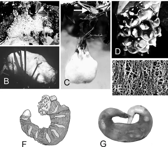
FIG. 5.—Ovipositional modes. (A) Bubble nest ofLimnodynastessp. (Limnodynastidae) (B) foam nest ofPhysalaemus pustulosus(Leiuperidae), (C) foam nest ofChiromantis xerampelina(Rhacophoridae) with attendant parent (arrow), (D) arboreal clump of Hyalinobatrachium fleischmanni (Centrolenidae) on the underside of a leaf, (E) electron micrograph of outer jelly layer ofXenopus laevis(modified from Bonnell and Chandler, 1998), (F) sac ofBatrachuperus karlschmidti(Hynobiidae) with embryos shaded for emphasis (modified from Liu, 1950), and (G) sac of Hynobius kimurae(Hynobiidae; Southern Illinois University H-07590).
Thus, one must examine clumps closely to determine their actual morphologies.
Most clumps involve relatively few eggs. In contrast, many ranids lay eggs in large, aquatic clumps that are open at least at oviposition and deposited as a single unit. Adjacent jellies are adherent. Even after the jellies swell or sag enough to obliterate the interstices, a regularly lobate surface is different from the smooth to irregular surface of a mass (see below). Some aquatic and terrestrial clumps commonly have one surface attached to a sub- strate or surrounded by a folded leaf (e.g., Phyllomedusa hypochondrialis, Pyburn, 1971) and may result from multiple oviposition bouts (e.g.,Pachymedusa dacnicolor, Bagnara et al., 1986). Other aquatic clumps are typically rounded in top view, horizontally oblong, and commonly surround a piece of aquatic vegetation. Old, aquatic clumps with embryos nearing hatching may break free from their supports and float to the surface, often with many bubbles trapped in the jelly;
the bubbles and multitiered structure distin- guish them from surface films.
Mass.—In a mass of eggs (Fig. 3E), each egg has an individual jelly layer(s) that appears to be embedded in a jelly matrix (i.e., the eggs and their jelly layers are like marbles embed- ded in a volume of gelatin, the matrix; see below). Colored water poured over a mass flows only over the surface, which may be irregular but is never regularly lobed like that of a melded clump. The matrix may be moderately sparse or voluminous relative to the volume of the ova, which gives the impression of the ova either being uniformly distributed within the mass (e.g., Pseudacris) or absent from the periphery of the mass (e.g., Ambystoma). The material forming the matrix is not extruded separately from the outer layers of jelly surrounding the ova (see below).
The egg jelly of most species ofAmbystomaor Pseudacrisis watery and usually falls apart if handled. In contrast, masses of Ambystoma gracileandA. maculatumhave a voluminous, dense matrix and retain their shape when removed from the water. Most masses start to disintegrate at or immediately after hatching, but the dense masses of these twoAmbystoma often persist for more than a month after the embryos hatch (Regester et al., 2005; Regester
and Whiles, 2006). Most masses are vertically oblong and usually attached to vegetation, and females of Ambystoma tigrinum and others may deposit a clutch among several, widely- spaced masses (Gopurenko et al., 2006).
The fact that a mass may involve an entire or a partial clutch reveals something about its formation. The inner jelly layer(s) around each ovum is distinct, but the more volumi- nous outer layer is indistinct, seemingly homogeneous, lacks a defining outer mem- brane, and melds imperceptibly with neigh- boring jellies. Thus, the outer egg jellies form the seemingly homogeneous matrix of the mass. The widths of the hydrated outer layers determine the distribution and spacing of the ova within a mass. If the width of the outer jelly layer is thin, eggs appear uniformly distributed throughout the mass (e.g., Pseu- dacris); if the layer is thick, peripheral eggs appear more widely spaced and particularly isolated from the margin of the mass (e.g., Ambystoma).
Sac.—Sacs (Fig. 5F, G) are unique to hynobiid salamanders. They are turgid sacs that enclose all ova once the layers around individual eggs are formed by the penultimate zones of the oviduct (Greven, 2002, 2003).
Females usually extrude two unfused sacs per ovulation event, one from the ovisac of each oviduct. The sac is usually shaped like a curved or spiraled sausage with a pointed distal end, which emerges first and serves as the pedicel.
According to Liu (1950; also Sato, 1992) the wall of the sac ofBatrachuperus karlschmidti is striated longitudinally, the embryos lie at 90u to the axis of the sac, and hatchlings escape by forcing off the distal cap. Hasumi (1996) noted that it took about 18 h for the sac to form inHynobius nigrescensafter all eggs arrived in the ovisac. The secretory mechanics that form a sac around a group of eggs needs further study.
Floating Arrangement
A single mode of floating eggs is described.
Film.—A film is usually a single sheet (i.e., a two-dimensional or planar tier) of either
‘adherent’(i.e., outer jelly surface sticky so that adjacent eggs adhere to each other; some hyline and pelodryadinine hylids and ranid frogs) or ‘coherent’ (i.e., outer jelly surface not
sticky so that adjacent eggs merely cohere and can be pushed apart easily; many microhylid frogs) dark or black eggs that float at or above the water surface of a puddle or pond. Some pelodryadinines deposit what appear to be multi-tiered surface films (RA, personal ob- servation). The statement by Schlu¨ter and Salas (1991) that the ‘‘entire clutches [of Chiasmocleis ventrimaculatus and Cteno- phryne geayi] are held together by a thin layer of viscous jelly’’ needs verification; the
‘‘layer of viscous jelly’’ they observed may actually be the meniscus distorted by the egg jellies. Even light rain causes surface films to sink, and eggs wetted by rain usually do not float again. Films also may sink when the embryos approach hatching. In either case, a sunken film appears as a sheet draped over vegetation or lying on the bottom. A film may consist of part or all of the eggs in a clutch.
Films appear as single groups of eggs in species (e.g., some microhylids) whose am- plectant pairs do not move around much between ovipositional bouts. In other species (e.g., some hylids and other microhylids) the amplectant parents may move around the pond or even between puddles with each egg- laying bout, splitting the clutch among multi- ple ovipositional groups of eggs (5‘rafts’). In most cases, egg films remain afloat until after the embryos hatch, but they may sink at an earlier stage if they are physically disturbed or rained upon.
Outer layers of jelly deposited in water are somewhat hydrophobic at the moment of oviposition. If eggs that typically are deposited below the water happen to be extruded above the water surface, part of the outer jelly surface may remain unwetted, causing some of the eggs to float (e.g.,Scaphiopus holbroo- kii; RA, personal observation). Although these are not films, one must conclude that species that produce films must have some behavior whereby the vents of the parents are tempo- rarily lifted above the water surface. In hylids and microhylids, this is accomplished by the amplectant pair tipping head-downward for a few seconds while eggs are extruded (e.g., Pyburn, 1967; Webb, 1971). In at least some North American ranids, the female arches her back downwards, briefly placing her vent above the water surface (Aronson, 1943). In
either case, extruded eggs, presumably sur- rounded with either hydrophobic fluids or flimsy jelly layers, spread outwards as they fall upon the water surface. Eggs in such films may rest entirely on the water surface (some microhylids), be partially submerged (other microhylids), or be submerged so that the upper surface of the outer jelly lies at the water surface (some hylids and ranids). Eggs that rest on the surface have asymmetrical egg jellies that likely facilitate floatation (e.g., species ofKaloula, Liu, 1950).
Eggs of Hyla chrysoscelis are laid as multiple rafts of single-tiered, adherent films.
The rafts may drift together and adhere, and if a breeze occurs, all films may end up on one side of a pond and not at the actual ovipositional sites. In freshly laid eggs, the top part of the upper hemisphere of the outer jelly layer is at the water surface but not flattened; the ovum is centered at the radius of the outer layer. An amorphous, very watery gel that is not visible except by careful examination in the laboratory lies external to what appears as the outer jelly layer. This adherent gel is more or less equidimensional around the more discernible next inner layer in submerged eggs. Observations through a microscope suggest that pressure caused by the weight of the egg sinking through the surface distorts this flimsy gel toward the top of the outer jelly layer to form a flat, hydrophobic, surface that affords flotation for the egg.
Eggs of Gastrophryne carolinensis are oviposited as coherent films and lack the rim-like protrusion of the outer jelly that is found in several Old World microhylids. The eggs float with about the upper fifth of the outer jelly hemisphere above the water surface (Fig. 2C). The jellies apparently lose turgidity after a few hours, and the top of the outer jelly then lies flattened and nonwetted at the surface. The ovum remains centered within the jelly so that its upper surface lies slightly below the plane of the water surface.
Wright and Wright’s (1949; also Noble, 1927) notations that the jellies of G. carolinensis and Hypopachus variolosus are flat-topped spheres apparently was based on observations of eggs a number of hours after oviposition.
This nonstructural configuration apparently is
a function of surface tensions and hydropho- bic characteristics of the jelly. Jellies of submerged eggs are spherical.
Froth Nests
Froth nests are mixtures of oviductal secretions (Bhaduri, 1932; Kabisch et al., 1998) and air that have groups of eggs dispersed through them. Such nests are pro- duced by certain hylids (Haddad et al., 1990), hyperoliids (Amiet, 1974), leptodactylids (e.g., Ho¨dl, 1990; Schlu¨ter, 1990; Shepard and Caldwell, 2005), limnodynastids (e.g., Little- john, 1963; Tyler and Davies, 1979), micro- hylids (e.g., Glaw and Vences, 1992; Haddad and Ho¨dl, 1997), and rhacophorids (e.g., Coe, 1974; Fukuyama, 1991; Liu, 1950); and see Duellman and Trueb (1986). Suggested func- tions for froth nests include: escape from the aquatic environment (Heyer, 1969), protec- tion of eggs and embryos from desiccation and thermal damage (Gorzula, 1977; Heyer, 1969;
Ho¨dl, 1986), floatation for aquatic eggs (Haddad and Ho¨dl, 1997), protection from aquatic predators and cannibals (Ho¨dl, 1990;
but see Drewes and Altig, 1996 and Menin and Giaretta, 2003), and enhanced oxygena- tion of the eggs and embryos either from being held near the meniscus (Haddad and Ho¨dl, 1997) or from the air trapped in the bubbles per se. There are two types of froth nests: foam and bubble.
Foam nests.—Foam nests typically are formed when one or both parents whip oviductal secretions with the hind legs to trap numerous small air bubbles. The foam of Scinax rizibilis is produced by the pair jumping onto the jelly. The elaborate se- quence of kicking and wiping behavior (e.g., Heyer and Rand, 1977; Ho¨dl, 1990), which enhances the mixing of sperm and eggs, varies among species. This behavior likely evolved from general movements used by the parents to stabilize or maintain their positions in the water (e.g., Rabb, 1973; Pipa pipa). Ho¨dl (1990; Physalaemus ephippifer), Liu (1950;
Polypedates leucomystax, and Coe (1974;
Chiromantis rufescens) indicated that the foam-forming material is distinct from and released prior to egg extrusion. In contrast, Heyer and Rand (1977,Leptodactylus penta- dactylus, Physalaemus pustulosus), Ryan
(1985,Physalaemus pustulosus, Fig. 5B), and seemingly Schlu¨ter (1990,Edalorhina perezi) pointed out that it is a jelly-ova mixture that is whipped into foam. Further detailed observa- tions are needed in all cases. Perhaps the ova with their jelly layers and an oviductal secretion(s) that is whipped are released at the same time. Simultaneous release of eggs and secretions has been reported for Poly- pedates bambusicola by Liu (1950). Coe (1974) described and illustrated a discrete structure called a ‘foam gland’ that formed from three large oviductal folds along the posterior sections of the oviduct inChiroman- tis rufescens; secretory cells lining the lumen reportedly contained ‘foam parent material.’
Similar structures present in foam-nesting Leptodactylusspp. (RWM, personal observa- tion) will be reported elsewhere.
Foam nests are formed in aquatic, arboreal, subterranean, and terrestrial sites, and are generally the result of a single reproductive bout. Schlu¨ter (1990) reported up to three nestings by single pairs of Edalorhina perezi (Leiuperidae) in a single night in captivity.
Whether this happens in other species is not known. Males ofE. perezialso are reported to smooth the surface of the foam between egg laying bouts (Schlu¨ter, 1990). In most species, a foam platform is built before the ova are released. As eggs are extruded, the male catches them in a ‘basket’ formed by his hind feet, presumably fertilizes and moves them dorsally onto his back, and then pushes them into the foam with his hind legs (e.g., Physalaemus ephippifer, Ho¨dl, 1990). In those species studied, eggs are released in several bouts and scattered throughout the mass of foam, and trophic eggs are sometimes de- posited in foam nests (Prado et al., 2005). In arboreal cases the outer portion of the foam dries to a crust (Fig. 5C; Coe, 1974). Bossuyt and Milinkovitch (2000) suggested that arbo- real foam nests of AsianPolypedatesspp. could be an initial step toward direct development on land or trees (see Altig and Crother, 2006).
Actions of hatchlings and enzymes eventually degrade the inner portion of the foam, and the hatchlings stream out into the water or drop into the water in arboreal species of Chiro- mantisspp. andPolypedatesspp. (Rhacophor- idae; Liu, 1950; Coe, 1975).