The following investigations are a preliminary attempt leading to a more intensive monograph of the genus Gnathotrichus Eichhoff. Regarding the position of the genus Gnathotrichus in the family Scolytidac, Eichhoff states.
NO. IO MORPHOLOGY OF GNATHOTRICHUS — SCHEDL 7
Descriptio materiae Filch ab Eichhoff in Ratione Tomicinorum data est longe accuratissima. Pro-thorace latior quam lato, dimidio longiore, cylindraceo, basi truncato, lateribus rectis, parallelis, apice acute rotundatis, angulis posterioribus distincte rectis; supra valde cylindraceum convexum, inflexum, posterius antrorsum linea mediana, transversa, elevatum, notatum, antice rarius, rugis imbricatis, tenuissime pubescentibus, posterius glabris, subniid, sinistrum, infra oculum, punctum subtilissimum. omnium.
NO. IO MORPHOLOGY OF GNATHOTRICHUS — SCHEDL 9
Very common in sapwood of dead and dying pine and spruce trees, logs and stumps; widespread. Excavates several branching galleries from a single-entrance burrow, with broods living in short side chambers in sapwood and heartwood of wounded, dying, and recently felled pine and spruce.
NO. 10 MORPHOLOGY OF GNATIIOTRICHUS — SCHEDL II
GNATHOTRICHUS RETUSUS LE
The main food of the larva, and an important food of the adult, is a special fungus called Ambrosia, which grows in a dense, glistening layer on the walls of the tunnels and cradles. These small, black, round, branching tunnels in the forest are characteristic of the Wood Beetles or Ambrosia Beetles.
NO. 10 MORPHOLOGY OF GNATHOTRICHUS SCI1EDL 13
GNATHOTRICHUS SULCATUS LEG
NO. 10 MORPHOLOGY OF GNATHOTRICHUS SCIIEDL 1 5
GENERAL APPEARANCE, VESTITURE, COLOR, AND SIZE The general form of all three species is slender in both sexes,
NO. 10 MORPHOLOGY OF GNATIIOTRICHUS — SCHEDL 17
The sculpture of the pronotnm and elytra, which is very useful in distinguishing the species in many other genera of the Scolytidae, does not vary to any extent in this genus. Secondary sexual characteristics. Secondary sexual characteristics were mainly found in the development of the hairs on the antennae, the number of fully developed tergites and in the number of spiracles.
THE HEAD
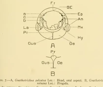
10 MORPHOLOGY OF GNATIIOTRICHUS SCHEDL 21Gula.— The gula is bounded by the two very closely placed gular- Gula.— The gula is bounded by the two very closely placed gular- Stltures (Figs. 6, 7, Gus) and expands anteriorly into form. the pre-regula. Epicranium.—The remaining lobes of the skull, situated between the epicranial suture, the gula, and the foramen, bear the connection.
THE APPENDAGES OF THE HEAD THE ANTENNAE
Females (fig. 8, A) bear, moreover, on the anterior end of the club some very long hairs. BB- — The outer side of the club with small wrinkles, the inner side with very few hairs and a little.
THE MOUTHPARTS
AA —Septae wider laterally, indistinct and narrow medially; external lateral side of bill smooth or with very few, sparse wrinkles; club stronger. The palpifer (c) which is only a topographical area of the stipes, is not limited by lines or sutures.
28 SMITHSONIAN MISCELLANEOUS COLLECTIONS VOL. 82 The lacinia bears on the anterior portion of the free dorsal margin
NO. 10 MORPHOLOGY OF GNATHOTRICHUS SCHEDL 20,
Anterior margin very weak, shallowly cut or evenly rounded; however, this does not appear to be constant among individuals of the same species. On the ventral side there is a row of setae (d) on each side near the anterior margin. The sculpture of the ligule on the ventral side is very similar in all three species.
Gn, matcriarius and sulcatus have the corresponding area smooth with slight signs of transverse folds on the sides.
THE THORAX
52shape is elongated; it is subparallel with two more or less distinct elongate conshapeis; it is subparallel with two more or less distinct contractions when viewed from the dorsal aspect. No specific differences have been found either in the shape or in the number of these setae, which vary in number from three to seven in each row. The dorsal plate, or tergum, the ventral plate, or the sternum, and the dorsal plate, or tergum, the ventral plate, or the sternum, and the lateral region, or the pleuron.
The prosternum is about half as long as the metasternum and about one-third the length of the pronotum, making the pleural region of the prothorax shaped like a trapezium.
THE PROTHORAX
This increased difference served as one of the main characters that placed this genus near Pityophthorus and allied genera. The lateral limits of the pronotum are not clearly defined, but near the postero-lateral angle is a longitudinal ridge which may be regarded as a remnant of the pleuro-notal suture. Pleural area.— The propleural area (fig. 14) appears as a continuous plate in the form of a trapezium, the base of which is.
The episternal area is completely covered by setae of serrations that appear on the anterior half of the pronotum.
34 SMITHSONIAN MISCELLANEOUS COLLECTIONS VOL. 82
THE MESOTHORAX
The scutum (Fig. 16, Stu) is represented as two slightly chitinized lobes, which are fused anteriorly with the prescutum. A short distance behind the clavicle is a well-developed prealar process that covers the third axillary of the elytra. A narrow band in front of the episternum, bounded externally by a suture, and its continuation towards the ventral posterior corner of the episternum may represent the preepisternum.
82 Sternellar area.— Sternellar area produced strongly posterior- Sternellar area.— Sternellar area produced strongly posterior-.
THE METATHORAX
NO. 10 MORPHOLOGY OF GNATHOTRICHUS — SCHEDL 37
Dorsally, the extreme angle of the episternum with the pleural suture (which probably also contains elements of the epimeron) is produced in the parapterum (e), or coracoid process, and the wing process, or clavicular process (d). A more flexible, partially membranous sclerite is inserted between the pleural suture and the lateral margin of the metanotum. Posteriorly, it is subdivided by a branch of the pleural suture that separates the postepimeron from the epimeron proper.
The area between the anterior margin of the hypopleurite and the dorsal lobe of the pleural suture is deeply impressed.
NO. IO MORPHOLOGY OF GNATHOTRICHUS SCHEDL 41
THE ABDOMEN
These bands or ridges resemble the parapsids of the metathorax both in structure and position. The surface of the uncovered part of the first sternite bears numerous hairs, which are arranged in concentric rows surrounding the metacoxa. The median line of the commissure, which gives, with certain modifications, the spiculum ventrale opportum, is especially well defined in Gn.
The part where the spiculum joins the seitlichen Ansatzlappen (Verhoeff), seitlichen Lappen (Fuchs) can be called the radixspiculorum; more or less chitinized, sometimes membranous bands extending anteriorly,.
NO. IO MORPHOLOGY OF GNATHOTRICHUS SCHEDL 45
THE SPIRACLES
THE LEGS
46 SMITHSONIAN MISCELLANEOUS COLLECTIONS VOL. 82 a marginal flange and is visible in both the fore- and the
NO. 10 MORPHOLOGY OF GNATIIOTRICHUS SCHEDL 47 the heavily chitinized wall of the coxa which is covered by a thin
10 MORPHOLOGY OF GNATIOTRICHUS SCHEDL 47the strongly chitinized wall of the coxa, which is covered by a thin. For the same purpose, a hook-like process is used on the postero-median angle of the basicosta. The shape of the condyle is the same in all three pairs of legs; the small differences that appear on the plate are due to the different angles from which the drawings are made.
The femur (Fig. 26, F) is the strongest segment of the leg and is approximately equal in length to the tibia (Ti).
NO. 10 MORPHOLOGY OF GNATHOTRICHUS — SCHEDL 49
THE WINGS
MESOTHORACIC WINGS OR ELYTRA
The articulatory elements of the elytra itself mainly consist of the projected costa (Co) and subcostal veins (Sco) and the costal (a) and the subcostal heads (b). The second axilla (ax2) articulates at its base with the prealar process (a) of the prescutum. The apex of the second axil forms a heavily chitinized clamp into which the elytra fit as a tongue.
It begins on the inner surface of the second axillary and ends on the outer surface of the elytra.
NO. IO MORPHOLOGY OF GN ATHOTRICHUS SCHEDL 53 author and therefore this statement is merely an attempt to explain
METATHORACIC WINGS OR HIND WINGS
54 SMITHSONIAN MISCELLANEOUS COLLECTIONS VOL. 82
NO. 10 MORPHOLOGY OF GNATHOTRICHUS — SCHEDL 55 extends to the folding hinge on the wing gradually increasing in
The horn forms with the scapular arm an axilla (m) in which the anterior projection (n) of the second axilla rests. The margin towards the apex (q) of the wing fits into the lateral groove (r) of the second axilla. Second armpit. The second axillary plate (fig. 30, ax2) or subscapular plate has the shape of an equilateral triangle with the base forward.
Lateral emarginaiion.- The lateral emarginaiion (Hopkins) (fig. .. 17, p) is a protrusion on the scutum at the lateral edge of the scutellar lobe in which the posterior inner lobe of the scapular plate and the scapular hooks are implanted.
58 SMITHSONIAN MISCELLANEOUS COLLECTIONS VOL. 82 The testes consist of two oval structures which are closely connected
The dorso-caudad portion of the inner lids was called the Endplatten by Lindeman, the laminae dorsales by Fuchs and the dorsal plates by Hopkins. The caudad portion of the laminae ventrales, thecaput (Fuchs), is variously modified, and sometimes bears a beak-like projection dorsally called therostrumby Fuchs. The area of the pallidium, from which the peduculi penis arise, the radix (Fuchs), is not characterized by a heavier chitinization.
Enclosed parts.- The enclosed parts are a short part of the ductus ejaculatorius, the preputial sac and chitinous reinforcements of the latter.

NO. 10 MORPHOLOGY OF GNATHOTRICHUS SCHEDL 63
64 SMITHSONIAN MISCELLANEOUS COLLECTIONS VOL. 82 same level as the cement glands ; it has the shape of a pipe and bears
NO. 10 MORPHOLOGY OF GNATHOTRICHUS SCHKDL 65
In Gnathotrichus, both of these plates are separated by a median suture (a) which is clearly visible on the masticatory plate and indicated by a row of hairs (b) on the anterior plate. Cephalad, they are bordered by several (8-12) longer teeth (c) which vary greatly in shape and which are directed towards the center of the proventriculus. To distinguish them from a similar arrangement of teeth occurring in Gnathotrichus and other genera at the caudal end of the masticatory plate (f), it is proposed to call the cephalad closing teeth, the latter the caudad closing teeth.
In Gnathotrichus the following fixtures are not present. a) Hackenzaehne (Nuesslin), or apical teeth of the anterior plate; the indication of a row of short, often curved teeth on the apical edge of the anterior plate.
NO. IO MORPHOLOGY OF GNATHOTRICHUS SCHEDL 67 (d) Abdachungszaehne (Nuesslin), masticatory teeth which have
68 SMITHSONIAN MISCELLANEOUS COLLECTIONS VOL. 82
NO. 10 MORPHOLOGY OF GNATHOTRICHUS SCHEDL 69
JO SMITHSONIAN MISCELLANEOUS COLLECTIONS VOL. 82 The structure and general appearance of the larva are shown in
THE CHITINOUS SKELETON THE HEAD
Laterally, this structure is slightly bent forward and this part carries the dorsal joint of the mandibles. Towards the occipital foramen it extends into another border from which the connecting membranes of the maxilla and maxilla emerge. The entogular plate extends into the foramen below the occipital apodeme, giving the open space of the foramen the shape of a triangle, the sides of which are broadly rounded.
IO MORPHOLOGY OF GNATIOOTRICHUS – SKULL 73Other topographical areas of the skull are not defined by.
NO. IO MORPHOLOGY OF GNATIIOTRICHUS — SCHEDL 73 Other topographical regions of the cranium are not defined by
Thelabium (Lab) of the larva differs greatly in structure from that of the adult. Indications are that the labium of the larva as well as that of the adult will become more and more important as the bearer of taxonomic characters in the Scolytidae. It is slightly chitinized, laterally connected to the maxillae and bears three pairs of setae (d), the same number and in a similar ar-.
82mentum does not extend as far as origin of palpi as i.
76 SMITHSONIAN MISCELLANEOUS COLLECTIONS VOL. 82 mentum does not extend as far as the origin of the palpi as in the
NO. IO MORPHOLOGY OF GNATHOTRICHUS — SCHEDL JJ THE THORAX
Nine pairs of spiracles are present, eight of which are located on the epipleurites of the first eight abdominal segments. The ninth trachea is located on the same sclerite of the prothorax, very close to the mesothorax.
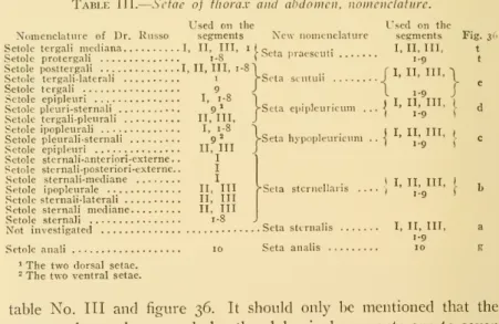
THE THORACIC AND ABDOMINAL SETAE
NO. IO MORPHOLOGY OF GNATHOTRICIIUS— SCHEDL 79
THE ALIMENTARY CANAL
The cautlad widened part, which is also surrounded by strong muscles, may correspond to the united tumor and proventriculus. Midgut.- The midgut occupies the largest area of the entire digestive system of the larva. The latter is clearly wider than the rest of the hindgut, and the muscles surrounding it are much more strongly developed.
THE PUPAE
82 SMITHSONIAN MISCELLANEOUS COLLECTIONS VOL. 52
NO. IO MORPHOLOGY OF GNATHOTRICHUS — SCHEDL 83
No remains of the larva's sternal bristles are visible on any of the legs, as found by Hopkins. In tergite six, setae e3 and e4 have the same shape and appearance as e2 in the previous tergites. Even in the young pupa, the eighth pleurite is more similar to that of the adult than to the larva.
The tail spine (Hopkins) most likely represents the only external remnant of the ninth abdominal segment of the larva and.
BIBLIOGRAPHY
Verhoeff, C, Vergleichende Studien über die Abdomensegmente und die Kopulationsorgane der männlichen Coleoptera, ein Beitrag zur Kenntnis ihrer natürlichen Beziehungen. Verhoeff, C, Vergleichende Untersuchungen der Abdomensegmente, insbesondere der Legebohrer der weiblichen Coleoptera, ein Beitrag zu ihrer Phylogenie.
NO. IO MORPHOLOGY OF GNATHOTRICHUS SCHEDL 87