Studying persistent social state change has been challenging because of the difficulty of precise, time-resolved presentation of the social cues. Although we show that persistent internal state change can be induced by transient stimulation of the sensory cues in Chapter II, the circuit implementation of such persistence is not clear.
INTRODUCTION
Persistent internal states influencing social behaviors in fruit flies
Interestingly, activation of P1 interneurons in single male flies induces sustained arm extensions that last longer than P1 stimulation ( Inagaki et al., 2014a ). Transient activation of P1 neurons in pairs of male flies evokes persistent aggression (Hoopfer et al., 2015).
Optogenetics and functional imaging to map brain function
Activation of neurons using optogenetics has another advantage that could not be achieved by other genetically encoded actuators such as TrpA1 ( Hamada et al., 2008 ): thermogenetic activation. Most optogenetic tools are activated by a broad spectrum of visible light and are difficult to manipulate two different populations of neurons with different effectors (Klapoetke et al., 2014).
Summary
Introduction
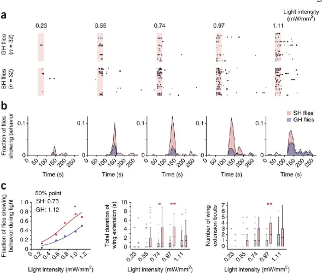
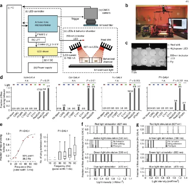
Here we demonstrate that expression of ReaChR in adult central nervous system (CNS) neurons enables rapid and temporally precise control of behavior in freely moving adult Drosophila. Using this optogenetic method, we separated the control of wing extension, a male-specific courtship behavior, into either probabilistic, state-dependent, or deterministic, command-like components.
Result
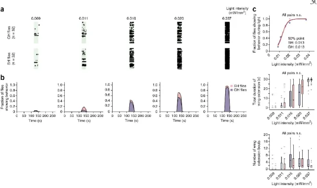
In contrast to the results observed in P1 neurons, activation of pIP10 neurons triggered robust wing extension in a deterministic manner (Fig. 3a). As pIP10 neurons are thought to be functionally downstream of P1 neurons (von Philipsborn et al., 2011) (Figure 3b), we investigated whether the activation of wing extension ReaChR via these descending neurons was also sensitive to social experience.
Discussion
Main figures
The properties of box plots are as in Figure 2.1f. f, g) Fraction of flies showing characteristic behavior at different photostimulation intensities and wavelengths, in animals expressing hb9-GAL4 (f) or fru-GAL4 (g) and UAS-ReaChR. Properties of box plots are as in Figure 2.1f; “+” indicates outlier data larger than the top whisker.
Supplementary figures
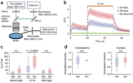
Supplementary Figure 2.2: Expression of ReaChR in the brain and ReaChR-based activation of behavior at different frequencies. a) Expression and trafficking of ReaChR (top) and C1V1(T/T) (bottom) in P1 neurons in adult flies. Note that both opinions are expressed in cell somata (white dashed box), but only ReaChR is transported to the arborizations. Both opsins are visualized with a citrine or YFP tag immunostained with anti-GFP antibody.
Note that regardless of photostimulation conditions (see also Figs. 3,4 ), ReaChR-based activation of P1 neurons triggers stochastic and sustained wing extensions. The data were fitted by a sigmoidal function to calculate the 50% point. blue points) are the same as red points used in Fig.
Materials and methods
A photodiode force sensor (S130VC, Thorlabs) was placed at the location of the behavioral chamber, but in the absence of the chamber. To filter out autofluorescence from the brain and light from the amber stimulation LED (for ReaChR activation), we used a 500/20 nm (center wavelength/bandwidth) bandpass filter (Chroma) in the emission path to pick up the GCaMP3 fluorescence track. To reduce the bandwidth of the LED output, we connected the optical fiber to a fiber optic filter holder (World Precision Instruments) equipped with 589/10 nm (center wavelength/bandwidth) bandpass filter (Edmund optics).
One side of the optical fiber was custom made to be a bare tip (Thorlabs) and was immersed in the saline image bath and placed 430 μm away from the brain. The distance between the heat source and the labellum was set to be 540 μm using a micrometer.
Drosophila HB9 is expressed in a subset of motoneurons and interneurons, where it regulates gene expression and axonal pathfinding. Joint control of Drosophila male courtship behavior by movement cues and activation of male-specific P1 neurons. Temporal dynamics of neuronal activation by Channelrhodopsin-2 and TRPA1 determine behavioral outcome in Drosophila larvae.
Light-induced activation of distinct modulatory neurons induces appetitive or aversive learning in Drosophila larvae.
Summary
Introduction
In mice, transient activation of agouti-related peptide (AgRP)-expressing neurons in the arcuate nucleus of the hypothalamus promotes persistent food seeking, an effect mediated by neuropeptide Y (NPY) signaling ( Chen et al., 2016 ; Chen et al., 2016 ., 2019). Transient activation of steroidogenic factor 1 (SF1) neurons in the dorsomedial/central part of the ventromedial hypothalamus (VMHdm/c) promotes persistent defensive behavior (Kunwar et al., 2015). Artificial stimulation of P1 neurons in solitary males can trigger a rapidly emerging courtship song ( Pan et al., 2011 ; von Philipsborn et al., 2011 ; Inagaki et al., 2014 ).
Similarly, the effect of P1 activation to drive aggression between males lasts for minutes after compensation of photostimulation (Hoopfer, 2016; Watanabe et al., 2017). In contrast to these persistent behavioral effects, optogenetically evoked physiological P1 activity, measured via calcium imaging in live, head-fixed flies, returns to baseline in tens of seconds (Inagaki et al., 2014; Hoopfer et al. ., 2015) (although it has been reported to persist in brain explants (Zhang et al., 2018a)).
Result
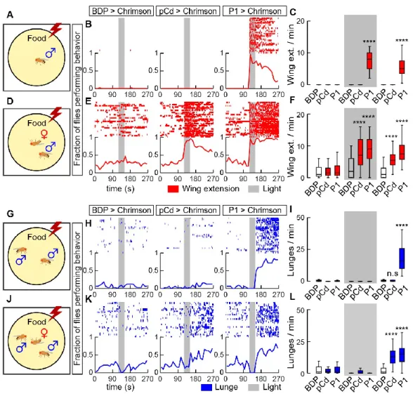
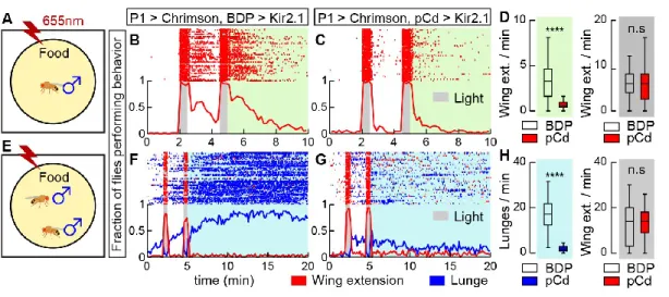
In solitary males (Figure 3.2A), silencing pCd neurons dramatically reduced persistent wing extension induced by Chrimson activation of P1 cells (Figure 3.2B vs. 2C, green shading; 2D). Interestingly, optogenetic activation of pCd neurons in solitary flies had no apparent effect, in contrast to optogenetic activation of P1 neurons ( Inagaki et al., 2014 ; Clowney et al., 2015 ; Hoopfer et al., 2015 ) ( Figure 3.3A, B). To test this, we examined the effect of pCd stimulation on the behavioral response of males to female cues (Figure 3.3D).
But in the presence of a dead female, which produced increased baseline aggression in male flies (Lim et al., 2014), activation of pCd neurons significantly increased flies' aggressiveness after photostimulation, an effect not observed in photostimulated controls ( Figure 3.3J-L). Among pCd neurons persistently activated by P1 stimulation (Figure 3.7C1), only half responded to cVA alone (defined as >2σ above baseline; Figure 3.7C2).
Discussion
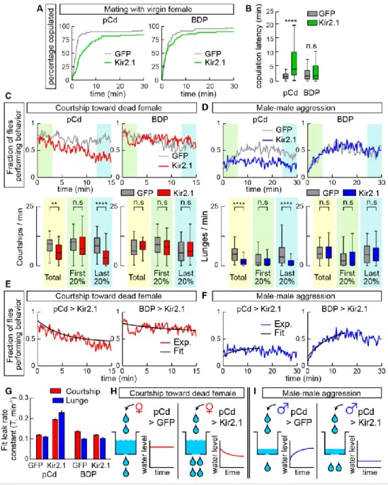
Whether such mechanisms also operate in mammalian systems where female exposure promotes aggressiveness (Remedios et al., 2017) remains to be determined. Recently, Zhang et al reported. a role for pCd neurons in a recurrent circuit with NPF neurons that accumulate mating drive under conditions of extended sexual deprivation in Drosophila males (Zhang et al., 2019). Consistent with our results, Zhang et al found that constitutive silencing of pCd neurons partially reduces mating behavior.
However, they also reported that acute activation of these neurons has no effect on courtship, and that behavioral effects are only observed after 12 hours of continuous thermogenetic stimulation of these cells (Zhang et al., 2019). Whether NPF neurons are involved in this aggression-promoting function of pCd neurons remains to be determined; we previously reported a weak effect of NPF neuron stimulation to enhance aggression ( Asahina et al., 2014 ), whereas another group reported that silencing NPF neurons increased aggression ( Dierick and Greenspan, 2007 ).
Main figures
Lower: fraction of flies performing wing extensions (red line) or lunges (blue line) in 20-second epochs. Inhibition of pCd neurons increases copulatory latency and reduces the endurance of naturally occurring social behavior. Inhibition of pCd activity with Kir2.1 increases the integrator leak rate constant. pCd neuronal activity is required for physiological persistence.
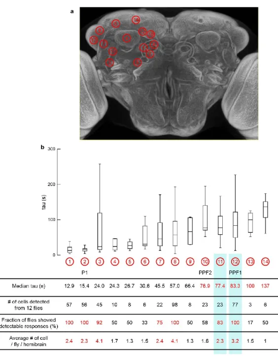
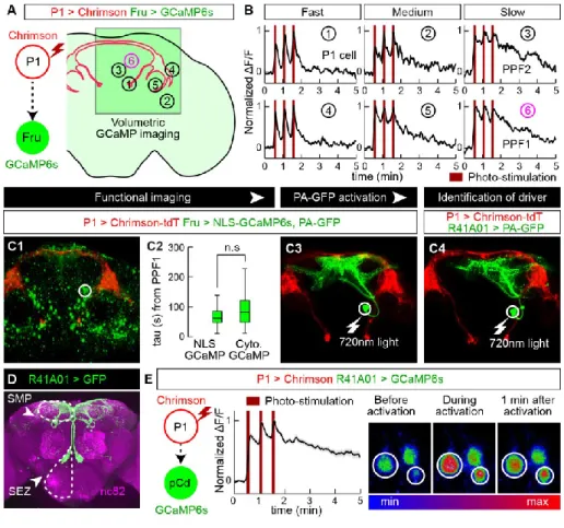
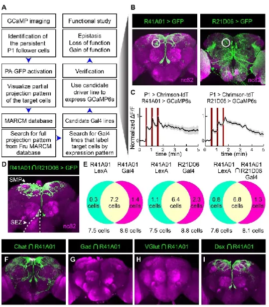
If pCd neurons simply inherit persistence passively from the center (left), persistence should rebound after transient pCd silence. If persistence does not recover, this implies that pCd activity is required for the center to maintain persistence (right). C) Representative two-photon image showing cell body locations of pCd and PPF2 neurons expressing Fruitless>GCaMP6s in vivo.
Supplementary figures
Traces represent the normalized ∆F/F response to P1 stimulation (dark red lines, 3 repetitions of 5 s stimulation, 10 Hz, 10 ms pulse width, 25 s interstimulus interval) and were obtained from cell bodies within white circles. listed in (B). Integration of repeated P1 input to pCd neurons associated with Figure 1. A, C, E) Normalized GCaMP response of pCd neurons to optogenetic stimulation of P1 neurons. Co-registered images showing the somatodendritic region of pCd neurons and the presynaptic region of P1 neurons.
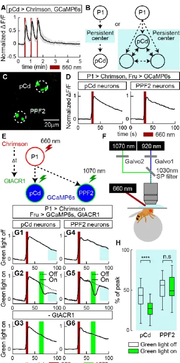
Multiple cycles of P1 stimulation and GtACR1 activation, and inhibition of pCd neurons following P1 stimulation labeled by a pCd-specific driver, related to Figure 5. A) Representative GCaMP fluorescent images of pCd (top) and PPF2 neurons (bottom) at different time points following Chrimson-mediated P1 stimulation (wide-field LED actuation at 660 nm), and cell-restricted GtACR-mediated pCd or PPF2 inhibition (2-photon spiral scan actuation at 1070 pm). Multiple cycles of P1 stimulation with or without GtACR1 activation did not change the initial responses of pCd and PPF2 neurons to P1 stimulation.
Materials and methods
Salt was added to the upper side of the plate to submerge the fly's head. Conditions within the imaging setup were kept similar to growth conditions (25°C and 55% humidity). 20 minutes after the second spiral scan cycle, 3-dimensional images with a resolution of 1024 X 1024 pixels were acquired.
All behavioral assays except mating assay (Figure 4A,B) were performed in 8-well acrylic chamber (16 mm diameter x 10 mm height, modified from ref. Inagaki et al., 2014), and the side of each well was coated with an Insect -a-Slip (Bioquip products). Temperature probe (Vktech) was inserted into one side of the chamber to accurately monitor the chamber temperature.
Genetic and neuronal mechanisms controlling the sex-specific interaction between sleep and sexual behavior in Drosophila. Behavioral responses to a repeated visual threat stimulus express a persistent state of defensive arousal in Drosophila. Anterior hypothalamic neural activation and neurochemical associations with aggression in pair-bonded male prairie voles.
Drosophila visually induced initiation, an innate courtship-like subsequent pursuit, is mediated by a central excitatory state. A circuit node that integrates convergent inputs from neuromodulatory and social behavioral neurones to control aggression in Drosophila.
FUTURE DIRECTIONS
- Persistent P1 follower cells besides pCd neurons that are identified by functional connectomics screening. connectomics screening
- Mechanism underlying persistent neuronal activity
- Other unsolved problems: conflicts between observations
- Limitations and future improvements
- Figures
- References
Direct activation of pCd neurons (Figure 5A) did not induce persistent pCd activity, indicating that the persistent neuronal activity induced by P1 stimulation is not due to intrinsic cell properties of pCd neurons. First, in Chapter 3, we showed that activation of pCd neurons does not cause persistent pCd activity (Figure 5A). This result indicates that in the presence of the female signal, activation of pCd neurons may be sufficient to cause sustained neuronal activity.
Second, although the decay constant (~80 s) of pCd activity induced by P1 activation is comparable to that obtained from P1-induced persistent courtship, the effect of P1 activation on aggressive behavior lasts much longer (~10 min) than neuronal activity of pCd neurons. But also with a woman's belly. touch, stimulation of pCd neurons did not produce persistent activity comparable to P1 stimulation.