Abstract
Introduction
Obesity has been known as a risk for a lot of metabolic diseases including type 2 diabetes mellitus (T2DM), hypertension, coronary heart disease, and certain forms of cancer for a long time. Still, the prevalence of obesity keep increasing and become the 21st century public health threat, which in the end will be a burden for public health and economic consequences.(1) Lifestyle intervention including energy-restriction, exercise, and behavioural therapy have been always the main strategies to manage obesity. These treatment result in clinically relevant weight loss (WL) up to 10% in the short term.(2) Somehow, only 15% individuals
can maintain the WL for one year after lifestyle intervention.
(3) Therefore, regain weight to the initial weigh or even more become a challenge and considered as a relapsing condition.(4,5)
Growing evidence suggests that in obese, the energy homeostasis system was dysregulated. That’s how the same calorie intake will yield different output in obese individuals compare to the lean. Thus, we need to elucidate how to reset this disorder. This will not be simple since many forces such as genetic, developmental, and environmental affect the energy homeostasis system. Energy balance can no longer be sees as the sum of energy intake (EI) versus energy expenditure (EE). The interaction of macronutrition substrate (carbohydrate, protein, and fat), hormonal and
Obesity: A Multi Perspective of Physiology and Neurobiology Energy Regulation
Anna Meiliana
1,2,3,, Nurrani Mustika Dewi
2,3, Andi Wijaya
2,31Department of Pharmacology and Clinical Pharmacy, Faculty of Pharmacy, Universitas Padjadjaran, Jl. Raya Bandung-Sumedang Km 21, Jatinangor 45363, Indonesia
2Prodia Clinical Laboratory, Jl. Supratman No 43, Bandung 40114, Indonesia
3Prodia Education and Research Institute, Jl. Kramat Raya No. 150, Jakarta, 10430, Indonesia
Corresponding author. E-mail: [email protected]
Received date: Dec 12, 2023; Revised date: Jan 5, 2024; Accepted date: Jan 8, 2024
R E V I E W A R T I C L E
B
ACKGROUND: World Health Organization has reported four million people die every year due to obesity comorbidity, and the prevalence of obesity is keep increasing, especially after COVID-19. Obesity has been defined as a chronic disease involving adipose tissue dysfunction which leads to metabolic diseases and psychosocial consequences. The review article will highlight some recent researches regarding the new conceptual framework that integrates both metabolic drives, as well as to summarize the numerous discussions about the current understanding of hypothalamic control of food intake and energy homeostasis.CONTENT: Obesity apparently is not simply regulated only by food and exercise. Hypothalamus takes part in
controlling energy intake and expenditure via appetite regulation. Hedonic control in cortical and subcortical brain areas process cognitive, reward, information, and executive function. Managing metabolic adaptation, browning the white adipose tissue, and preserving lean mass can be another strategy to safely manage obesity.
SUMMARY: Obesity need to be managed in a multimodal strategy including neurophysiology and physiology approach, together with environment support. Thus, a weight regain can be prevented. Commitment from both scientific and regulation point of view can shed a light to eradicate obesity.
KEYWORDS: adipocyte, appetite, nutrition, obesity, physical activity, reward, satiety
Indones Biomed J. 2024; 16(1): 1-22
Obesity Pathogenesis and The Compensatory Theory of Relapse
excessive weight gain. When we have excess calories, the brain by adipocyte-autonomous processes will impose it on the passive adipocytes, and save the calories in adipocyte tissue (AT). Some debates arisen on how far the adipocyte- autonomous processes can activate the passive adipocytes, and partitioning calories to yield a higher fractional deposition of calories as fat. This theory leads to an isocaloric diet, where individuals consume a diet in which caloric intake and EE are matched, means the ingested calories will match the amount expenses by the lean mass, and there will be energy balance.(1)
Recent studies show that diet composition (the specific types and quantities of micronutrition including carbohydrates, sugars, and fatty acids) contributes different caloric content, and the varied composition of diet itself may powerfully affect palatability and hedonic motivation of feeding. Somehow there is still a debate about how significant the contribution of the diet composition itself compared to dietary’s secondary metabolic consequences (e.g., effects of insulin on circulating nutrient levels).(1) Weight change is also affected by interindividual variability of energy metabolism. Some determinants associated to individuals’ susceptibility to weight gain including: First, inter-individual variance of energy metabolism, which is the total of EE and non-protein respiratory quotient (RQ).
Age, genetic, gender, ethnicity, glucose tolerance, body composition and size will also take part in EE variability.
Second, individual variety in short-term adaptation in respond to acute changes of EI, or so-called adaptive thermogenesis.
There are two metabolic phenotypes in respond to these, which is the thrifty phenotype that efficiently save energy either in energy surplus (overfeeding) or deficit (fasting), and the spendthrift phenotype that maintains higher EE in fasting condition while expend more energy in overfeeding situation. Third, individual variability in thermogenesis capacity in respond to many stimuli such as food intake, heat or cold exposure, physical work, emotional state, or pharmacological treatment. This is associated with the individual’s brown adipose tissue (BAT) activation. Lastly, the great variability in causal link between EI and EE or known as energy sensing. Some individuals may sense to increase or decrease their EE than is truly needed by consuming more (or less) food than required.(12)
Our body was initially designed to survive, especially in the scarce of food. Thus, WL will induce physiological adaptations to bring back the homeostasis both appetite control system and EE. This normalization to lower body weight significantly induce a reduction in total energy expenditure (TEE), by declining resting and non-resting enzymatic processes, and oxidation rates will determine
how much the energy accumulation in our body. The surplus of energy will be deposited as fat mass, and the deficit will otherwise burn the fat mass.(6) Some studies showed different result by sustained caloric restriction (CR) depend on how the calories distributed between fat mass and lean mass. Most pharmacotherapies also face the problem of lean mass loss.(6–8)
While this review aims to explore the multi mechanisms involved in obesity and give the perspective of managing obesity personally beyond current appetite regulation, but the primary goal of this review is to give spotlight for some recent researches regarding the new conceptual framework that integrates both metabolic drives, whether generated by real or perceived nutrient needs, as well as hedonicdrives to eat, which is generated by factors other than nutrient needs), given the numerous discussions summarizing current understanding of hypothalamic control of food intake and energy homeostasis.(9,10)
In a general way, obesity is defined as an excess body fat mass. Reliable body fat mass analysers require sophisticated tools which are limited (magnetic resonance imaging (MRI) or dual energy X-ray absorptiometry). As a consequence, people use simpler measurement to define obesity such as body mass index (BMI) or waist circumference (WC).
WHO classified BMI <18.5 kg/m2 as underweight, BMI 18.5-24.9 kg/m2 as normal weight, while obese adults BMI can be subclassified further into class 1 (BMI 30 to <35 kg/m2), class 2 (BMI 35 to <40 kg/m2), and class 3 (BMI
>40 kg/m2).(11)
In the most basic levels, obesity pathogenesis was explained as the exceed amount of calories intake compared to EE. Thus, obesity creates negative paradigms associated to laziness, self-indulgence, lack of will power, etc. While pathogenesis of obesity actually is far more complex than that, which integrates genetic, developmental, molecular, behavioural, environmental, and socioeconomic factors.
This is the reason why obesity is so difficult to treat.(1) In the evolutionary physiology, centuries ago when food was scarce, human body was designed to preserve energy in the form of body fat as a factor of survival. Now, as the food are very easy to get, our body seems to keep the survival mode and utilize calories in efficient ways.
Thus, our body appears to be biologically predisposed to
Neurobiology of Nutrition and Obesity
Going back in time, the struggle for nourishment led to the formation of human biological structure and function. For autonomic, endocrine, and peripheral cellular processes to result in an internal energy-saving state, the regulatory system must be effective. Nutrient-depletion signals thus strongly activate neural mechanisms that modulate appetitive, ingestive, and foraging behaviors. To maintain this regulatory system, powerful effector mechanisms for metabolism and EI are needed, as well as redundant and complex nutrient sensing and monitoring mechanisms, a EE. There will be alteration in fat mass (FM) and fat-
free mass (FFM), as result of adaptive thermogenesis or metabolic adaptation which can be sustained up to 6 years.
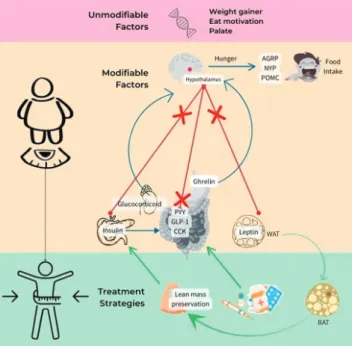
This phenomenon has been associated to the risk of relapse, while EE reduction with the risk of weight regain in the long term still become controversial issues if this metabolic adaptation are exaggerated because there is no evidence of metabolic adaptation in the level of resting metabolic rate (RMR). WL shown consistent data to upregulate the hunger hormone ghrelin, and downregulate the satiety hormone glucagon-like peptide 1 (GLP-1), total peptide YY (PYY), and cholecystokinin (CCK).(13–19) These increase the drive to eat which sustained in the long term, even after partial weigh regain.(13,20)
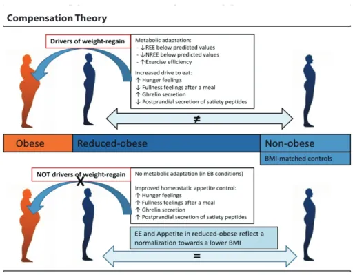
One of the most challenge in managing obesity is the weight regain, or relapse, as described in Figure 1, named the Compensatory Theory. Physiological adaptation occurred in WL regards as the effort of body to fight back against WL and to bring back the weight to its original state by increasing orexigenic drive to eat and reduced EE. The Compensatory Theory propose that there is a physiology difference in EE and homeostatic appetite markers between reduced-obese subjects and non-obese, although they have same BMI. Therefore, when the body sense a negative energy balance, metabolic adaptation is likely due, though there are not enough evidence this phenomenon cause weight regain.
Therefore, a new theory (the Normalization Theory) was coined. The Compensatory Theory states there no
Compensation Theory
Normalization Theory
Figure 1. The difference between Compensation Theory and Normalization Theory in obesity management. REE: resting energy expenditure; NREE: non-resting energy expenditure; EE: energy expenditure; BMI: body mass index.
(5) (Adapted with permission from Oxford University Press).
metabolic adaptation or improved appetite control in reduced-obese state, since either reduce-obese and non- obese has identical homeostatic appetite markers and EE physiology, and a normalization towards WL accompanied by EE reduction and hunger increased.(5)
WL usually followed by some physiological mechanisms such as changes in sympathetic activity. Some studies showed the impact of low sympathetic activity in skeletal muscle and AT to weight gain, but if the impact seems not significant in short-term. Somehow the long-term modulation effect needs to dig further. Another alteration including insulin sensitivity, gut microbiota, and brain signal can modulate the weight regain in long-term or relapse.
Understanding the metabolic differences among individuals lead to personalized therapy and prevention for obesity.(12)
flexible integrative mechanism, and the ability to learn from and adapt to changing internal and external conditions.
The hypothalamus was identified as the primary brain region in mid-1900s classical studies that regulated energy balance and food intake, to determine when we need to eat and what nutrient we need. Electrical stimulation on the ventromedial hypothalamus or electrical stimulation of the lateral hypothalamus produced either weight gain or appetite, thus given the terms "satiety center" and "feeding center", respectively. Recent research from the Genome- Wide Association Study (GWAS) demonstrated the participation of some genes expressed in the central nervous system (CNS) that associated with BMI were located in the hypothalamus.(21)
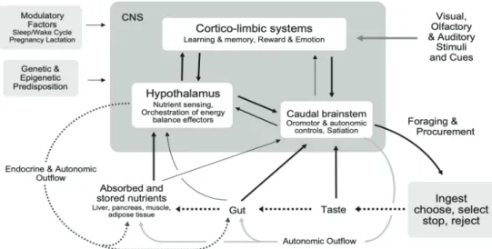
The most significant advance over the first finding is the understanding that the hypothalamus is closely linked to internal and external world representations, rather than functioning independently. Therefore, it can be viewed as the ultimate integrator of dietary data from the internal milieu as well as the external environment. The hypothalamus can be considered the central processor in the regulation of appetite, together with the hindbrain and the corticolimbic system. Since the hindbrain or brainstem has all the necessary components to detect sensory information mediated by vagal afferents and circulating substances, as well as to produce motor output related to food ingestion, digesting, and absorption, it is primarily concerned with meal-size management.(22) But the brainstem alone is unable to adapt food intake to outside demands, like anticipating a food shortage, on its own.(23)
The interaction between nutrient and the nervous system happens in a variety of ways. At the very beginning of the ingestive process, human recognize the cues for potential food in the environment through visual, olfactory, and auditory signals. These inputs generate gustatory and retronasal olfactory signals that are specific to nutrients, and by comparing this signal to the stored information from previous experience, human will decide to accept or reject the food. Food is processed by the gastrointestinal tract (GIT) when it is swallowed, which involves a series of signals produced by chemo- and mechano-sensors.
Through primary afferent nerves and/or hormones or so-called the gut-brain axis, these signals provide the brain with information about the quantity and qualitative characteristics of incoming nutrients. Some of the more recently identified hormones, like GLP-1 and PYY, have the potential to suppress food intake over the long term and energy balance, which makes them appealing targets for medication development.(24)
After ingested, the micro- and macro-nutrient might affect the neurological system. Micronutrient and other particular substances found in food will refer as functional food to impact general structure and function of the nervous system. While macronutrient enters the pathways of energy metabolism and storage interact with the neurological system directly or through the production of peptide hormones to lead the brain to sense the availability of fuels both in the short and long term. These nutrient-sensing pathways integrate with the brain's integrative circuits, autonomic, behavioural, and endocrine output systems to form the homeostatic system, which governs energy balance by controlling intake and expenditure as described in Figure 2. The homeostatic can be biased other factors, including as gender-related reproductive cycles, circadian and circannual rhythms, and relative life span stages.(25) Finally, it should be noted that all the aforementioned interactions between internal and external signals and the nervous system are susceptible to epigenetic modification and genetic predisposition.
The brainstem and hypothalamus are closely linked to the corticolimbic system, which comprises the hippocampus, amygdala, basal ganglia, and vast cortical areas. This system provides the emotional, cognitive, and executive support for ingestive behavior. There is growing recognition that the neurobiology of economics, reward, and decision-making plays a significant role in determining how much food is consumed.(26) The three sections exhibit bi-directional interconnections and, to a certain degree, reflect an evolutionary hierarchy. For an organism to make informed judgments regarding its dietary choices, it is imperative that it possesses precise and reliable information pertaining to the availability of nutrients both inside its internal environment and in the external surroundings.
Sensors inside the body keep track of what nutrients each cell, muscle, and organ need and let the brain know when they're running low. Then the organism must know where to find certain nutrients and be able to balance its need for these nutrients with the time and work needed to get them in order to plan its long-term nutrition.
Choosing which food can be helpful food or potentially dangerous food are key survival behaviors, and caudal brainstem areas play an important part in these processes.
The basic representations of gustatory input in the rostral portions of the nucleus tractus solitarius (NTS) and taste receptor cells on the tongue and palate as the most significant for guiding food intake and selection. The gustatory and trigeminal systems serve as gatekeepers at the alimentary canal's entrance. The four traditional taste modalities are
Figure 2. Neural control of food intake and energy balance.(29) (Adapted with permission from International Life Sciences Institute).
innate detectors for acceptable foods (sweet), dangerous or poisonous foods (bitter and sour), and particular needs (salt, water). There is also growing evidence supporting a preference for fat, namely polyunsaturated fatty acids.
(27,28) Thus, many of the features of ingested meals can be sent to the caudal brainstem via sequential and simultaneous neuronal and hormonal routes. Macronutrient content, caloric density, osmolarity, and possible toxicity are among them.(29)
Every single cell in the body possesses an evolved fuel- sensing machinery made up of nutrition sensors, including mechanistic Target of Rapamycin (mTOR) and AMPK (adenosine monophosphate-activated protein kinase). Then the critical question is how individual cells' nutritional status is transmitted to other cells, tissues, organs, and most importantly, the brain.
Larger lipids, or chylomicrons, are absorbed by the intestinal lymph system, skip the liver, and enter the bloodstream directly. In contrast, absorbed glucose, proteins, and tiny lipids are first transported to the liver through the hepatic portal vein prior entering the general circulation. All of the body's tissues subsequently use or store the nutrients that are in circulation; the brain, in one way or another, helps to coordinate this nutritional flux and keeps an eye on the organism's overall and maybe specific tissue nutritional status.
The other aspect of energy balance regulation that can be controlled by the brain, at least in part, is EE, in addition to EI. Each of the several elements that make up whole-body EE (basal metabolism, thermic action of food, thermogenesis to regulate body temperature, and physical activity) has its own brain regulators and effector pathways.
It has been hotly debated whether excess energy, for instance,
after a bigger than usual meal, is automatically burned off by enhanced basal metabolism and thermogenesis, in the same way that excess water is eliminated by the kidneys.
The discovery of the hormone leptin, that is produced by AT in proportion to adipose mass and acts in the brain to reduce food intake, activate EE, and affect development and reproduction, was the main factor driving this expansion.
Leptin serves as an efficient means of communicating to the brain the state of the body's energy reserves. It is now understood that AT is not an inert organ but instead secretes a wide range of endocrine and paracrine hormones, commonly referred to as adipokines, which affect immunological response, metabolism, and other end points. These signals connecting the communication between the brain and other tissues including AT (30), myokines, the hormones that appear to act on the brain, are secreted by muscle in response to exercise or metabolic stress, and the liver via metabolic hormone fibroblast growth factor-21 (FGF21).
In addition to promoting changes in EE, food intake and selection, glucose, and lipid homeostasis and growth, FGF21 acts on peripheral tissues and the brain.(31) All the macronutrients (glucose, amino acids, and fatty acids); gut hormones (ghrelin, GLP-1); and numerous other hormones (leptin, insulin, and FGF21) have comparatively easy access to the brain and are involved in energy balance regulation and food intake control.(32)
The identification of brown fat-like adipocytes, also called beige or brite adipocytes, within traditional white fat storage depots has increased interest in the function of fat tissue in regulating energy balance and in using it as a tool to prevent and cure obesity. The ideal circumstances and cues for beiging white fat depots are being studied assiduously by researchers in the hopes of effortlessly increasing EE.
Until a new pill of this kind becomes accessible, we will have to use the antiquated method of burning off extra EI, which is exercise.
Since the body does not aim to maintain constant levels of each end point, EI and spending are strictly speaking managed but not regulated processes. On the other hand, energy homeostasis is the phrase used to describe how body weight is controlled to maintain consistent levels through the management of intake and expenditure. While eating behavior receives most of the focus, poor regulation of body weight and energy balance, not poor eating controls, is the primary cause of obesity. Obesity can undoubtedly result from consuming too much food, but only if the regulatory system does not make up for the higher EI by either raising EE proportionately or limiting food intake at following meals. Body weight can be protected by a healthy regulatory system up to a certain extent, which is frequently defined as a small range. Both humans and animals have behaviorally established the concept of setpoint regulation.
Adaptive adjustments in food intake and EE quickly restore body weight to pre-perturbation values when periods of either under- or overfeeding cause artificial perturbations in body weight.(33)
EE, Fat Oxidation and Lean Mass Preservation
Beyond appetite management, EE, fat oxidation, and lean mass preservation are significant factors that determine WL and WL maintenance. In addition to being risk factors for weight gain, poor EE and decreased fat oxidation also lead to resistance to WL. A booster of EE, fat oxidation, or both is therefore expected to contribute to better body weight management and prefer FM loss over lean mass loss in any pharmaceutical treatment.(34)
Variability in the WL and its maintenance can be affected by variations in physical activity and adherence to the prescribed treatment, although these are not the ultimate. Homeostatic regulation refers to the process by which body weight, or more accurately, body energy stores, are maintained in a stable state by activating intricate physiological processes in response to perturbations in energy balance. A higher body weight is frequently defended by changes in the homeostatic control of both EI and EE because obesity patients typically have changed body weight homeostatic regulation.(1) As a result, EE is significantly decreased concurrently with WL. Lean mass, or the metabolically active tissues, is declining, and this
is mostly responsible for the EE drop brought on by WL.
Fifteen percent to 40% of the WL implies losses in lean mass, even if lifestyle changes and WL treatments already on the market can result in significant FM decreases. For every kilogram of lost muscle mass, the amount of energy used during rest is decreased by roughly 13 kcal/day.(6–8)
EE is suggested as the main driver of EI (35), as therapies that only alter EE typically produce less-than-expected WL because changes in EE are typically followed by adaptation in EI.(36) Somehow, in the long-term, low levels of TEE, appear to be associated with appetite dysregulation, where hunger is actually increased and the satiety is poor, finally lead to hyperphagia. Human body can effectively match EI to expenditure better at high energy flow. According to this theory, the desire to eat does not decrease proportionately even though EE is kept low enough. On the contrary, a strategy which has been demonstrated to support effective WL maintenance is the maintenance of high TEE through increasing voluntary or spontaneous physical activity (or addressing inactivity). As such, the reduced EE brought on by WL may, in part, counteract the effects on EI that appetite-regulating medications have.(37,38)
The majority of EE is produced in mitochondria by oxidative phosphorylation, which produces ATP.
Naturally, mitochondrial proton leak about 20-30% of resting EE which majority in liver and muscle. This makes the efficiency of ATP conversion to energy varies in part of the energy consumption from the ATP generation and releases it as heat. TEE and the response to diet-induced WL somehow, are linked to such variations in mitochondrial efficiency. The adenine nucleotide translocases (ANT), ANT1 in muscles and ANT2 in other tissues, and uncoupling protein 1 (UCP1) in BAT are linked to the basal component of the mitochondrial proton leak. The inducible part of the leak is caused by activating ANT, UCP1, UCP2, and UCP3, with UCP3 being the most plentiful UCP in skeletal muscle. Muscle-specific overexpression of UCP3 result in WL in mice, but in other species the contribution of both UCP2 and UCP3 to thermogenesis is assumed to be minimal. Instead, it's possible that UCP2/3's primary roles include controlling reactive oxygen species (ROS), promoting the transport of fatty acids inside mitochondria, and adjusting hormone. On the other hand, ANT1 may be a viable pharmaceutical target for raising EE because it catalyzes over half of the basal mitochondrial proton leak in muscles. Nevertheless, chemical mitochondrial uncouplers have been the main focus of pharmacologically increased proton leak and decreased mitochondrial efficiency thus far.(39)
Recent data shows that lean body mass also contributes to the motivation to eat and the achievement of energy balance, despite the AT feedback signals, such as leptin, provide the biochemical connection between daily energy intake and long-term energy needs. It is crucial to differentiate between two aspects of FFM's involvement in the drive to eat: first, its comparatively passive role on EI, which is mediated by energy-sensing mechanisms that translate FFM-induced energy needs to EI, and second, its more active role in the drive to eat, which is mediated by feedback signals between FFM deficit and EI. Lower FFM will result in lower EE. The loss of FFM after dieting or sedentary should therefore be seen in conjunction with the body's attempt to restore FFM through overeating in order to prevent weight gain and fatness. Thus, treatment for obesity should not only focusing on WL but also to maintain effectively. Current therapies including calorie restriction and appetite suppressant showed promising result for WL, but to maintain this success, further EE and/
or fat oxidation regulation with preservation of lean mass is needed. Furthermore, by targeting EE increase during WL can increase the rates of WL.(40)
The capacity to react or adjust to conditional variations in metabolic demand is known as metabolic flexibility.
This broad idea has been used to emphasize the metabolic inflexibility of obesity and T2DM due to insulin resistance and the mechanisms controlling fuel selection between fatty acids and glucose. Recent studies on exercise physiology identified plausible processes that underlie modified fuel metabolism in individuals with diabetes and obesity. With the aim of preventing and treating metabolic disease, advances in omics technology have further driven basic and clinical-translational research to further examine mechanisms for enhanced metabolic flexibility in skeletal muscle and AT.
One of the main causes of metabolic inflexibility that can occur in a variety of tissues and organs is insulin resistance both in skeletal muscle and liver. There has been a thorough study of the molecular mechanisms behind insulin resistance. These mechanisms include poor mitochondrial fatty acid oxidation and excess buildup of lipid metabolites, including ceramides and diacylglycerol.(41–43)
Skeletal muscle accounting for 60-80% of the increase in glucose metabolism as response to insulin, then
Metabolic Flexibility and Metabolic Adaptation
skeletal muscle become one of the causal role of insulin resistance.(44,45) When there is no corresponding rise in insulin release from the pancreatic beta cells, the quantity of glucose entering the muscle cells and adipocytes from the bloodstream is decreased, together with a lessened suppression of hepatic glucose production would raise blood glucose levels. According to the reasoning, diabetes arises when and if beta cells are unable to secrete more insulin in a suitable amount to compensate for this insulin resistance.
Through a finely regulated mechanism of absorption, esterification, and release of FFAs (also known as triacylglycerol (TAG) cycling), white adipose tissue (WAT) buffers circulating free fatty acids (FFAs) for peripheral tissues, including the liver and skeletal muscle. Glycerol kinase is among many other requirements for this process.
It is noteworthy that BAT also experiences TAG cycling, but unlike WAT, which is tied to TAG cycling primarily with supply and storage for peripheral tissues, BAT's metabolic flexibility is more closely associated with TAG cycling linked with combustion within the cell. A normal-weight healthy lady can have the same amount of WAT as an obese man with T2DM, despite the fact that both the absence and excess (in obesity) of WAT are linked to metabolic problems.
Therefore, obesity-driven metabolic problems are not solely caused by the WAT bulk, emphasizing the significance of healthy and metabolically adaptive WAT.(46)
Energy demand and EE can rise sharply in response to physical exercise. When compared to RMR, rigorous exercise can raise EE up to 25-fold. For many years, researchers have focused on the physiology and biochemistry of fuel choices during exercise on youthful, normal-weight individuals usually have a high degree of metabolic flexibility when choosing their fuel. The desire to enhance athletic performance has also played a major role in the development of these theories and the research efforts made to better understand fuel metabolism during exercise. There are different routes and processes involved in metabolic flexibility. Therefore, fuel selection, EE, or metabolic flexibility could be effective targets for therapy, to the degree that a specific target could be activated perhaps can be a solution for obesity and diabetes. Whether changing fuel choices without also raising energy demands can be beneficial in the context of obesity or nutrient overload is a major point of contention in this as simulate in Figure 3.(47) Our body stored most energy in the form of fat, and less fraction in the form of protein and carbohydrate. Somehow in energy balance, almost 100% of carbohydrate is used for daily turnover (intake and oxidation), thus change in carbohydrate proportion will soon change the rate of
oxidation, not as in fat or protein. Fat have a role as buffer in any surplus or deficit in EI.(40)
Usually, obese people struggle with their body weight before achieving significant WL by therapies, and this is upsetting. Most of them also regain their initial body weight within five years of starting treatment, with 30%
to 50% of the body weight is typically restored within a year (48), even with bariatric surgery. Overall, one of the main obstacles to treating obesity is often preventing body weight regain (relapse). Combined, alterations in muscle mass, organ size, and FM contribute about 60% of the EE decreases in response to WL, leaving the 40% of the EE decline unexplained.(8) The decrease in energy required to maintain weight-bearing activities at a lower weight and the decreased thermic effect of meals due to a lower EI should be considered. However, an additional ~40% can be accounted by higher energy efficiency, or the amount of energy used per unit of metabolic mass; this process is referred to as metabolic adaptation, adaptive thermogenesis, or metabolic slowdown. Decreases in plasma leptin concentrations, reduced thyroid and sympathetic nervous system (SNS) activity, enhanced mitochondrial coupling of oxidative phosphorylation, and increased mitochondrial biogenesis are some of the possible mechanisms underlying metabolic adaptation. Reductions in total daily EE of 6%
to 10% are typically attributed to metabolic adaptation to WL. This adaptation has been observed in response to a variety of behavioral interventions, such as lifestyle changes, medication, and surgical therapies for obesity.
Metabolic adaptation is thought to be a significant obstacle to the maintenance of WL and can last for several years
Figure 3. Short-term substrate balance in response to perturbation of energy balance.(40) (Adapted with permission from The Obesity Society).
following WL. However, there is still a lack of data that clearly connects the level of long-term metabolic adaptation to WL recovery. Pharmacological tactics should target at preserving lean mass or inhibiting metabolic adaption to offer extra advantages for managing EI and resulting in greater and longer-lasting WL.(40)
Adipose Tissue in Control of Metabolism
AT consist of adipocytes, pre-adipocytes, macrophages, endothelial cells, fibroblasts, and leucocytes. AT plays a significant role in the control of metabolism throughout the body. By acting as a fuel reserve, AT regulates lipid mobilization and retains body heat. Neutral TGs are effectively deposited as excess energy in AT via the lipogenic route. But neutral TG accumulation in adipocytes enlarges lipid droplets, leading to the growth of AT and eventual obesity. On the other hand, when food is scarce, there is a stimulation of EE, or when the storage of neutral TGs exceeds the capacity of adipocytes, TGs retained in adipocytes are hydrolysed via the lipolytic pathway into glycerol and fatty acids. The blood can then carry the free glycerol and fatty acids from AT and allow them to enter the muscle, liver, and other organs, influencing lipid distribution and the overall energy balance of the organism.(49)
Due to its delicate actions at the organ and systemic levels, AT is essential for controlling glucose homeostasis and energy levels across the entire body. AT regulates the body's lipid mobilization and distribution and stores energy in the form of lipid. AT on the other hands also functions
as an endocrine organ in addition to a passive fuel reserve.
By circulating and transmitting information to other metabolically active organs like the brain, liver, muscle, and pancreas through endocrine processes, these bioactive substances produced by AT modify systemic metabolism.
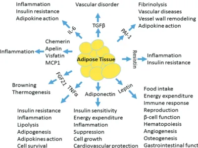
(50–52) Among these are the adipokines (cytokines made by AT), which are linked to obesity and metabolic diseases associated with obesity. These include leptin, adiponectin, visfatin, resistin, apelin, vaspin, hepcidin, chemerin, omentin, and many more with their function as in Figure 4.(53,54) Adipokines primarily exert their effects by attaching to their specific receptors on target cell membranes and initiating specific intracellular signalling cascades. A substantial body of research has established a connection between the onset of obesity and related illnesses and abnormalities in the synthesis, assembly, secretion, and signalling transduction of adipokines.(55,56)
The most common cell type in AT are adipocytes, commonly referred to as fat cells. origins, morphologies, mitochondrial abundance, and expression of thermogenic genes are all different between white, brown and beige adipocyte. The predominant adipocyte type in WAT is white, with a size range of 25–200 μm, a unilocular lipid droplet, few mitochondria, and a low rate of oxidative stress. Accordingly, white adipocytes have a large capacity to store TGs, which are energy molecules, and they shield vital organs like the liver and muscles from lipotoxicity.
Subcutaneous adipocytes differ from visceral adipocytes in rodents and humans in terms of their developmental origins and metabolic characteristics, even if white adipocytes originate from resident cells of mesenchymal origin in white fat.(57–59) Subcutaneous WAT is protective against the development of obesity and associated metabolic diseases in
Figure 4. The physiological functions of adipokines. IL-6: interleukin-6; TGF-b:
transforming growth factor-b; PAI-1: plasminogen activator inhibitor-1; TNFa: tumor necrosis factor-a; FGF21: fibroblast growth factor 21; MCP1:
monocyte chemoattractant protein-1.(56) (Adapted with permission from Society for Endocrinology).
mice, but visceral fat, which includes mesenteric, gonadal, epicardial, retroperitoneal, omental, and peri-renal fat pad, is considered to be more harmful. Transplanting subcutaneous, but not visceral, AT enhances insulin sensitivity and glucose tolerance in animals, providing evidence in favor of this.
The distribution of fat rather than total FM is probably a major factor in the development of obesity and related disorders.(60)
Originally, it was believed that brown adipocytes were a lineage deriving from Myf5+ progenitors that resembled skeletal muscle, but recently has the complexity of adipocyte identification and origin been taken that Myf5+ precursors are not the only source of brown adipocytes. While cervical BAT is partly formed from Myf5− precursors, peri-renal and peri-aortic BAT are entirely from Myf5− precursors, while interscapular and subscapular BAT are derived from the Myf5+ lineage. Furthermore, it has been demonstrated that interscapular and retroperitoneal WAT include Myf5+ precursors, which can differentiate into some white or beige adipocytes. Brown adipocytes are specialized cells that release heat as a by-product of stored energy and have a multilocular shape, an abundance of mitochondria, and an enrichment of UCP1.(61) The inner mitochondrial membrane contains UCP1, which separates ATP generation from fuel oxidation. Brown adipocyte clusters are primarily found in abdominal locations, such as the peri-renal region of baby infants, and the interscapular and peri-renal regions of rodents, where they are highly vascularized and innervated.
Recent researches using 18F-fluorodeoxyglucose (FDG) positron emission tomography-computed tomography (PET-CT) scans have found the adult human BAT in the supraclavicular and lower neck regions. Uncertainty surrounds the properties of the recently discovered brown
fat in adult humans. Since human brown fat expresses the beige marker CD137, TMEM26, and TBX1, and because cAMP stimulation significantly increases UCP1, it has characteristics with mouse beige adipocytes.(62) On the other hand, a different study revealed that brown markers, such as miR-206, miR133b, LHX8, and ZIC1, are also expressed in the supraclavicular BAT, indicating the presence of both beige and brown adipocytes in adult people.(63)
With multilocular shape and UCP1 positive expression, beige adipocytes are a unique subset of brown-like thermogenic adipocytes that mostly develop from Myf5− progenitor cells as white adipocytes.(62) The majority of beige adipocytes are located in subcutaneous white fat, with a minor amount being present in visceral fat.
The process known as browning or beiging of WAT involves the recruitment and activation of beige adipocytes, which is notably induced by cold stress or by a β3-adrenoceptor agonist that imitates cold stress.(64) When combined, beige adipocytes can be differentiated from other sources, transdifferentiated from mature white adipocytes, or produced from beige progenitor lineage. It is yet unknown what molecular processes underlie trans differentiation and the commitment of the beige progenitor lineage.
Adipogenesis, the process by which committed preadipocytes differentiate into mature adipocytes, is crucial for the formation of adipose tissue and maintaining systemic energy balance. The primary regulator of adipogenesis is peroxisome proliferator-activated receptor (PPAR)γ, a member of the nuclear receptor superfamily.
(51) A deficit of PPARγ leads to lipodystrophy because it is unable to activate adipogenic programs in fibroblasts, but overexpression of PPARγ is adequate to induce adipocyte differentiation. Furthermore, adipogenesis is regulated by PPARγ-dependent mechanisms by other factors or pathways, such as pro-adipogenic factors like C/EBPs and Krüppel-like factors (KLFs) and anti-adipogenic factors like GATA transcription factors. Moreover, PPARγ is essential for differentiation maintenance and also adipogenesis.(51)
By lipogenesis and lipolysis, respectively, AT as an energy-storing organ releases fatty acids and stores TGs. Overall, eating activates the lipogenic pathways and increases the AT's TGs capacity store, whereas fasting activates the lipolytic pathway, which facilitates the breakdown of TGs and the release of fatty acids from the AT.
Lipogenesis includes TG production and de novo fatty acid synthesis from acetyl-coenzyme A (acetyl-CoA). The rate- limiting enzyme of lipogenesis, acetyl-CoA carboxylase (ACC), is expressed when glucose is present. It reduces de
novo lipogenesis and enhance fatty acid β-oxidation, and also triggers the release of pancreatic insulin. Additionally, glucose supplies its own metabolite, acetyl-CoA, as the substrate for the de novo synthesis of fatty acids.
The catabolic process known as lipolysis, in contrast to lipogenesis, releases free fatty acids and glycerol when TGs held in adipocytes are broken down. Fasting triggers lipolysis, which provides free fatty acids (FFA) for oxidation in response to energy requirements in other organs and glycerol for hepatic gluconeogenesis. To be noted, glycerol can be utilized as a substrate for gluconeogenesis in the liver, but not fatty acids. Fatty acids can be further broken down in the liver through a process known as ketogenesis to form a class of compounds known as ketone bodies that supply energy to the brain when there is a high fatty acid content and reduced availability of carbohydrates. It has been demonstrated that a number of hormones control the lipolytic pathway. Reduced insulin levels in the suppress lipogenesis and activate lipolytic pathway during fasting.
Moreover, increased levels of circulating glucagon during fasting are invariably linked to the induction of the cAMP- dependent protein kinase A (PKA) pathway and adipocyte lipolysis.
Through its substantial effects on energy storage, endocrine function, and adaptive thermogenesis, AT plays a major role in regulating systemic metabolic homeostasis.
Obesity and illnesses associated to it are linked to the AT dysfunction as a causative factor. In order to identify new and promising therapeutic targets for the prevention and treatment of illnesses connected to obesity, it is crucial to comprehend the biology and pathology of AT. Specifically, a wealth of data regarding the recently identified thermogenic and endocrine functions of AT strongly implies that targeting AT specifically as a therapeutic method is both viable and workable.(56)
Brown and beige fat provide a novel approach to combat obesity and related illnesses by dissipating energy in the form of heat. The expression of UCP1 in beige fat is extremely low under thermoneutral conditions in rodents, in contrast to brown fat, which has relatively high thermogenic activity and enrichment of UCP1 in this condition. This low expression level of UCP1 in individual beige adipocytes may also contribute to the low number of beige adipocytes.(62) However, exposure to cold or administration of β3-adrenoceptor or PPARγ agonists
Browning of WAT
significantly increases the expression of UCP1 in beige fat.(62)
Over the past ten years, many studies have been conducted to try browning the WAT as anti-obesity strategy.
It is hypothesized that the browning process is caused by a complex interaction of hormonal components. These variables include hormones secreted by other metabolically active organs and chemicals generated locally inside AT.
Norepinephrine is the most important and well-researched stimulator of adaptive thermogenesis. It is released from sympathetic nerve endings and binds to brown adipocyte surface β-adrenergic receptors to trigger a variety of thermogenic processes, such as upregulating intracellular lipolysis and mitochondrial oxidation, stimulating circulating triglyceride uptake, and inducing UCP1 transcription. Though in knock-out mice models for BAT activation both β1- and β2-adrenergic receptor activations can compensate for the loss of the β3-adrenergic receptor, but still the β3-adrenergic receptor is the most significant adrenergic receptor implicated in BAT activation.(65)
Thyroid hormones are thought to be non-sympathetic activators of BAT because they function through the thyroid receptor. Comparing animal studies to euthyroid controls, it was shown that hypothyroid mice had much lower interscapular BAT activities whereas hyperthyroid mice had significantly higher BAT activities. In BAT, thyroid hormones have the ability to directly and indirectly promote the transcription of the UCP1 gene.(66)
In response to cold stimulation, the body will attempt to maintain homeostasis through non-shivering (skeletal muscles) and shivering (BAT activation) thermogenesis. It's interesting to note that it was also discovered that exposure to cold stimulates the release of the brown adipokine FGF21 from BAT and the myokine irisin from muscle. In adaptive thermogenesis, irisin and FGF21 both encourage the browning of white adipocytes.(67,68) These outcomes show how muscle, BAT, and WAT work together to coordinate cold-induced adaptive thermogenesis.(69)
The primary regulator of glucose metabolism, insulin plays a vital function in facilitating the uptake of glucose from the bloodstream into the skeletal muscle, liver, and AT.
Furthermore, insulin plays a critical role in both the inducible beige adipocyte and classical brown adipocyte stages of development. It was discovered that beige adipocytes' ability to differentiate was impaired by insulin shortage, but this inhibitory impact could be overridden by more potent stimuli such adrenergic activation. In conjunction with leptin, insulin acts on pro-opiomelanocortin (POMC) neurons to cause browning.(70)
Since the GIT is the organ responsible for breaking down food and absorbing nutrients and energy, it is able to detect changes in nutrient status and emit a variety of hormones to keep the body's energy balance. Numerous GI hormones can control feeding behavior and metabolic processes involving the activation of BAT and the browning of WAT. They can also send signals of hunger or satiety to the CNS and SNS via the gut-brain axis. Generally speaking, orexigenic gut hormones frequently suppress BAT activity, whereas anorexigenic gut hormones typically boost BAT.
A lot of work has gone into trying to figure out which brain circuits control the sympathetic outflow to BAT. By targeting the high-order neurons in the CNS and secreting different neuropeptides like orexin, melanin-concentrating hormone (MCH), cocaine- and amphetamine-regulated transcript (CART), and corticotropin-releasing hormone (CRH), the LH and PVN work in tandem with the ARC to serve as a metabolic integrator and regulator. In fact, sympathetic fibers that originate in the hypothalamus extensively innervate both BAT and WAT.(71)
It is plausible to assume that these neural circuits have a similar involvement in the regulation of WAT browning.
Numerous investigations have demonstrated that although stimulation of POMC neurons stimulates WAT browning, activation of AgRP neurons in the hypothalamus slows the browning process. These two opposing effects show that whole-body metabolism, including fat browning, is regulated by AgRP/NPY and POMC neurons in the ARC, which may also sense the body's energy state.(71,72)
Research has demonstrated that after being exposed to cold, inguinal WAT expresses more FGF21. Both paracrine and autocrine processes play a role in the production of FGF21 in WAT, which leads to the local upregulation of PPARγ coactivator-1 alpha (PGC-1α), a co-activator of peroxisome proliferator-activated receptor, and an increase in thermogenesis. A protein called PGC-1α is involved in regulating a number of outcomes in skeletal muscle after exercise, such as enhanced energy and glucose metabolism.
(73) It's interesting to note that PGC-1β is also activated by exposure to insulin or irisin; these hormones have a definite interaction with FGF21 during exercise. Shivering intensity is positively correlated with irisin-induced activation of extracellular signal-related kinase (ERK) and p38 mitogen- activated protein kinase (p38 MAPK).(68,74) There is a direct correlation between exercise intensity and FGF21.
These PGC-1β inducers have the effect of promoting adaptive thermogenesis by causing WAT to brown.
Following FGF21 action, PPAR-gamma activation in WAT and the irisin effect's induction of the MAPK and ERK
pathways constitute the primary mechanism. Pre-adipocytes undergo development into mature white adipocytes as a result, at which point they become suitable for browning.
(75,76)
Leptin serves as an excellent model for how WAT communicates energy status signals to the hypothalamus in the central nervous system, which in turn influences whole- body metabolism and directly influences the browning of white adipocytes. Browning is further promoted by bone morphogenetic protein-4 (BMP-4), an additional significant adipose-derived component. The particular conversion of mesenchymal progenitors to beige adipocytes is stimulated by transgenic production of BMP-4, indicating a significant function of BMP-4 in browning.(77)
The hormone catecholamines is involved in the control of browning. It's interesting to note that alternatively activated macrophages (M2) in AT have been observed to produce norepinephrine for BAT activation in addition to sympathetic nerve synapses.(78) Another important organ that releases adrenaline and norepinephrine into the bloodstream for systemic effects on several organs, including AT, is the adrenal gland. Moreover, a number of observational studies showed that individuals with pheochromocytomas have higher BAT FDG uptake activity, which decreases after removal.(79,80) Therefore, by use of adrenergic receptors, these catecholamine- releasing cells and organs may as well control BAT activity and WAT browning. Some hormonal factors secreted from different organs and tissues play roles in browning WAT were described in Figure 5.
In conclusion, pharmacological and nutritional stimulation of beige cells is a viable anti-obesity and anti- diabetic method to treat metabolic disorders, considering the scarcity of BAT in human adults. Furthermore, finding nutritional components that promote WAT browning also seems like a good strategy. According to available data, hormones also have a significant impact in mediating the browning effects of numerous dietary components. For instance, capsaicin and capsinoids with well-established WAT browning properties can stimulate Ca2+ influx by activating the transient receptor potential cation channel, subfamily V, member 1 (TRPV1), which in turn activates the Ca2+/calmodulin-dependent protein kinase II and AMP- activated kinase. This, in turn, stimulates the deacetylation of PPARγ and PRDM16 by sirtuin-1, which in turn stimulates the synthesis of BMP8b and UCP1.(81) Similar to this, eicosapentaenoic acid and docosahexaenoic acid, which are rich in ω3 polyunsaturated fatty acids (PUFAs) and fucoxanthin, can also cause UCP1 expression in WAT
by upregulating the expression of β3-adrenergic receptors, which in turn increases WAT sensitivity to adrenergic stimulation in adipocytes. The WAT browning levels may not only be determined by hormonal pathways, but they may also indicate the energy condition of the entire body.
For example, despite the fact that exercise has been shown to raise EE and browning of WAT (68), a study comparing the BAT volume and activity of athletes and non-athletes revealed that the former tended to be less active. This shows that when there is an energy deficit, as occurs with prolonged exercise, brown fat may adjust by decreasing.(82)
Obesity is the single biggest risk factor for premature disability and death, yet the prevalence of obesity is still astronomically high. Constant body adiposity is maintained by CNS systems that adjust EI and expenditure in response to variations in body energy reserves; key regulators of these systems are the adipocyte-derived hormone leptin and its receptor (LepR). Similar to insulin resistance, a number of mechanisms have been proposed to disrupt the action of leptin and obstruct the systems that regulate body energy homeostasis in order to promote or maintain obesity. However, it is unclear how important a role each of these mechanisms plays in this process. These mechanisms include feedback inhibition, inflammation, gliosis, and endoplasmic reticulum stress. Adipokine leptin governs a number of bodily processes, including hunger, weight, the maturity of the reproductive axis, and the neuroendocrine responses to fasting. It is also widely accepted that leptin controls glucose homeostasis in the brain. However, common obesity may augment (rather than impair) LepR signalling, indicating that lesions other than the initial LepR signal must be the cause of any obesity-associated problems in leptin function. Additionally, it's feasible that some of the modifications in hypothalamus function linked to obesity are mediated by elevated LepR signalling.(83)
The synthesis of leptin and the control of the body's stored energy is largely dependent on AT. Gaining insight into how leptin acts on the CNS to regulate AT, and how the sympathetic nervous system (SNS) influences AT metabolism, offers a new way to address the obesity issue.
The brain controls the metabolism of BAT, WAT, and Beige AT (BeAT), and in turn, these tissues communicate with the brain via sensory innervation and hormone release regarding the state of energy storage. The cloning of Lep3
Hormonal Factors in
The Control of EI and EE
Brain
Enriched environment
POMC AgRP
Hypothalamus
Thyroid Heart
Liver
Pancreas
Gastrointestinal AcidBile
Figure 5. Hormonal factors in the control of browning of white adipose tissue. POMC: proopiomelanocortin; AgRP: hypothalamic agouti-related peptide; BDNF: brain-derived neurotrophic factor; GH: growth hormone; NPY: neuropeptide Y; CRH: corticotropin- releasing hormone; T4: thyroxine; T3: triiodothyronine; DIO2: iodothyronine deiodinase 2; FGF: fibroblast growth factor; WAT: white adipose tissue; VEGF: vascular endothelial growth factor; BMP-4: bone morphogenetic protein-4; GLP-1: glucagon like peptide-1; CCK:
cholecystokinin.(72) (Adapted with permission from Walter de Gruyer GmbH).
sparked a renewed interest in this reciprocal relationship between the brain and adipose depots. The proof that mature humans have functional brown adipocytes has also sparked fresh research on BAT's neuronal control. Although leptin's function in controlling energy balance is widely recognized, its significance in the neuronal regulation of BAT, WAT, and BeAT remains unclear. There are numerous thorough and current reviews available on the leptin-sensitive neurons that maintain energy balance or the neuronal control of AT.(29,84–90)
Ghrelin affects systemic metabolism by triggering orexigenic neuronal circuits. It has been linked to a wide range of central and peripheral actions, including promoting gastric acid secretion and gut motility, regulating sleep,
taste perception and reward-seeking behavior, controlling glucose metabolism, inhibiting brown fat thermogenesis, reducing stress and anxiety, preventing muscle atrophy, and enhancing cardiovascular processes like vasodilatation and cardiac contractility.(91–93)
Early on in the study of ghrelin, it was proposed as a meal initiation or hunger hormone, informing the CNS of the GI fuel level to adjust food intake and EE. This function is supported by the fact that ghrelin is generated in the gastric fundus's oxyntic glands (94), and the elevated blood levels of the hormone correlate with higher feelings of hunger (95).
Ghrelin’s receptor is found in the hypothalamus neurons that control appetite and satiety. This conventional and constrained understanding of ghrelin as a hunger hormone,
however, has recently come under scrutiny. A growing body of research indicates that ghrelin plays a more intricate function in controlling hunger and metabolism and suit better to be called as satiety hormone.(96)
GLP-1 is a complex hormone with a wide range of pharmacological applications. GLP-1 has a wide range of metabolic actions, including modulating the proliferation of rodent beta cells, increasing natriuresis and diuresis, delayed stomach emptying, inhibiting food intake, glucagon secretion suppressing, and stimulating insulin secretion in a glucose-dependent manner. GLP-1 also affects learning and memory, reward behavior, palatability, and cardio- and neuroprotective benefits in addition to reducing inflammation and apoptosis. GLP-1 receptor agonists, biochemically altered for increased potency and prolonged action, are effectively used in clinical practice to treat T2DM, and a number of GLP-1-based pharmacotherapies are being evaluated in clinical settings to treat obesity.
Pancreatic beta-cells co-synthesize and co-release insulin, the pancreatic hormone amylin. For a very long time, it was believed that the only amylin that was suitable for controlling metabolism came from the pancreas. But new research indicates that amylin is also expressed in the CNS, namely in regions like the lateral hypothalamus that are involved in metabolic regulation. Amylin reduces EI, modifies nutrient consumption, and increases EE to control nutrient fluxes globally. The role of amylin as a satiation signal has received the most research attention. In fact, long-term amylin administration lowers total EI, which ultimately causes a decrease in body weight. These results served as the foundation for the creation of amylin analogs, which could be a novel method of treating overweight in obese people.(97–99)
Activating BAT and/or generating browning in WAT may have therapeutic implications because of its high metabolic activity and capacity to oxidize fats and glucose, making it a desirable target for obesity and T2DM therapy.(100) Additionally, adipokines (also known as batokines) secreted by brown and beige adipocytes have endocrine, paracrine, and autocrine activities that may have positive metabolic consequences (such as enhancing insulin sensitivity).
(101) Exercise is supposed to decrease BAT activity and WAT browning since it promotes EE and heat production.
Exercise-induced WAT browning, via some hormones
Endocrine Mechanisms Connecting Exercise To BAT Metabolism
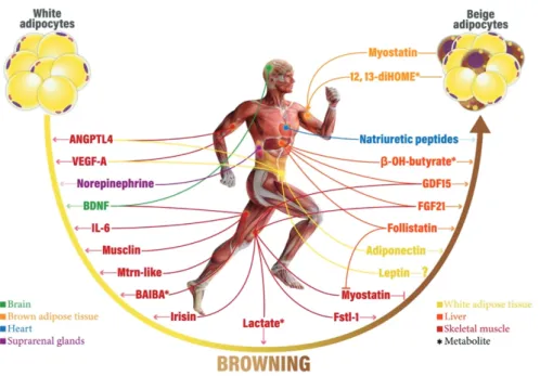
secretion at least in rodents, even though the impact of exercise on traditional BAT is still debatable.(102) Figure 6 describe how exercise can induce secretion of different metabolites to help browning the WAT, while on the contrary the brown and beige adipocytes also secrete signaling factors like myostatin and 12,13-diHOME that can influence skeletal muscle metabolism.(103)
Heart-secreted natriuretic peptides (NPs) are primarily responsible for controlling diuresis, natriuresis, and vasodilatation in order to maintain blood pressure.
(104) Moreover, NPs have a role in the stimulation of lipolysis in WAT (105) and the oxidation of fat in human skeletal muscle (106). Additionally, NPs in BAT and WAT browning, specifically atrial NP (ANP) and B-type NP (BNP), facilitate energy dissipation. Exercise causes the heart muscle to contract, which in turn triggers the release of NPs. ANP levels have been shown to rise in circulating levels in a number of investigations following acute moderate- and high-intensity endurance exercise in various groups. Similarly, in healthy males, both acute and chronic endurance exercise result in an increase in plasma BNP levels.(107–109)
One of the master transcriptions factors that exercise upregulates in skeletal muscle is PGC-1α. The protein fibronectin type III domain-containing 5 (FNDC5) is expressed more when PGC-1α is active. Following cleavage, FNDC5 is released into the bloodstream as irisin, which attaches to adipocyte surfaces to promote WAT browning and increase UCP1 expression, at least in mice.(110) Following acute exercise, a number of human investigations have demonstrated an increase in circulating serum irisin and FDNC5 gene expression in skeletal muscle. For example, in both trained and untrained healthy adults, a 50-min cycling bout at 80% of maximum oxygen consumption (VO2max) was able to enhance circulating irisin 10 min after exercise. The activation of irisin secretion may be significantly influenced by the intensity of exercise.
(111) Indeed, thermogenic stimulation increases the release of FGF21 in mouse brown adipocytes. WAT browning is induced by FGF21 via PGC-1α activation.(112) FGF21 can stimulate BAT thermogenesis and UCP1 expression by autocrine, paracrine, and endocrine actions. It's interesting to note that in healthy men, there has been a reported positive correlation between circulating FGF21 and BAT volume.(113)
Exercise can cause a 100-fold rise in circulating IL- 6. The primary factors that mediate the IL-6 response to acute exercise are exercise intensity and duration, muscle injury, and the type of muscular contraction (eccentric or
Figure 6. Endocrine mechanisms relating exercise to BAT metabolism and WAT browning in humans.
ANGPTL-4: angiopoietin-like 4;
VEGF-A: vascular endothelial growth factor A; BDNF: brain-derived neurotrophic factor; IL-6: interleukin-6;
BAIBA: β-Aminoisobutyric acid;
Fstl-1: follistatin-like 1; FGF21:
fibroblast growth factor 21; GDF15:
growth differentiation factor 15;
12,13-diHOME: 12,13-dihydroxy- 9Z-octadecenoic acid.(103) (Adapted with permission from Springer Science+Business Media).
concentric). Meteroin-like protein production and secretion are stimulated by PGC-1α expression in skeletal muscle, which is a splice variant of the gene encoding PGC-1α.
Meteorin-like binds to its receptor in AT and stimulates M2 macrophages in an eosinophil-dependent manner, releasing IL-4 and IL-13. This, in turn, causes WAT browning and the expression of genes encoding the mitochondrial and thermogenic programs through the release of norepinephrine.
In response to cold, skeletal muscle as well as beige and brown adipocytes generate meteorin-like substances.
Musclin is a blood-stream-generated peptide synthesized by skeletal that can interact to certain common receptors since it has some structural similarities with NPs.(114) In skeletal muscle, myocilin stimulates mitochondrial biogenesis. Furthermore, it has been proposed that musclin contributes to browning since it is a PPARγ agonist.(115)
A member of the transforming growth factor-β (TGF-β) superfamily, growth differentiation factor-15 (GDF15) is a protein whose receptor is mostly expressed in the brain and in WAT.(87) While the liver is the primary source of GDF15 in circulation, it is also expressed in the skeletal muscle, BAT, and WAT, among other tissues. In reaction to thermogenic exercise, brown and beige adipocytes produce GDF15, which targets BAT macrophages and reduces local inflammation.(116) Exercise has been shown to raise GDF15 circulating levels following both a high-intensity (117) and a moderate-intensity (118) session. In young males of normal weight, an increase in plasma GDF15 during recovery and immediately following a 60-minute aerobic workout (67%
of VO2 max) was reported.
Another member of the TGF-β superfamily, growth differentiation factor-8, or myostatin, was identified as a myokine early in the 1990s.(119) Since the primary function of myostatin is to limit muscle growth, its reduction significantly promotes muscular growth. Loss of myostatin activity leads not only to hypertrophy of the muscles but also to reduced fat accumulation and browning of the WAT.
The activation of the AMPK enzyme and the subsequent induction of PGC-1α and FNDC5 cause the induction of WAT browning via myostatin inhibition.(120) Myostatin appears to be a key player in the inhibition of WAT browning.
Exercise, whether acute or chronic, affects myostatin expression and levels in the blood, though the kind and intensity of the exercise appear to have an impact as described in Figure 6.(121) Human myostatin circulating levels are lowered by prolonged exercise.(122) On the other hand, myostatin levels in the blood rise sharply following high-intensity exercise. Significantly, the myostatin action on BAT provides evidence that exercise stimulates the release of both browning inhibitors and pro-browning chemicals.(121)
Skeletal muscle, the liver, and other tissues such WAT and BAT can all release follistatin after acute high exercise such as cycling or sprinting.(123) Follistatin neutralizes the biological activity of numerous TGF-β superfamily members, such as activins and myostatin, by binding to them.(123) Consequently, it has been determined that follistatin-mediated inhibition of myostatin signaling plays a key role in muscle development, differentiation, and metabolism. Follistatin probably stimulates muscle growth and BAT development in addition to inhibiting myostatin
activity by directly triggering precursor cells and Myf5 expression.(124)
Exercise together with an enriched environment, such as one with mazes and toys, cause mice to release BDNF, which causes WAT to brown in both situations.
(125) PGC-1α and FNDC5 expression appear to be partially mediating the effects of BDNF.(126) While the acute effects of strength training are yet unknown, numerous studies have demonstrated an increase in BDNF circulating levels following both moderate and high-intensity aerobic exercise across diverse populations.(127–130) There is debate regarding the impact of exercise on circulating levels of adiponectin. While some studies, indicate an increase solely in trained people, others claim that adiponectin plasma levels remain unchanged following exercise.(131–133) On the other hand, in obese young females, continuous endurance exercise may raise adiponectin levels.(134)
There is still debate, however the leptin response to exercise appears to be consistent throughout the data published in the literature. Exercise of any intensity or high appears to have no effect on leptin levels, or to slightly lower them. Following a session of aerobic and resistance training, circulating VEGF-A has been found to rise in both men and women, while some studies did not find any effect.
Although the research is still in its early stages, it's possible that VEGF-A secretion brought on by exercise also plays a role in BAT activation and/or WAT browning. Exercise in humans also regulates the synthesis of ANGPTL4 in addition to dietary status. Acute endurance exercise was found to raise circulation levels of ANGPTL4 in males in good health, with the liver serving as the primary source of secretion.(135)
β-Aminoisobutyric acid (BAIBA) is an amino acid that is not a protein that is generated by the highly active mechanism of valine catabolism in skeletal muscle.(136) Skeletal muscle cells release BAIBA in reaction to PGC- 1α activity.(137) BAIBA promotes the expression of thermogenic genes in WAT, which speeds up the browning process. Both human-induced pluripotent stem cells and white adipocytes produced from human pluripotent cell lines showed comparable effects. Because muscle absorption of β-hydroxybutyrate is higher than hepatic synthesis during exercise, circulating quantities of this compound are typically lowered.(138) However, elevated blood levels of β-hydroxybutyrate are frequently seen after vigorous exercise or during extended exercise. It is noteworthy that the amount of training and nutrition appear to have an impact on how the ketone bodies react to exercise.(139)
Drugs acting on both reducing food intake and increasing EE may have a more potent effect to produce synergistic WL. Increasing thermogenesis appears to have positive metabolic effects that are at least somewhat independent of WL. Indeed, BAT is highly effective in eliminating circulating glucose, fats, and lipoproteins rich in triglycerol.
As a result, in human, an acute increase in BAT activity can have an immediate impact on glucose and lipid homeostasis, which is anticipated to enhance metabolic health over time.
(140,141)
Overall, improvements in metabolism can result from browning of WAT, and BAT activation in both body weight- dependent and body weight-independent ways; however, the evidence for antidiabetic effects seems greater than for the treatment of obesity in humans. Browning of WAT and BAT activation, however, might be helpful as an adjuvant therapy to assist prevent weight rebound or in conjunction with other weight-reducing medications to accomplish even more weight reduction.
Therefore, many efforts have been done to explore new agents that can induce WAT browning for example via januse kinase inhibitors. Novel medicines working through PPAR may target post-translational modifications (PTMs) as an alternative to traditional agonism. In states of obesity and insulin resistance, there is an increase in the phosphorylation of PPAR at serine 273 by cyclin-dependent kinase 5 and extracellular signal-regulated kinase, which leads to a dysregulation of adipocyte gene expression. Inhibitors of this phosphorylation included the well-known anticancer medication Gleevec, which was found to modulate WAT browning in mice. A cyclin-dependent kinase 5 inhibitor called Roscovitine can similarly cause WAT browning in mice by preventing PPAR phosphorylation at serine 273 but have not been proved in human. It's interesting to note that the phosphodiesterase inhibitor sildenafil, which is used to treat erectile dysfunction and pulmonary arterial hypertension, short-term application causes overweight subjects' WAT to begin to form beige adipocytes; however, this effect does not seem to be mediated through a direct action on AT.(142)
Aside from cold stimulation, mirabegron is arguably the most sophisticated and powerful pharmacological activator of human BAT found to date. Humans treated with chronic mirabegron administration have the ability to stimulate BAT's thermogenic potential and somewhat