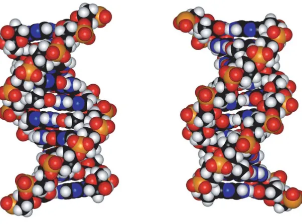
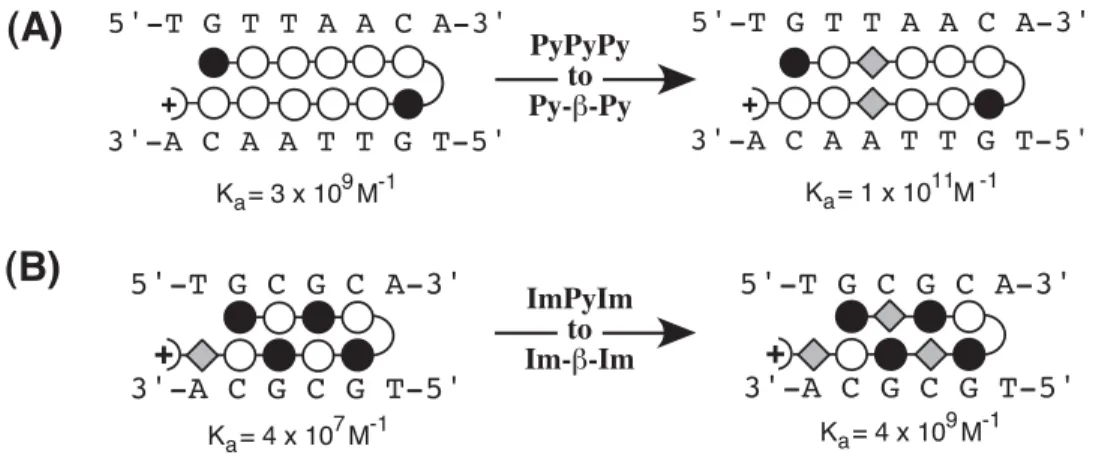
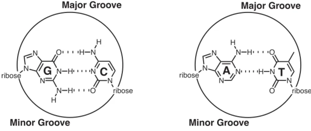
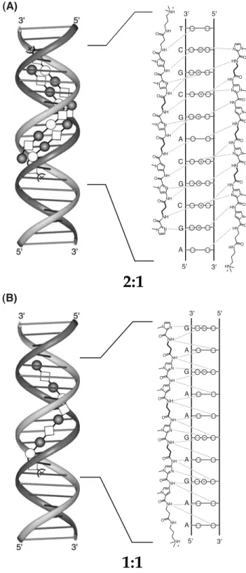
The minor groove of the A,T tracts is both narrow due to the helix twist of the base pairs and relatively deep due to the lack of protruding G-NH2 (Fratini et al., 1982). Subsequent footprinting experiments revealed that Im-Py polyamides tolerate G•C base pairs but show little sequence specificity (Lown et al., 1986). 10 Antiparallel polyamide subunits assembled in a 2:1 complex can be covalently linked, head-to-tail, to form hairpin polyamides with increased binding affinity and sequence specificity (Mrksich et al., 1994).
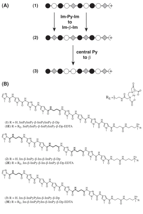
Polyamides composed of multiple linked Py and Im residues are curved relative to the DNA helix (Kelly et al., 1996). In a recent discovery, Laemmli and coworkers reported the use of Py-Im polyamides to induce phenotypic changes in Drosophila melanogaster (Janssen et al., 2000a; 2000b). Attempts to improve this recognition code using new heterocyclic amino acids, such as furan, thiophene, thiazole, and hydroxythiophene, have been presented (Marques et al., in preparation).
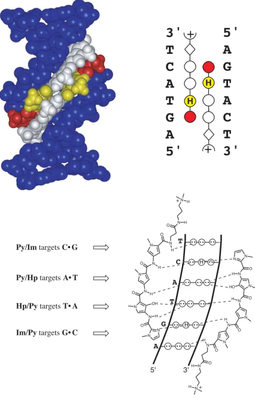
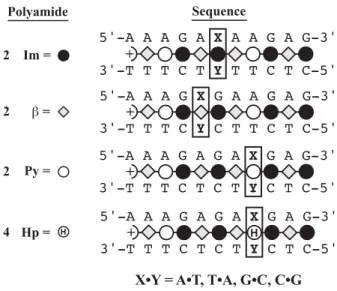
The first direct evidence is provided for hydrogen bond formation between Im-N3 and G-NH2 in the 1:1 motif, confirming the original lexitropsin model (Urbach et al., 2002). Replacement of β with α-(R)-acetamido-γ-aminobutyric acid forces an 82,000-fold reversal of specificity toward the hairpin motif (Urbach et al., in preparation).
The Importance of β -Alanine for Recognition of the Minor Groove of DNA 1
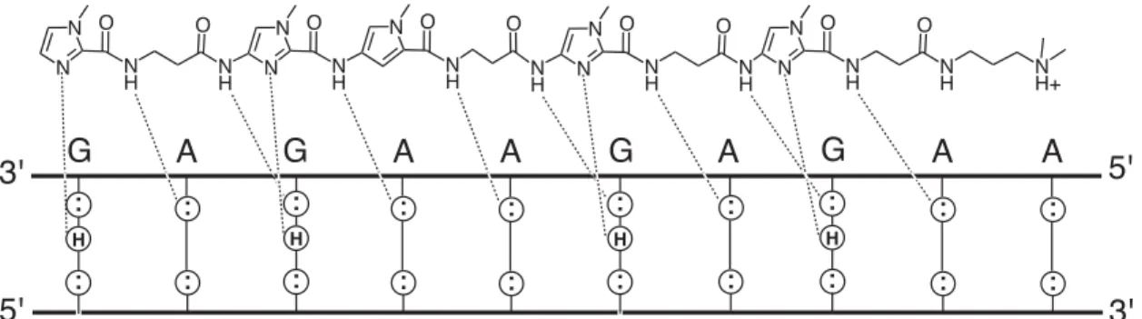
Solid lines are best fits to Langmuir binding isotherms obtained by a nonlinear least-squares algorithm as described in the experiment. However, 2 E and 3 E reveal cleavage only at the 5'-end of the site, 5'-AAGAGAAGAG-3', indicating a single orientation and 1:1 polyamide:DNA binding stoichiometry, similar to the finding of Laemmli (Janssen et al., 2000). Quantitative DNase I footprinting titration experiments (Brenowitz et al., 1986; Trauger and Dervan, 2001) were performed to determine the apparent equilibrium dissociation constants (KD = concentration of polyamide bound at half saturation) of 1-3 at each target site (Figure 12 -B).
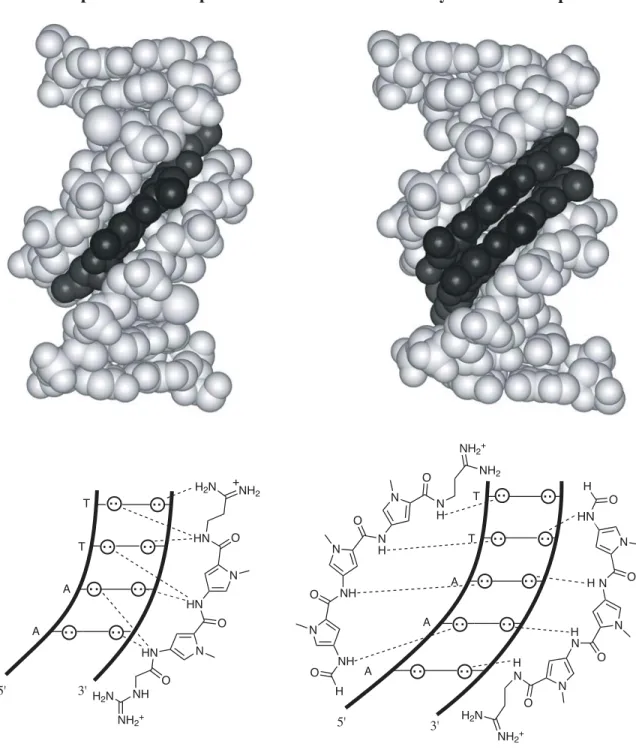
In the study described in the next section, Im, in sequence context of polyamide 2, is shown to bind all four base pairs (within a factor of 2) (Urbach and Dervan, 2001). From the crystal structure of the 1:1 netropsin:DNA complex, we understand the molecular mechanism by which Py specifies A•T/T•A > G•C/C•G (Kopka et al., 1985). This 1:1 recognition code for Py choosing A•T/T•A > G•C/C•G must now be modified to include the sensible placement of β for A•T/T•A recognition to reset the curvature in the 1:1 motif.
There are several implications for adding a 1:1 motif to the current 2:1 targeting of mixed G, C/A, T DNA sequences in the minor groove. We face the added complexity of the same molecule binding very different DNA sites depending on the stoichiometry.
Toward Rules for 1:1 Polyamide:DNA Recognition 2
This specificity is largely attributed to the unique functionality that each heterocycle presents at the bottom of the minor groove. The ring atom closest to the bottom of the DNA minor groove was examined for partial charge. The high-resolution solution structure presented later in this thesis reveals an important register of amide NH groups with the purine N3 and pyrimidine O2 groups at the bottom of the DNA minor groove (Urbach et al., 2002).
This phenomenon is covered in more detail in the discussion of high-resolution NMR structure. A homogeneous distribution of contacts is observed, which unambiguously defines the position of the ligand within the minor groove. 62 and it suggests that part of the driving force behind the polyamide DNA registry is the correct alignment of amide NH groups relative to the DNA base pairs.
Hydrogen bond frequency was tested using the CARNAL module of the AMBER 6.0 software package (Kollman et al., 2000). Strong NOEs are observed from methylene protons in each β-residue to the proximal A-H2 proton on the floor of the minor groove, as observed in the 2:1 motif (de Clairac et al., 1999). Based on the structure presented here, we believe that polyamide orientation is controlled by a combination of the inherent geometry in the amide-.
Inspection of relative trace sizes on DNase gels further supports the MPE results. Inspection of the DNase gel reveals that 2 binds with very high affinity to the 5′-AAAAGAAAAG-3′ region. The observed hydrogen bonds between Im-N3 and G-NH2 provide the first direct evidence of the lexitropsin model as originally predicted in the 1:1 motif (Kopka et al., 1985).
Alternatively, the hairpin binding mode is favored by incorporating an α-(R)-amino-substituted γ-linker, i.e., the Herman turn, to connect the flexible polyamide subunits. 1999) NMR characterization of the aliphatic β/β linkage for recognition of A·T/T·A base pairs in the minor groove of DNA. 1995) NMRPIPE – a multidimensional spectral processing system based on UNIX tubes. 1998) Stereochemical control of the DNA binding affinity, sequence specificity and orientation preference of chiral hairpin polyamides in the minor groove.
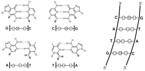
1992) Antiparallel side-by-side dimeric motif for sequence-specific recognition of the minor groove of DNA by the designed peptide 1- methylimidazole-2-carboxamide netropsin. 1998) Recognition of the four Watson-Crick base pairs in the DNA minor groove by synthetic ligands. The binding model shown at the top gives DNA base numbers used in the assignments, as well as the orientation and binding site of the ligand.
Due to the similarity of the correlation assigned in each plot, the assignments are usually indicated by the DNA residue number, and the type of correlation is indicated in the plot title.
Intraresidue DNA to DNA Constraints
Intramolecular Ligand to Ligand Constraints
Intermolecular Ligand to DNA Constraints
Parameters are plotted along the ordinate for each DNA base step along the abscissa. Mean values over the final ensemble of 12 structures are connected by solid lines, and y -axis error bars indicate one standard deviation from the mean.