Localization of DNA-Binding Polyamides In Living Cells
Thesis by Timothy Patrick Best
In Partial Fulfillment of the Requirements for the Degree of
Doctor of Philosophy
California Institute of Technology Pasadena, California
2005
(Defended 22 July, 2004)
© 2005 Timothy Patrick Best
All Rights Reserved
…for my parents…
Acknowledgements
The foundation for any successful effort is its most important part, bearing the weight of the work built upon it. My foundation has been the support of my family:
aunts, uncles, and cousins all; my grandmother, Olive Sonner; my sister, Jennifer. Most especially I would like to thank my parents, Ralph and Sharron Best; they have always been, and continue to be, my example. To those people whose words and deeds interested me in science, I offer my thanks: the writings of Carl Sagan; from Corydon—
Larry Hauswald and my first teacher of chemistry, Dennis Lopp; from Butler University—Joe Kirsch, Shannon Lieb, Dave Hall, and especially my advisor and friend Anne Wilson.
I would like to thank my research advisor, Peter Dervan, for his enthusiasm and for providing the opportunity to work in a rich academic environment with such stellar co-workers. I would like to thank the members of my committee, Linda Hsieh-Wilson, Carl Parker, and Scott Fraser for their support and guidance. I would also like to thank Dr. Fraser for providing the use of his microscope facilities. I would like to extend my appreciation to Harry Gray for providing an opportunity for weekly escape to the Rathskellar after Friday seminars, and to his wife for excellent pecan pie.
The high quality of my collaborators and co-workers at Caltech has provided an exemplary environment in which to learn about both chemistry and the world: I would like to thank the coffee-time club of postdoctoral fellows Anna Mapp, Inger Kers, Ulf Ellervik, Leonard Prins, Phillip Weyermann, Christoph Briehn, and Dorte Renneberg;
French lessons from Pierre Potier; German from Alex Heckel and Hans-Dieter Arndt; my first teacher in the lab and perennial fantasy baseball foe Bobby Arora; and Bogdan
Olenyuk, who has always been a source of good chemical knowledge, good conversation, and good help.
I appreciate the fellowship of many students in the Dervan lab, past and present:
Dave Herman, Merideth Howard, Nick Wurtz, John Chevillet, Adam Urbach, Amanda Cashin, Victor Rucker, Jason Belitsky, Shane Foister, Eric Fechter, Michael Marques, Jim Sanchez, Nick Nickols; the Freeballers (Ray Doss, Justin Cohen, Carey Hsu, Ryan Stafford, Adam Poulin-Kerstien); my newest co-workers Jim Puckett, Julie Popowski, and Sherry Tsai; and my parter in crime Ben Edelson. I have also had good co-workers outside of the Caltech community: Zhengxin Wang, Gerd Blobel, and Aseem Ansari.
As the eminent philosophers McCartney and Lennon put it, I’ve gotten by with a little help from my friends. The Corydon crew: John McCollum, Jared Bachman, Joe Harmon, Jeremy Schoen, Matt Burnham, Jay Hanaver, Micky Emily and Melody Mathes; from Butler: Mike and Kathleen Julius, Sarah Bohl, Pete Wibbenmeyer, Jill Carter, and Sherry Nichols. Caltech has been no exception, as I have found many good people here with whom to share the time: the Wednesday lunch crew—Jeremy Heidel, Steve Spronk, Julie Casperson, and Swaroop Mishra; the house on Mentor—James Peterson and Greg Drummond; the gamers—Micol Christopher, Mat Matuszewski, Meg Wessling, Carlos Mochon, James Chakan, Charlene Ahn, and Geoff Swift; Jenny Roizen, for many interesting conversations and her good company; my roommate and friend Neal Oldham; and Ted Corcovilos, whom I would also like to thank for helping me to edit this text.
To anyone whom I have forgotten and who deserves my thanks: Cheers!
Abstract
Regulation of the processing of genes into nucleic acids and proteins is a substantial goal in medicine. Small molecules that could enter cells, localize to the nucleus, and bind chromosomal DNA sequence-specifically and with high affinity would be important tools for gene regulation. Pyrrole-imidazole polyamides are small molecules that bind the minor groove of DNA in a sequence-specific fashion according to a set of pairing rules, and with affinities rivaling natural transcription factors. Several in vitro experiments have shown that by directly competing with transcription factors for binding sites in gene promoter regions, polyamides can act to inhibit transcription of those genes. Polyamides bearing transcription activation domains can bind to promoter regions, recruit the transcriptional machinery to the gene, and activate transcription in vitro. Attempts to reproduce these results in vivo were largely unsuccessful, perhaps due to poor cellular trafficking properties of polyamides and polyamide-peptide conjugates.
It was found that polyamides bearing the Bodipy fluorophore localize primarily to the cytoplasm of cells, or were excluded from cells altogether. In attempts to overcome this quality, peptides shown to improve cellular trafficking were appended to the polyamides. These peptides were generally not successful at inducing uptake, and were in many cases toxic to the cells. Small molecules were also appended to polyamides, likewise to improve uptake properties, but met with limited success. Surprisingly, the addition of a fluorescein or fluorescein-like fluorophore to polyamides permit them to localize to the nuclei of all cell lines tested, in a molecular content- and shape-dependent manner. This technology has been applied to several in vivo experiments, including the inhibition of androgen receptor binding to its cognate element in gene promoter regions.
Table of Contents
Page
Acknowledgements……….iv
Abstract………...…vi
Table of Contents………...…vii
List of Figures and Tables………..ix
Chapter 1 Introduction to DNA Recognition by Minor Groove-Binding Polyamides………..……….1
Chapter 2 Transcription Activation with Polyamide-Polyproline-Peptide Conjugates………..………24
Chapter 3 Polyamide-Peptide and Polyamide-Small Molecule Conjugates for Cellular Uptake Studies………43
3A Polyamide-Peptide Conjugates for Cellular Uptake Studies……….44
3B Polyamide-Small Molecule Conjugates for Cellular Uptake Studies………..……..65
Chapter 4 Nuclear Uptake of Polyamide-Fluorophore Conjugates in Mammalian Cell Lines………...84
4A Polyamide-FITC Conjugates for Cellular Uptake Studies……….85
4B Influence of Structural Variation on Nuclear Localization of
DNA-Binding Polyamide-Fluorophore Conjugates…….………...106
4C DNA-Binding Characteristics of Polyamide-Fluorophore
Conjugates………145
Chapter 5 Inhibition of Transcription on the Androgen Response Element
with Polyamides and Polyamide Conjugates………...155
List of Figures and Tables
Chapter 1 Page
Figure 1.1 DNA base pairs………...….3
Figure 1.2 Structural features of the DNA double helix………...…4
Figure 1.3 X-ray crystal structures of DNA recognition by proteins………...….5
Figure 1.4 X-ray crystal structures of minor groove-binding small molecules……....7
Figure 1.5 Minor groove recognition by polyamides……….…..….9
Figure 1.6 Hydrogen-bonding model of the hairpin motif………..10
Figure 1.7 Other covalently-linked polyamide motifs………12
Figure 1.8 Hairpin polyamides as transcriptional inhibitors………...14
Figure 1.9 Hairpin polyamides as transcriptional activators………...15
Figure 1.10 X-ray crystal structure of a hairpin polyamide bound to the nucleosome core particle………16
Figure 1.11 Cellular uptake of a fluorescent polyamide………...17
Chapter 2 Figure 2.1 Activation of gene transcription by artificial transcription factors………27
Figure 2.2 Poly-L-proline-based molecular rulers………...28
Figure 2.3 Structures of polyamide-activation peptide conjugates……….28
Figure 2.4 Synthesis of polyamide-peptide conjugates………...29
Figure 2.5 In vitro transcription reactions with polyamide-polyproline-peptide conjugates………..30
Figure 2.6 Summary of four independent in vitro transcription reactions showing the relative potency of each compound in comparison
to PAPro6-AD………31
Figure 2.7 Quantitative DNase I footprinting titration of polyamide-
polyproline-peptide conjugates………..32 Figure 2.8 Synthesis of polyproline-dye conjugates………...34 Figure 2.9 FRET data for polyproline helices……….35
Table 2.1 Summary of equilibrium dissociation constants for polyamide-
polyproline-peptide conjugates………..33
Chapter 3
Figure 3.1 Localization of polyamide–Bodipy conjugates in live cells………..46 Figure 3.2 Confocal microscopy illustrates cellular localization of a
polyamide-Bodipy conjugate in live human cell lines………..…….47 Figure 3.3 Localization of peptide-polyamide–Bodipy conjugates in live
Cells as determined by confocal microscopy………...………..49 Figure 3.4 DNA-binding characteristics of tail-linked polyamide-peptide
conjugates………..…50 Figure 3.5 Localization of N-methyl-linked peptide-polyamide–Bodipy
conjugates………..52 Figure 3.6 DNA-binding characteristics of N-methyl-linked polyamide-
peptide conjugates……….53
Figure 3.7 Effect of molecular shape on uptake and DNA-binding properties……...54 Figure 3.8 N-terminal imidazole-linked peptide-polyamide-Bodipy
conjugates for nuclear uptake studies………55 Figure 3.9 Synthesis of a representative compound in the N-terminal-linked
series………..56 Figure 3.10 Localization of the N-terminal-linked series in live cells………..57 Figure 3.11 N-terminal imidazole-linked polyamide-peptide conjugates
for footprinting studies………...59 Figure 3.12 DNA-binding characteristics of N-terminal imidazole-linked
polyamide-peptide conjugates………...60 Figure 3.13 Disulfide-linked polyamide-peptide prodrugs………...62 Figure 3.14 Synthesis of disulfide conjugates………...64 Figure 3.15 Bodipy-polyamide-chlorambucil conjugates for cell uptake studies…….66 Figure 3.16 Bodipy-polyamide-small molecule conjugates for cell uptake studies…..67 Figure 3.17 Synthesis of CHL conjugates……….69 Figure 3.18 Synthesis of DHT conjugate………..70
Chapter 4
Figure 4.1 Structures of compounds used in uptake experiments………...89 Figure 4.2 Cellular localization of polyamide-fluorescein conjugates………90 Figure 4.3 Uptake profile of polyamide-fluorescein conjugates in 13
mammalian cell lines……….91
Figure 4.4 Quantitative DNase I footprinting titration of polyamide-
fluorescein conjugates………...92
Figure 4.5 Energy dependence of nuclear localization of a polyamide-
fluorescein conjugate……….94
Figure 4.6 Conjugates to test the effect of dye composition on cellular
localization………...110 Figure 4.7 Uptake profile of dye composition compounds………...111 Figure 4.8 Conjugates to test the effect of next generation rings on cellular
localization………...…114 Figure 4.9 Uptake profile of next generation ring compounds……….115
Figure 4.10 Conjugates to test the effect of extended hairpins on cellular
localization……….…..…117 Figure 4.11 Uptake profile of extended hairpin compounds…………..…….………118 Figure 4.12 Conjugates to test the effect of increasing size on cellular
localization………...…119 Figure 4.13 Uptake profile of large hairpin compounds……….120
Figure 4.14 Conjugates to test the effect of β-alanine tails on cellular
localization………...…122 Figure 4.15 Uptake profile of β-alanine tail compounds………...….123
Figure 4.16 Conjugates to test the effect of the H2Nγ-turn on cellular
localization………...125 Figure 4.17 Uptake profile of compounds possessing the H2Nγ-turn ………..126
Figure 4.18 Conjugates to test the effect of other polyamide motifs on cellular
localization………...127 Figure 4.19 Uptake profile of compounds based on other polyamide motifs……...128 Figure 4.20 Conjugates to test the effect of addition of short peptides on
cellular localization……….……….130
Figure 4.21 Synthetic scheme for short peptide-polyamide-FITC conjugates………131 Figure 4.22 Uptake profile of short peptide-polyamide-FITC conjugates………..…132 Figure 4.23 Chemical structures of tail-linked polyamide-FITC conjugates
for DNase I footprinting……….…….……….146 Figure 4.24 Fooprinting gels for compounds 1-3………147 Figure 4.25 Fooprinting gels for compounds 4-6………149 Figure 4.26 Chemical structures of polyamide-fluorophore conjugates with
alternate motifs……….150
Figure 4.27 Footprinting gels of alternate motif polyamide-fluorophore
Compounds………..151
Table 4.1 Mass spectra of hairpin polyamide-FITC conjugates………..103 Table 4.2 Mass spectra of large panel of polyamide-fluorophore conjugates…….139 Table 4.3 Binding characteristics of polyamide-FITC conjugates compared
to their parent polyamides………...……….148 Table 4.4 Comparison of the binding characteristics of alternate motifs for
polyamide-FITC conjugates……….152
Chapter 5
Figure 5.1 X-ray crystal structure of androgen receptor binding DNA…………....158 Figure 5.2 Polyamides for AR-ARE inhibition……….159 Figure 5.3 DNase I footprinting of polyamides for AR-ARE inhibition….……….160 Figure 5.4 In vitro transcription assay of AR-ARE inhibition polyamides………...161 Figure 5.5 Polyamide-DHT conjugates for AR-ARE inhibition………...…163 Figure 5.6 Synthetic scheme for polyamide-DHT conjugates………..…164 Figure 5.7 Mechanism of androgen-mediated AR activation………...165 Figure 5.8 DNA-binding and transcription inhibition with DHT conjugates……...166 Figure 5.9 Polyamide-FITC conjugates for AR inhibition………168
Chapter 1
Introduction to DNA Recognition by Minor Groove-Binding Polyamides
Background and Significance
DNA is the medium of information storage by which content from the large-scale combinatorial experiment of evolution has been stored and refined over time.1 This information is retained in lengths of discrete linear polymers of deoxyribonucleic acids, or genes, which serve as instructions for the synthesis of an organism’s complement of ribonucleic acids and proteins. Faithful reading, duplication, and utilization of genetic material are the essential steps by which living organisms are created, grow, and multiply. This privileged placement at the root of all processes necessary to the maintenance and propagation of life highlights DNA as an attractive target for a plethora of diagnostic and therapeutic applications. The recently completed initiative to record the complete human genome provides a wealth of knowledge necessary to take advantage of DNA as a target in human medicine. This achievement provides a blueprint for the ground floor of living systems. The human genome describes more than 30,000 putative genes, each a possible therapeutic or diagnostic target, or a subject of basic biochemical research.2,3 Illumination of the organizational parameters of the genome, the diversity of individual variation in genetic sequence, and the dynamics of genetic processing are among the next growing points of biochemical research. The means by which these central questions will be pursued are diverse; among them are molecules designed to bind predetermined DNA sequences and provide some analyzable effect. Minor groove- binding polyamides, programmed to strongly and selectively bind desired DNA sequences, are powerful tools that may be leveraged in a multitude of diagnostic and therapeutic applications.4
Structural Features of DNA
Genomic material is composed of two complementary, antiparallel polydeoxyribonucleotide strands intertwined in a double helical structure. The strands are associated by specific hydrogen bonding interactions between the nucleotide bases, adenine pairing with thymine and guanine pairing with cytosine (Figure 1.1).5 The sugar phosphate backbones of the paired strands define helical grooves, in whose floors the edges of the heterocyclic bases are exposed (Figure 1.2). The biologically relevant B- form of the DNA double helix is characterized by a wide and shallow major groove, and
Figure 1.1 DNA base pairs. (a) The chemical structure of the A·T and G·C DNA base pairs. (b) Space-filling models, based on crystal structure data, for the A·T and C·C base pairs, aligned in an analogous fashion to the chemical structures. “T” base is in blue, “A” base is in yellow, “G” base is in green, and “C” base is in wheat.
Nitrogen atoms at interface are in marine and oxygen atoms at interface are in red. (c) View of the A·T and G·C base pairs looking down into the minor groove, colored as in (b).7
a narrow and deep minor groove.6 The molecular surfaces and chemical features presented by a given DNA sequence, though essentially identical to first order, at the atomic scale are distinct. This differentiation provides a basis for the sequence-selective recognition of DNA by proteins and small molecules.
DNA Recognition Strategies in Natural Systems
Evolution has resulted in the employment of a diverse selection of structural motifs by proteins recognizing DNA. Combinations of electrostatic interactions with the negatively-charged sugar phosphate backbone and van der Waals interactions with the nucleobases in the floor of the helical grooves provide both the affinity and specificity by which proteins bind DNA (Figure 1.3).8-12 Specific interactions of proteins with DNA
Figure 1.2 Structural features of the DNA double helix. The phosphate backbone of the DNA is in slate, and the nucleic acid bases are in grey. The major and minor grooves are outlined.7
Figure 1.3 X-ray crystal structures of DNA recognition by proteins. (a) GCN4 recognizes DNA through homodimeric recognition of the major groove. (b) Zinc finger Zif268 through recognition of the major groove. (c) TBP recognizes DNA primarily through minor groove contacts, causing a severe bend to the DNA helix.
often require multiple proteins, or multiple copies of the same protein, to achieve the programmed effect. These interactions are dynamically rich phenomena, often proceeding through structural shifting of either or both the protein components and the DNA helix.
A majority of DNA-binding proteins rely on major groove contacts to provide sequence specificity, though a few, such as TATA-binding protein, utilize minor groove contacts. The engineering of proteins with novel DNA-binding properties is an ongoing effort in biochemical research. There have been some successes using phage display techniques to select zinc finger proteins that bind predetermined sequences. However, attempts to modify the DNA-binding qualities of these proteins in a rational manner have met with little success. This has shown that, to date, there is no known general recognition code linking target DNA base pair sequence with protein amino acid sequence.13
Though the complexity of protein structure makes the rational design of DNA- binding polypeptides quite challenging, other paradigms exist in nature for the recognition of DNA sequences. A number of small molecules have been discovered that bind DNA with high affinity and with some sequence specificity.14-16 Many of these compounds utilize the narrow, deep minor groove as their recognition domain on the DNA double helix. These natural products include calicheamicin, Hoechst 33258, distamycin, and the closely related netropsin (Figure 1.4). Among these, the polypyrrole compounds netropsin and distamycin are particularly attractive as lead compounds due to their relative chemical simplicity, small size, and, most especially, their modular nature.
The synthetic manipulation of the polypyrrole lead molecules into a sophisticated class of
Figure 1.4 X-ray crystal structures of minor-groove binding small molecules. (a) Calicheamicin is an enediyne-containing oligosaccharide that upon recognition of sequences such as TCCT, TCTC, and TTTT, causes a double-strand cleavage. (b) Hoechst 33258 recognizes A·T, T·A domains selectively. (c) Distamycin also recognizes A·T, T·A tracts selectively, and may bind as either a monomer, or as antiparallel dimers (as shown) within the minor groove.
heterocyclic oligomers has led to the investigation of the static and dynamic parameters governing their interaction with the DNA minor groove in great detail.17
The laboratories of Prof. Peter B. Dervan have utilized the distamycin scaffold to synthesize a new class of small molecules which are able to bind the minor groove of DNA with high affinity and specificity.4,18-20 This set of molecules is composed of N- methylimidazole (Im), N-methylpyrrole (Py), N-methyl-3-hydroxypyrrole (Hp), 3-chloro- thiophene-2-carboxamide (Ct), benzimidazole (Bi), and hydroxybenzimidazole (Hz) amino acid residues linked into crescent-shaped oligomers. As with distamycin, polyamides may bind the DNA minor groove as monomeric units or as antiparallel dimers, in a concentration-dependent manner. DNA association is driven by a combination of van der Waals interactions and specific hydrogen bonds. Side-by-side pairings of aromatic residues stack the polyamide heterocyclic rings against each other and the walls of the minor groove, positioning the polyamide backbone and aromatic 3- substituents to contact the edges of nucleotide bases in the minor groove floor. This arrangement allows polyamides to exploit the specific pattern of hydrogen bond donors and acceptors present along the edges of the nucleic acid bases, as well as the subtle variations in molecular shape in the floor of the minor groove, to recognize and bind to specific Watson-Crick base pairs (Figure 1.5).21
Exhaustive physical studies of the interaction of minor groove-binding polyamides with DNA using a variety of techniques, including DNase I footprinting, X- ray crystallography, multidimensional NMR spectroscopy, and fluorescence asssays, have yielded a set of guidelines. These pairing rules dictate that specific unsymmetrical pairings of Im with Hp, Hz, and Py residues, and of Ct with Py, in a cofacial
arrangement, underlie the binding characteristics of minor groove-binding polyamides.22 A pairing of Im with Py (symbolized Im/Py) targets a G·C base pair, while Py/Im targets C·G.23 The physical basis for this specific interaction is primarily due to a linear hydrogen bond formed between the N3 of the Im residue and the exocyclic amine of guanine.24 A pairing of Py with itself (Py/Py) is degenerate for both T·A and A·T. In order to specify between adenine and thymine DNA residues, either the Hp or Hz residues may be employed. Hp/Py and Hz/Py target T·A, while Py/Hp and Py/Hz target A·T. The selectivity of Hp is derived from both hydrogen bonding interactions with the thymine O2, and shape-selective recognition of the asymmetric cleft between the T·A Figure 1.5 Minor groove recognition by polyamides. An X-ray crystal structure of a DNA helix bound by homodimeric polyamides.26 The polyamides and the DNA base pairs they recognize are represented in the ball-and-stick model. A black circle represents Im, a white circle represents Py, a red circle containing an “H” represents Hp, a diamond represents β-alanine, and an arc with a “+” represents Dp. The recognition of minor groove contacts in a T·A base pair by Hp/Py and the recognition of a G·C base pair by Im/Py are shown at right, from the same crystal structure. DNA bases in the polyamide binding site are colored as in Figure 1.1.
versus A·T base pair.21,25,26 The selectivity of Hz is thought to be due to the same structural and chemical reasons, though no X-ray crystal structures or NMR structures have been determined thus far.27 A pairing of Ct with Py at the N-terminal cap position has been shown to target T·A selectively over A·T, G·C, or C·G, it is thought through projection of the 3-chloro substituent into the floor of the minor groove.28
The Hairpin Motif
The ability of linear polyamides to dimerize within the DNA minor groove suggested the possibility of creating a covalent linkage between the antiparallel strands, reducing the entropic cost of association with DNA. This hairpin motif, connecting the N-terminus of one polyamide strand with the C-terminus of the second strand through an aliphatic γ-aminobutyric acid residue (γ), provides ligands with affinities and specificities
Figure 1.6 Hydrogen-bonding model of the hairpin motif. (a) H-bonding and pairing rules for classic Im/Py/Hp polyamides. (b) H-bonding and pairing rules for hairpin polyamides incorporating new aromatic residues Ct, Bi, and Hz. Im residues are in bold black, Py residues are in regular black, Hp residues are in red, Bi residue is in violet, Hz residue is in blue, Ct residue is in yellow.
N N
N O
H
N O
O N H N O NH
N O
N H
N N
N O H
O N
O H N
N
N O
H
N
O N H N H
O
N O
H
NH NHR
A
A
A A T
T
T
T C
C G
G H
H H
H
N-Terminus
C-Terminus
5' 3'
5' 3'
R = Ac, H
γ-Turn = T,A·A,T
Py/Im = C·G
Py/Hp = A·T
Hp/Py = T·A
Im/Py = G·C
β-Tail = T,A·A,T
S N O
H N
Cl O
N H
N N
N O H
N
HN
N
O N H
N H
O
N O
H
NH NHR
N N
N
O N H N N
H O
O A
A
A T T
T
A
T C
A T
G H
5' 3'
H
H
N-Terminus
C-Terminus R = Ac, H
γ-Turn = T,A·A,T
Bi/Im = C·G
Py/Py = T,A·A,T
Py/Hz = A·T
Ct/Py = T·A
β-Tail = T,A·A,T
(a) (b)
rivaling DNA-binding proteins.29,30 Hairpin polyamides utilize the same hydrogen bonding and van der Waals interactions to govern DNA binding, exhibiting the same orientational preference as unlinked dimers, aligning N→C with respect to the 5’→3’
direction of the adjacent DNA strand (Figure 1.6).31,32
Exceptional cases have been observed in which some hairpins in some DNA contexts will bind in a reverse fashion, N→C with respect to the 3’→5’ direction of the adjacent DNA strand. There are also examples of hairpins binding DNA in an unfolded conformation as a single extended strand. Both of these issues are resolved upon introduction of a chiral amine moiety on the α carbon of the γ-turn residue.33 Both turn residues exhibit selectivity for A·T and T·A base pairs over G·C and C·G base pairs, presumably due to steric clashes with the guanine exocyclic amine.30,33
Similarly, the aliphatic β-alanine (β) and N,N-dimethylaminopropylamine (Dp) residues often found at the C-terminal tails of polyamides are selective for A·T and T·A base pairs over G·C and C·G. Though imparting additional selectivity to polyamide binding, the tail residues are not critical to DNA binding, and can be replaced or removed.34 Often, the β-alanine residue is useful as an internal residue within the polyamide strand, paired against either Im, Py, or other β residues.35,36 This utility arises due to the supercurvature of polyamide strands with reference to the curvature of the DNA minor groove. Though the rise per residue of polyamides correlates closely with the pitch of the B-form DNA helix, beyond five consecutive aromatic residues the shape of the polyamide is no longer complementary to the DNA minor groove.37 Internal β- alanine residues, which are inherently more flexible than aromatic residues, allow the
relaxation of the curvature of polyamide strands, allowing DNA sequences longer than five base pairs to be recognized effectively.
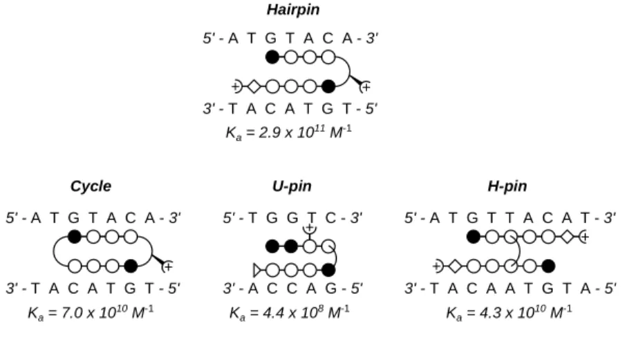
Despite the successes of the hairpin polyamide motif, the set of sequences that the hairpin motif can target is limited by the T,A selectivity of the aliphatic turn moiety. In order to enlarge the set of targetable DNA sequences, other polyamide motifs have been developed, employing a variety of strategies to covalently-link individual polyamide strands. These new motifs include cycles38, H-pins39, and U-pins40 (Figure 1.7).
Gene Regulation with Polyamides
By utilizing the pairing rules, minor groove-binding polyamides may be synthesized to bind predetermined DNA sequences, including those within the promoter regions of genes. Proper placement of polyamides in the promoter sequence may allow them to interfere with the association of transcription factors, leading to inhibition of transcription. It has been demonstrated that polyamides are able to inhibit the binding of certain zinc finger proteins (including Zif268, Figure 1.3b) that bind DNA without any
Cycle U-pin H-pin
5' - A T G T A C A - 3'
3' - T A C A T G T - 5'
5' - T G G T C - 3'
3' - A C C A G - 5'
5' - A T G T T A C A T - 3'
3' - T A C A A T G T A - 5' Hairpin
5' - A T G T A C A - 3'
3' - T A C A T G T - 5' Ka = 2.9 x 10¹¹ M-1
Ka = 4.4 x 10⁸ M-1 Ka = 4.3 x 10¹⁰ M-1 Ka = 7.0 x 10¹⁰ M-1
Figure 1.7 Ball-and-stick schematic for covalently linked polyamide motifs.
minor groove contacts, presumably by an allosteric mechanism.41 However, minor groove-binding ligands can also co-occupy DNA while certain proteins, such as GCN4 (Figure 1.3a) occupy the major groove.42 GCN4 binding has been inhibited successfully by hairpin polyamides bearing “positive patches” targeting protein-phosphate contacts,43,44 and by polyamide-intercalator conjugates.45
Better targets for polyamides have been minor groove-binding transcription factors, such as TBP (Figure 1.3c) and LEF-1. Several transcription factors and promoters have been successfully targeted by hairpin polyamides, including the TFIIIA zinc finger46,47 and the HIV-1 promoter,48,49 resulting in inhibition of RNA polymerase II transcription of targeted genes in vitro (Figure 1.8).
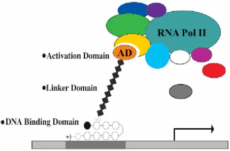
It is also possible to selectively activate transcription by synthesizing a polyamide, designed to bind in the promoter region of a gene of interest, to which has been appended a moiety that recruits the transcriptional machinery to the promoter (Figure 1.9). Polyamides bearing a viral peptide known to activate transcription have been used to increase transcription of diagnostic genes more than 30-fold over basal levels.50-52 Since polyamides can target a wide variety of sequences with high affinity and specificity, this approach has great potential as a general method of gene-specific activation by small molecules. Though the principle was proven, little was determined about the physical parameters of the DNA/polyamide/transcriptional machinery interaction—especially the optimal distance between the DNA and the activation domain.
The successful use of polyamides in model systems has shown their promise as diagnostic and therapeutic agents in more complex environments. For a polyamide to have a DNA-mediated effect in an organism, it must be able to enter the organism’s cells,
traffic to the nucleus, and bind chromosomal DNA. The fundamental repeating unit of chromatin is the nucleosome, which consists of the nucleosome core particle (NCP) and 20-80 base pairs of linker DNA. The NCP is built from two superhelical turns of DNA (147 base pairs) wrapped around a core of eight histone proteins to form a disc-shaped tightly-packed unit.
Figure 1.8 Hairpin polyamides have successfully inhibited the binding of several proteins to DNA in solution. Transcription factor binding sites are shaded.
HIV-1 Promoter
5'-G A G C T G C A T C C G G A G T A C T A C A A A G A C-3'
3'-C T C G A C G T A G G C C T C A T G A T G T T T C T G-5'
Xenopus tRNA Promoter Scanning (-39 to -8)
5'-C C C A T C C A A G T A C A T C G A A T T C A T G A C-3'
3'-G G G T A G G T T C A T G T A G C T T A A G T A C T G-5'
HIV-1 Promoter Scanning
5'-G A G C T C G T C C T C A G A T G C T G C A T A T A A-3'
3'-C T C G A G C A G G A G T C T A C G A C G T A T A T T-5'
Acaete-scute Promoter
5'-G C A T C G T A G C T C G T C A C G C G A C A G G G C-3'
3'-C G T A G C A T C G A G C A G T G C G C T G T C C C G-5'
HER2 Promoter
5'-G G G C T G C T T G A G G A A G T A T A A G A A-3'
3'-C C C G A C G A A C T C C T T C A T A T T C T T-5'
HTLV-1 Promoter
5'-C T C A G G C G T T G A C G A C A A C C C C T C-3'
3'-G A G T C C G C A A C T G C T G T T G G G G A G-5'
designed sequence
5'-G A G G C T G G A T G G C C T T C C C C A T T A-5'
3'-C T C C G A C C T A C C G G A A G G G G T A A T-3'
designed sequence ARE-1
5'-C C A T G G T T G C T G A C T A A T T G T T A T-3'
3'-G G T A C C A A C G A C T G A T T A A C A A T A-5' HIV-1 LTR (+8 to +32)
5'-T C T G G T T A G A C C A G A T C T G A G C C T-3'
3'-A G A C C A A T C T G G T C T A G A C T C G G A-5' +
5S RNA promoter
5'-C G G G C C T G G T T A G T A C T T G G A T G G G A G-3'
3'-G C C C G G A C C A A T C A T G A A C C T A C C C T C-5'
HIV-1 Promoter
5'-T C A G A T G C T G C A T A T A A G C A G C T G C T T-3'
3'-A G T C T A C G A C G T A T A T T C G T C G A C G A A-5'+ +
+ + +
+ +
+ +
+ +
+ +
+
+
+ +
PR R +
+
TFIIA-TFIID
TBP
Ets-1 LEF-1
TBP
Deadpan
DNA gyrase
Tax CREB Tax
GCN4 TFIIIB or TBP
LSF LSF
5S RNA promoter
5'-C G G G C C T G G T T A G T A C T T G G A T G G G A G-3'
3'-G C C C G G A C C A A T C A T G A A C C T A C C C T C-5' +
5S RNA promoter (+63 to +89) [45,46]
5S RNA promoter (+63 to +89) [46]
HIV-1 Promoter [47]
HIV-1 Promoter [47,51]
HIV-1 Promoter Construct [49]
Xenopus tRNA Promoter Construct (-39 to -8) [48]
Achaete-scute Neural Promoter Construct [50]
HER2/neu Promoter [52]
HTLV-1 Promoter [53]
designed construct from pBR322 [54]
designed construct ARE-1 [56]
HIV-1 LTR (+8 to +32) [61]
0.03 nM
2 nM
0.05 nM
0.06 nM 0.05 nM
0.05 nM
0.4 nM
0.03 nM
0.07 nM 0.4 nM
0.1 nM
1 µM 1 µM
0.1 nM
2 nM 0.7 nM 0.9 nM
TFIIIA (zinc finger 4) TFIIIA (zinc finger 4)
Ets
The question of whether polyamides can bind chromosomal DNA is important because the vast majority of the DNA in a cell is locked in nucleosomes. In order to determine the binding characteristics of polyamides on the NCP, several X-ray crystal structures were completed of nucleosome core particles into which had been bound polyamides (Figure 1.10).53 The structures show that not only can polyamides bind nucleosomal DNA, they have some access to the internal regions of the nucleosome core particle, where they can co-localize with the histone proteins. The only sites which were found to be inaccessible to polyamide binding were those blocked by the presence of the tails of the histone proteins in the minor groove.
In order to access chromosomal DNA, polyamides must be able to localize to the nucleus of cells. The cellular trafficking characteristics of polyamides have been studied by attachment of a fluorescent dye to a small selection of polyamides, exposing the resulting conjugates to various mammalian and insect cell lines, and viewing their
Figure 1.9 Polyamide-peptide conjugates have successfully been used to activate transcription of a target gene in vitro. In this experiment, the natural transcription factor (TF) activators (green ovals) have been replaced by polyamide-activator conjugates, binding the appropriate gene promoter sites. These artificial TF’s interact with other transcription factors (purple ovals) and parts of the transcriptional machinery (yellow ovals), sometimes via mediating proteins (blue ovals) to recruit the transcriptional machinery to the target gene, initiating transcription.
subcellular localization by confocal laser scanning microscopy.54 These results showed that in a limited number of cell lines, primarily lines derived from mammalian T-cells, fluorescent polyamide conjugates could enter the cells and traffic to the nucleus (Figure 1.11). However, in most cell lines, these compounds either did not enter the cells, or entered the cells but were sequestered in the cytoplasm.
This raises the issue of how might the uptake profile of polyamides and polyamide conjugates be altered in order that they be useful in the context of living
Figure 1.10 X-ray crystal structure of a hairpin polyamide bound to the nucleosome core particle (NCP). Top-down (a) and side-on (b) views of several polyamide molecules bound to the NCP. Histone proteins are depicted as multicolored cartoons.
(c) View down the minor “supergroove” in which are bound two hairpin polyamides.
Figure 1.11 Cellular uptake of a fluorescent polyamide. (a) Chemical structure and ball-and-stick representations of a hairpin polyamide labeled with the fluorophore Bodipy. (b) CEM cells stained with Bodipy-polyamide. At left is a fluorescence-only image, and at right is the bright-field image. Polyamide is located in the nucleus.
organisms. Several methods of altering the subcellular localization of molecules have been developed for medicinal chemistry and diagnostic applications. The application of many of these methods to polyamides is the subject of the majority of this work. By creating polyamides or polyamide conjugates that are able to localize to the nucleus of living cells, technology advances one step closer to in vivo regulation of gene transcription by small molecules.
Figure 1.11 Cellular uptake of a fluorescent polyamide. (a) Chemical structure and ball-and-stick representations of a hairpin polyamide labeled with the fluorophore Bodipy. (b) CEM cells stained with Bodipy-polyamide. At left is a fluorescence-only image, and at right is the bright-field image. Polyamide is located in the nucleus.
Scope of This Work
This thesis describes work examining DNA-binding polyamides in biological systems. Chapter 2 is conceptually related to the transcription activation studies outlined as in Figure 1.9. In previous work, polyamide-activation peptide conjugates had been synthesized with either short or long flexible linkers separating the two active domains.
These compounds were efficient activators of transcription, though it was uncertain what effect the spacing between the activating domain and the DNA had on RNA Pol II recruitment and transcription activation. In order to answer this question, a series of polyamide-polyproline-activation peptide compounds were synthesized and analyzed for their ability to activate transcription. The polyproline domain is a rigid linker allowing discrete spacing studies to be undertaken. These experiments, though successful in vitro, were not successful in vivo. It was hypothesized that this result was due to poor cellular uptake properties.
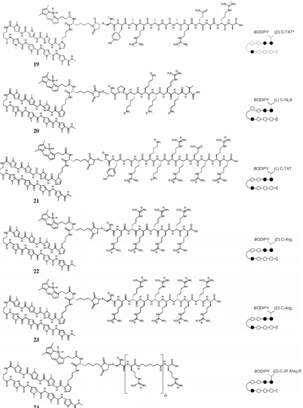
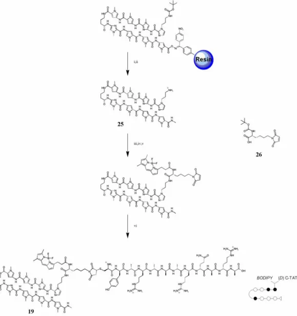
Chapter 3 details some attempts that were made to overcome poor cellular uptake and nuclear localization of polyamides. Chapter 3A recounts the addition of peptide domains shown in other systems to increase the uptake of conjugated small molecules and oligonucleotides to polyamides, along with the fluorophore Bodipy. Chapter 3B records the attachment of small molecules to polyamides in attempts to increase their cellular and nuclear localization. The subcellular localization characteristics of these compounds were determined with confocal laser scanning microscopy. Though largely unsuccessful, these experiments provided useful experience in microscopy and cell culture methods that were necessary to any successful uptake results.
Chapter 4 recounts the modification of polyamides with fluorescein and other structurally similar fluorophores, and the successful use of these compounds to stain nuclei of living cells. Chapter 4A records early efforts to determine the uptake characteristics of hairpin polyamides conjugated to fluorescein isothiocyanate. Chapter 4B presents a much wider view of the uptake profiles of several polyamide motifs conjugated to a variety of fluorophores. Chapter 4C is a first-pass overview of the DNA- binding characteristics of these fluorophore conjugates, showing that the fluorescent compounds, themselves, are appropriate in many cases for use in transcription inhibition experiments in vivo. Chapter 5 is a record of one such application, using polyamides and polyamide conjugates to inhibit the interaction of the androgen receptor with its cognate DNA-binding site.
References
1) Brown, T.A. Genomes; Wiley-Liss: New York, 1999.
2) Consortium, I.H.G.S. Nature 2001, 409, 860.
3) Venter, J.C. et. al. Science 2001, 291, 1304.
4) Gottesfeld, J.M.; Neely, L.; Trauger, J.W.; Baird, E.E.; Dervan, P.B. Nature 1997, 387, 202.
5) Dickerson, R.E.; Drew, H.R.; Conner, B.N.; Wing, M.; Fratini, A.V.; Kopka, M.L. Science 1982, 216, 475.
6) Saenger, W. Principles of Nucleic Acid Structure; Springer-Verlag; New York, 1984.
7) Leonard, G.A.; Hunter, W.N. J. Mol. Biol. 1993, 234, 198.
8) Pabo, C.O.; Sauer, R.T. Annu. Rev. Biochem. 1992, 61, 1053
9) Ellenberger, T.E.; Brandl, C.J.; Struhl, K.; Harrison, S.C. Cell 1992, 71, 1223.
10) Nikolov, D.B.; Chen, H.; Halay, E.D.; Hoffman, A.; Roeder, R.G.; Burley, S.K. Proc. Natl. Acad. Sci. U.S.A. 1996, 93, 4862.
11) Pavletich, N.P.; Pabo, C.O. Science 1991, 252, 809.
12) Kim, Y.; Geiger, J.H.; Hahn, S.; Sigler, P.B. Nature 1993, 365, 512.
13) Jantz, D.; Amann, B.T.; Gatto, G.J., Jr.; Berg, J.M. Chem. Rev. 2004, 104, 789.
14) Kumar, R.A.; Ikemoto, N.; Patel, D.J. J. Mol. Biol. 1997, 265, 187.
15) Quintana, J.R.; Lipanov, A.A.; Dickerson, R.E. Biochemistry 1991, 30, 10294.
16) Mitra, S.N.; Wahl, M.C.; Sundaralingam, M. Acta Crystallogr. D Biol.
Crystallogr. 1999, 55, 602.
17) Bailly, C.; Chaires, J.B. Bioconj. Chem. 1998, 9, 513.
18) Dervan, P.B.; Burli, R.W. Curr. Opin. Chem. Biol. 1999, 3, 688.
19) Dervan, P.B. Bioorg. Med. Chem. 2001, 9, 2215.
20) Dervan, P.B.; Edelson, B.S. Curr. Opin. Struct. Biol. 2003, 13, 284.
21) White, S.; Szewczyk, J.W.; Turner, J.M.; Baird, E.E.; Dervan, P.B. Nature 1998, 391, 468.
22) Wade, W.S.; Mrksich, M.; Dervan, P.B. J. Am Chem. Soc 1992, 114, 8783.
23) Mrksich, M.; Wade, W.S.; Dwyer, T.J.; Geierstanger, B.H.; Wemmer, D.E.;
Dervan, P.B. Proc. Natl. Acad. Sci. U.S.A. 1992, 89, 7586.
24) Kielkopf, C.L.; Baird, E.E.; Dervan, P.B. Rees, D.C. Nat. Struct. Biol. 1998, 5, 104.
25) Kielkopf, C.L.; White, S.; Szewczyk, J.W.; Turner, J.W.; Baird, E.E.; Dervan, P.B. Science 1998, 282, 111.
26) Kielkopf, C.L.; Bremer, R.E.; White, S.; Szewczyk, J.W.; Turner, J.M.; Baird, E.E.; Dervan, P.B.; Rees, D.C. J. Mol. Biol. 2000, 295, 557.
27) Renneberg, D.; Dervan, P.B. J. Am. Chem. Soc. 2003, 125, 5707.
28) Foister, S.; Marques, M.A.; Doss, R.M.; Dervan, P.B. Bioorg. Med. Chem.
Lett. 2003, 11, 4333.
29) Mrksich, M.; Parks, M.E.; Dervan, P.B. J. Am. Chem. Soc. 1994, 116, 7983.
30) Trauger, J.W.; Baird, E.E.; Dervan, P.B. Nature 1996, 382, 559.
31) deClairac, R.P.L.; Geirstanger, B.H.; Mrksich, M.; Dervan, P.B.; Wemmer, D.E. J. Am. Chem. Soc. 1997, 119, 7909.
32) White, S.; Baird, E.E.; Dervan, P.B. J. Am. Chem. Soc. 1997, 119, 8756.
33) Herman, D.M.; Baird, E.E.; Dervan, P.B. J. Am. Chem. Soc. 1998, 120, 1382.
34) Swalley, S.E.; Baird, E.E.; Dervan, P.B. J. Am. Chem. Soc. 1999, 121, 1113.
35) Turner, J.M.; Swalley, S.E.; Baird, E.E.; Dervan, P.B. J. Am. Chem. Soc.
1998, 120, 6219.
36) Wang, C.C.C.; Ellervik, U.; Dervan, P.B. Bioorg. Med. Chem. 2001, 9, 653.
37) Kelly, J.J.; Baird, E.E.; Dervan, P.B. Proc. Natl. Acad. Sci. U.S.A. 1996, 93, 6981.
38) Herman, D.M.; Turner, J.M.; Baird, E.E.; Dervan, P.B. J. Am. Chem. Soc.
1999, 121, 1121.
39) Olenyuk, B.; Jitianu, C.; Dervan, P.B. J. Am. Chem. Soc. 2003, 125, 4741.
40) Heckel, A.; Dervan, P.B. Chem. Eur. J. 2003, 9, 3353.
41) Nguyen-Hackley, D.H.; Ramm, E.; Taylor, C.M.; Joung, J.K.; Dervan, P.B.;
Pabo, C.O. Biochemistry 2004, 43, 3880.
42) Oakley, M.G.; Mrksich, M.; Dervan, P.B. Biochemistry 1992, 31, 10969.
43) Bremer, R.E.; Baird, E.E.; Dervan, P.B. Chem. Biol. 1998, 5, 119.
44) Bremer, R.E.; Wurtz, N.R.; Szewczyk, J.W.; Dervan, P.B. Bioorg. Med.
Chem. 2001, 9, 2093.
45) Fechter, E.J.; Dervan, P.B. J. Am. Chem. Soc. 2003, 125, 8476.
46) Gottesfeld, J.M.; Neely, L.; Trauger, J.W.; Baird, E.E.; Dervan, P.B. Nature 1997, 387, 202.
47) Neely, L.; Trauger, J.W.; Baird, E.E.; Dervan, P.B.; Gottesfeld, J.M. J. Mol.
Biol. 1997, 274, 439.
48) Dickinson, L.A.; Gulizia, R.J.; Trauger, J.W.; Baird, E.E.; Mosier, D.E.;
Gottesfeld, J.M.; Dervan, P.B. Proc. Natl. Acad. Sci. U.S.A. 1998, 95, 12890.
49) Dickinson, L.A.; Trauger, J.W.; Baird, E.E.; Dervan, P.B.; Graves, B.J.;
Gottesfeld, J.M. J. Biol. Chem. 1999, 274, 12765.
50) Ansari, A.Z.; Mapp, A.K. Curr. Opin. Chem. Biol. 2002, 6, 765.
51) Mapp, A.K.; Ansari, A.Z.; Ptashne, M.; Dervan, P.B. Proc. Natl. Acad. Sci.
U.S.A. 2000, 97, 3930.
52) Ansari, A.Z.; Mapp, A.K.; Nguyen, D.H.; Dervan, P.B.; Ptashne, M. Chem.
Biol. 2001, 8, 583.
53) Suto, R.K.; Edayathumangalam, R.S.; White, C.L.; Melander, C,; Gottesfeld, J.M.; Dervan, P.B.; Luger, K. J. Mol. Biol. 2003 326, 371.
54) Belitsky, J.M.; Leslie, S.J.; Arora, P.S.; Beerman, T.A.; Dervan, P.B. Bioorg.
Med. Chem. 2002, 10, 3313.
Chapter 2
Transcription Activation with Polyamide-Polyproline-Peptide Conjugates
The text of this chapter was taken in part from a manuscript coauthored with Paramjit S.
Arora, Aseem Z. Ansari, Professor Mark Ptashne (Sloan-Kettering Institute), and Professor Peter B. Dervan (Caltech).
(Arora, P.S.; Ansari, A.Z.; Best, T.P.; Ptashne, M.; Dervan, P.B. “Design of Artificial Transcriptional Activators with Rigid Poly-L-proline Linkers” J. Am. Chem. Soc. 2002, 124, 13067.)
Abstract
Typical eukaryotic transcriptional activators are composed of distinct functional domains, including a DNA-binding domain and an activating domain. Artificial
transcription factors have been designed wherein the DNA-binding domain is a minor groove DNA-binding hairpin polyamide linked by a flexible tether to short activating peptides, typically 16-20 residues in size. In this study, the linker between the polyamide and the peptide was altered in an incremental fashion using rigid oligoproline “molecular rulers” in the 18-45 Å length range. We find that there is an optimal linker length which separates the DNA and the activation region for transcription activation.
Introduction
Transcriptional activators typically bind near a gene and recruit the transcriptional machinery to a nearby promoter, thereby stimulating the expression of the gene. These activators comprise two regions: the DNA binding domain and the activating domain.
The former defines the promoter address in the genome where the activating region is to be delivered for recruitment of the transcriptional machinery.1,2 We have previously reported efforts to replace the natural protein activators with nonnatural components smaller in size than nature’s proteins. Artificial transcription activators comprised of three synthetic modules, a hairpin polyamide (PA) DNA-binding domain (DBD) and a short peptide activation domain (AD) (typically 16-20 amino acid residues in size) connected by flexible linkers which vary in length, have been shown to initiate transcription at targeted promoter sites in cell-free systems.3,4 Modeling studies on these polyamide-peptide conjugates with flexible linkers suggested a distance of 20-40 Å between the DNA and the activating region for transcription activation. We sought to determine whether the length of the linker that projected the activating region away from the DNA would have any effect on the degree of activation (Figure 2.1). In the present study, we replaced the flexible linkers between the DBD and AD with rigid oligoprolines of varying incremental lengths (18-45 Å). Our results suggest that there is an optimal window within which activating regions of the type used here can function efficiently.
Results
In this study, we replaced the flexible linker between the activating peptide and the hairpin polyamides with 6, 9, 12, and 15 L-proline residues (Pro6-Pro15) (Figure 2.2).
Poly-L-proline linkers were chosen as the “molecular rulers” for the present study since a stretch of proline residues forms a stable helical structure (the polyproline II helix).
Addition of each proline residue increases the length of this helix in a predictable manner, approximately 3 Å per proline residue. Thus, the oligoproline linker projects the activating region peptide away from the DNA-binding polyamide in an incremental manner spanning 18, 27, 36, and 45 Å (Figure 2.2).5 For the activating region we used two peptides, AH and VP2, that have been previously shown to function efficiently when tethered to a hairpin polyamide (Figure 2.3a).4 Therefore, three series were synthesized:
Figure 2.1 Activation of gene transcription by artificial transcription factors. The artificial activator is composed of three separate functional domains. The DNA binding domain consists of minor groove-binding pyrrole/imidazole polyamides. The DNA-binding domain is tethered to the activation domain (AD), a peptide, by a linker domain.
Figure 2.2 Poly-L-proline-based molecular rulers. (a) Structure of a poly-L-proline helix. (b) Predicted length of polyproline helices containing the indicated numbers of proline residues.
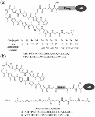
Figure 2.3 Structures of polyamide-activation peptide conjugates 1-5 prepared to explore the effect of linker length and flexibility on activation potential. (a) Polyamide-Pron-peptide conjugates for which the linker domain consisting of poly-L- proline helices serves as a molecular ruler. (b) Polyamide-peptide conjugates bearing a flexible linker.
PAPro6-15-AH (2a-d), PA-Pro6-15-VP2 (3a-d), and PA-Pro6-15 (1a-d) lacking the activation peptides as controls. Conjugates with flexible poly(ethylene glycol) linkers, 4 and 5, previously shown to activate transcription in vitro were included for comparison (Figure 2.3b).4
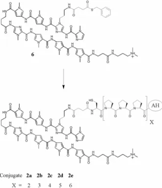
The eight-ring hairpin polyamides which target 5’-WGWWWW-3’ (W = A or T) and two peptides, AH and VP2, were synthesized by solid-phase methods.6 Each activating peptide contained an N-terminal cysteine for subsequent reaction with a thioester, 6, via the native ligation reaction to afford the desired conjugate (Figure 2.4).7 The ability of these conjugates, 1a-d, 2a-d, and 3a-d, to activate transcription in vitro was tested using yeast nuclear extract on a template bearing three palindromic binding sites upstream of the minimal core promoter driving the expression of a transcript that
Figure 2.4 Synthesis of polyamide-peptide conjugates. Thiolane 6 was converted to the targeted conjugates via the native ligation reaction.
lacks guanine residues.3,4 The results show that the linker plays a role in determining the ability of the activating region to stimulate in vitro transcription (Figure 2.5). This effect is not a function of the activating peptide, as both activating peptides tested showed similar profiles. Figure 2.5 also compares the two activating peptides attached to the polyamide via different proline linkers to conjugates 4 and 5 bearing the same activating peptides via flexible linkers.
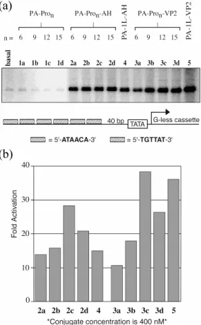
Figure 2.5 In vitro transcription reactions with the compounds listed in Figure 2.3;
the transcription reaction conditions are described in the Experimental Section. (a) Storage phosphor autoradiogram of the reactions which were performed with a 400 nM concentration of each conjugate. The template configuration showing the sequence and number of the polyamide binding sites is depicted below the gel. (b) Fold activation was determined by comparing the amount of transcription elicited by conjugates 2a-d, 3a-d, 4, and 5 with that of the basal level.
Our results indicate that the strength of the given activating peptide increases with every increment in the spacer length up to 12 residues (36 Å). An increase in the spacer length to 15 residues leads to a decrease in activation. These results suggest an optimal spacing of 36-45 Å between the DNA and the activating regions for efficient transcription. Figure 2.6 shows the relative potency of each compound in comparison to PA-Pro6-AD (2a, AD = AH; 3a, AD = VP2). Although the absolute fold activation mediated by all conjugates varies slightly between different experiments, the overall relationship between linker length and activation potential remains constant over four independent in vitro transcription reactions.
To determine whether any of the effects observed were due to inefficient or inappropriate binding of the conjugate to its site on the promoter, we measured dissociation constants for conjugates 3a-d using quantitative DNase I footprinting titrations.8,9 These studies revealed that conjugates bound with similar affinities and specificities to their sites on the promoter DNA (Figure 2.7 and Table 2.1). In control
Figure 2.6 Summary of four independent in vitro transcription reactions showing the relative potency of each compound in comparison to PA-Pro6-AD (2a, AD = AH; 3a, AD = VP2).
experiments polyamide-proline conjugates 1a-d lacking the activation transcription peptides did not activate.
Figure 2.7 Quantitative DNase I footprinting titration of conjugates 3a-d shows that all conjugates bound to their target sites with similar affinities. (Top) Storage phosphor autoradiogram of a quantitative DNase I footprinting titration of 3a-d on a 63 bp 5’-32P- labeled PCR fragment containing both the promoter region and 140 bp of the G-less cassette reporter. (a) Lane 1, undigested DNA; lane 2, A reaction; lane 3, G reaction;
lane 4, DNase I standard; lanes 5-14, 100 pM, 200 pM, 500 pM, 1 nM, 2 nM, 5 nM, 10 nM, 20 nM, 50 nM, and 100 nM 3a, respectively. (b) Lane 1, undigested DNA; lane 2, A reaction; lane 3, G reaction, lane 4, DNase I standard; lanes 5-14, 100 pM, 200 pM, 500 pM, 1 nM, 2 nM, 5 nM, 10 nM, 20 nM, 50 nM, and 100 nM 3b, respectively. (c) Lane 1, undigested DNA; lane 2, G reaction; lane 3, A reaction; lane 4, DNase I standard; lanes 5-14, 100 pM, 200 pM, 500 pM, 1 nM, 2 nM, 5 nM, 10 nM, 20 nM, 50 nM, and 100 nM 3c, respectively. (d) Lane 1, undigested DNA; lane 2, G reaction; lane 3, A reaction; lane 4, DNase I standard; lanes 5-14, 200 pM, 500 pM, 1 nM, 2 nM, 5 nM, 10 nM, 20 nM, 50 nM, 100 nM, and 200 nM 3d, respectively. (Bottom) Design of the plasmid DNA template used for the DNase I footprinting titration experiments. The promoter region contains six cognate binding sites for the hairpin polyamide upstream of a G-less cassette reporter.
The use of poly-L-proline linkers as molecular rulers was inspired by the seminal work of Stryer and co-workers, who utilized fluorescence resonance energy transfer (FRET) analysis to show that oligomers composed of up to 12 proline residues can retain a rigid helical structure.5 To ascertain that an oligomer composed of 15 proline residues does not deviate from the predicted structure, we repeated the FRET analysis on poly-L- proline linkers used in our experiments. To perform this analysis, we synthesized poly-L- proline linkers bearing Oregon Green (OG) energy donor and tetramethylrhodamine (TAMRA) energy acceptor moieties on each end (conjugates 7a-d, Figure 2.8). In the Stryer study, naphthyl and dansyl groups were used as the energy donor and the energy acceptor moieties, respectively. The different dyes were used in the present study because the Förster radius (the distance at which energy transfer is 50% efficient) for the dansyl-naphthyl pair (Ro = 27 Å) may be too low to allow for efficient FRET when the energy donor and the acceptor are spaced by the Pro15 linker (45 Å). The Förster radius (Ro) for the OG-TAMRA pair is predicted to be 55 Å.10 FRET measurements indicate that the oligomer composed of 15 proline units does retain a rigid structure. The plot of distance versus FRET efficiency provides a linear relationship between the conjugates, and importantly, the experimentally observed FRET efficiency is in good agreement with the predicted r-6 dependence (Figure 2.9).5 Thus, the FRET studies show that the
dependence of transcription activation on the number of L-prolines is not due to a change in the linker structure.
Discussion
In this study we examine the role of the distance separating the DBD and AD in our artificial transcription factors. This study was prompted by previous observations in Figure 2.8 Synthesis of polyproline-dye conjugates. (i) 20% hydrazine in ethanol.
(ii) Oregon Green-NHS ester, DMF, and DIEA. (iii) 20% TFA in CH2Cl2. (iv) TAMRA-NHS ester, DMF, and DIEA.
which we found that activating regions activated transcription to different degrees on the basis of the point of attachment as well as the nature of the linker separating them from the DNA binding polyamide.4 This dependence on spatial presentation led us to test the role of linker length in the degree of activation elicited by two acidic activating peptides.
The proline linker domain is conjugated to an internal pyrrole of the polyamide, rather than to the C-terminus, which allows the linkers to project out of the minor groove away from the DNA helix. Our data suggest that for the two acidic activating regions (AH and VP2) the strength of activation increases as the linker reaches a length of 36 Å. The increase, while modest, is reproducible and displays a clear trend (Figures 2.5 and 2.6).
The effects are muted in part due to three features that were incorporated into the experimental design: the use of multiple DNA binding sites to elicit robust activation, the presence of a flexible hinge tethering the proline linker to the polyamide, and the
Figure 2.9 FRET data for polyproline helices. (a) The emission spectra of OG-Pron- TAMRA conjugates. The conjugates were excited at 495 nm. (b) The dependence of the efficiency of energy transfer on distance is given in this plot of ln (E-1 – 1) vs. ln r.
The slope is 6.6, in good agreement with the predicted r-6 dependence.
unstructured nature of the activating peptides, which themselves may sample a significant amount of solvent space when fully extended.
We did not find a significant contribution of poly-L-proline linkers themselves to the level of activation in the absence of tethered activating regions. It has been reported that proline-rich (rather than poly-L-proline) activating domains can function to activate transcription even in yeast extracts, though this activation is not as robust as that elicited by acidic activators.11 Presumably the proline oligomer is not a sufficiently strong activator; its effects are therefore not detected in our studies. An alternative possibility is that the ability of proline-rich activating domains to elicit transcription may be more sensitive to their spatial location.
As the principle of recruitment implies, for a DNA-tethered activator to function efficiently, it must sample sufficient nuclear solvent space to bind and recruit various complexes that participate in transcriptional initiation to a given promoter. However, beyond a certain distance the activating region would not function to recruit, as the local concentration of the machinery at a given promoter would not be tremendously enhanced by binding a distant activating region. It has been shown that eukaryotic activating regions that are not tethered to DNA—even when presented at exceedingly high concentrations such that it would bind to its targets in the transcriptional machinery—do not improve the level of transcription from a given promoter. In fact, as expected, it
“squelches” the ability of a DNA-bound activator from functioning—presumably by binding the targets in the machinery.2,3,12 Thus, for efficient recruitment in the context of our in vitro studies with artificial activators on nonchromatinized templates using yeast