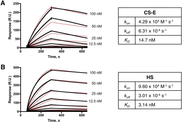
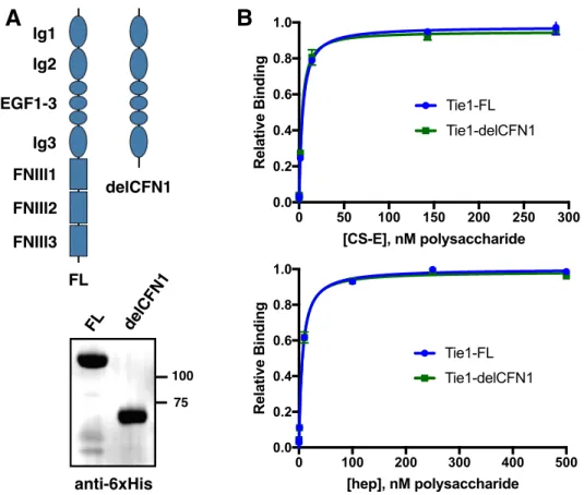
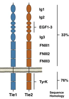
We localize the GAG binding site to the N-terminal region of Tie1, which may provide structural insight into the importance of this interaction for the formation of Tie1-Tie2 heterodimerization. Tie1 is generally considered to be an antagonist of Tie2 signaling by forming Tie1/Tie2 heterodimers. Experiments using shRNA-mediated knockdown of Tie1 showed that Tie2 activation by Ang1 is elevated after Tie1 depletion.25 However, a recent in vivo study showed that endothelial deletion of Tie1 in adult mice paradoxically leads to reduced Tie2 activation and downstream signaling. 26 instead suggests that the Tie1/Tie2 interaction is required to promote Ang1-mediated vascular responses. Solutions of Tie1-Fc at various concentrations were then flowed over the modified chip, followed by buffer alone, and the degree of response was measured over time.
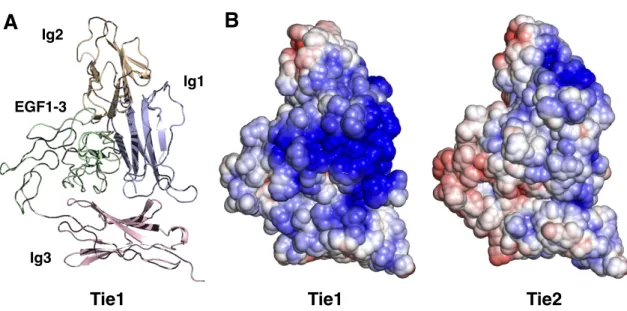
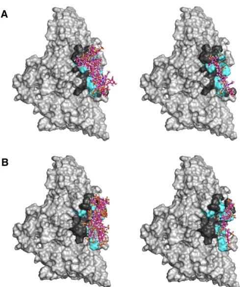
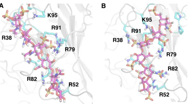
Both constructs maintain binding to GAGs, suggesting that binding occurs in the N-terminal domains of Tie1. To perform docking, we first produced a homology model of Tie1 based on the solved Tie2 crystal structure41 using the program SWISS-MODEL (Figure 3-7a).42. Interestingly, this region of Tie1 had previously been suggested to interact with a complementary electronegative surface of Tie2.25.
We tried the same experiment using single mutations, but neither showed a complete loss of Tie1.
Contextualization of Tie1-GAG Binding
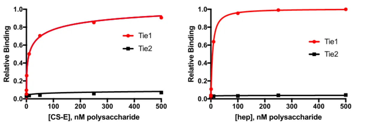
Furthermore, CS-E was not detected in HUVECs in this assay, strongly suggesting that HS was the physiologically relevant GAG for Tie1 binding at least in systemic endothelial cells. Cells were then incubated with Tie1-WT-Fc or Tie1-2A-Fc, and binding of the constructs was detected using a fluorescently labeled anti-human Fc antibody. Together, these results suggest that cell surface HS is required for Tie1 binding through the predicted Tie1-GAG binding interface and that loss of cell surface HS or the residues required for this interaction is sufficient to prevent Tie1 binding .
Untreated or heparinase-treated EA.hy926 cells were probed with Tie1-WT and visualized with an anti-human Fc antibody (red). Tie1-2A showed no cell binding, suggesting that HS binding is required for cell surface binding.
Towards a Functional Role of Tie-GAG Binding
The Tie1/Tie2 heterodimer has been previously documented to be the basis of Tie2-induced phosphorylation of Tie1;46,47, but this interaction has been observed using standard coimmunoprecipitation techniques. EA.hy926 cells were fixed, immunostained with (A) anti-Tie1 and anti-Tie2 antibodies or (B) anti-Tie1 antibody alone, stained with the PLA staining protocol, and visualized by microscopy. Tie1/Tie2 heterocomplexes are revealed as punctate red staining and can be normalized to cell number by DAPI (blue) nuclear staining.
Briefly, HUVEC or EA.hy926 cells were fixed, blocked, and probed with the appropriate Tie1 and Tie2 antibodies. As before, we will apply heparinase and glycan engineering techniques in this assay to further determine the effect of HS on Tie1/Tie2 heterodimerization.
Generation of a Tie1-GAG-Binding Deficient Mouse Model
We were pleased to observe the formation of punctate staining on the cell surface when both antibodies were used, whereas a control sample lacking one of the two primary antibodies showed minimal background. HS binds to a variety of angiogenic ligands and receptors; thus, understanding the consequences of the Tie1-GAG interaction caused by a global decrease in total HS sulfation (and thus total HS/protein binding) may be. In the absence of a repair template, endogenous machinery of the cell undergoes non-homologous end joining (NHEJ), producing random insertions and deletions (indels).
Two ssODN constructs were made that contained one of the two mutations flanked by approximately 60 nt of homologous sequence. Cas9 recognition of gDNA requires an NGG motif known as a PAM immediately at the 3' end of the complementary gRNA sequence. Loss of the NGG sequence prevents Cas9 from cleaving the sequence again after incorporating the mutation.
The zygotes were allowed to develop to the blastocyst stage, at which point they were subjected to genotyping to test for the inclusion of the desired mutations. Genotyping was accomplished by obtaining gDNA from blastocysts followed by PCR amplification of the desired sequence and Sanger sequencing (Figure 3-16). In both cases, the majority of zygotes progressed to the blastocyst stage (ssODN: 15/18; dsDNA: 15/21), suggesting that the injection solutions were nontoxic.
Interestingly, the sequencing results showed that the majority of blastocysts resulting from ssODN injection showed random indels, with no sample showing proper incorporation. We were pleased to see that 4/15 blastocysts from dsDNA injection showed correct incorporation of the mutations at both sites (Figure 3-16b). Two of the pups were produced from zygotes incubated with SCR7, while one was from untreated zygotes.
HS GAGs Bind to Angiopoietin Ligands and Potentiate Tie2 Signaling .1 ELISA
Ternary Complex Formation with Tie2
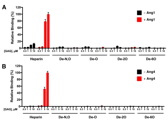
GAGs have previously been shown to both facilitate and hinder the formation of a ligand-receptor interaction, thereby exerting control over receptor activation and signaling. To quickly delineate whether HS binding to Ang1 or Ang4 affects their binding to Tie2, we used carbohydrate microarrays to directly observe. Binding of Tie2-Fc to the microarray in the presence of Ang1 or Ang4 suggests the formation of ternary carbohydrate-protein-protein complexes.
Here, Tie2-Fc, previously shown not to bind HS, was incubated with the HS microarrays in the presence or absence of Ang1 or Ang4. Tie2 immobilized on the microarray through its interaction with HS-bound Ang1/4 was visualized by a fluorescently labeled anti-human Fc antibody. Interestingly, the Tie2 signal increased dramatically in the presence of Ang1 or Ang4, suggesting that the Ang/HS interaction does not interfere with Ang/Tie2.
Moreover, the assembly of this ternary complex is dependent on the sulfation pattern and is only formed with the HS motif. This mode of ternary complexation has previously been demonstrated for both FGF2-FGFR1 and FGF8-FGFR366 and suggests that cell surface HS may enhance Ang/Tie2 signaling by bringing Ang ligands into close proximity to the Tie2 receptor.
Potentiation of Tie2 Signaling by Glycan Engineering
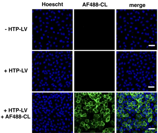
Resultant cells (+ HTP-LV) and parental cells (- HTP-LV) were treated with AF488-CL to visualize cell surface HTP. As expected, cells treated with HTP-LV showed strong surface labeling by AF488-CL, whereas parental cells showed no surface labeling. With HTP-expressing endothelial cells in hand, we decided to test the effect of cell surface HS on Ang/Tie2 signaling (Figure 3-20).
To achieve this, EA.hy926-HTP cells were first treated with heparinase I/III to eliminate endogenous HS and then grown to confluence. Cells were then treated with either 5 µg/ml HS-CL or desulfated (deS) HS-CL for 2 h at 37 °C in serum-starved medium (0.5% fetal bovine serum). EA.hy926 cells expressing HTP were treated with de-HS-CL or HS-CL and stimulated with Ang1, Ang2, or Ang4.
Phosphorylation of Akt at Thr308, which is dependent on 3-phosphoinositide-dependent kinase 1 (PDK1), was then measured by Western blotting as a readout for downstream Tie2 signaling. Strikingly, we saw that for both Ang1 and Ang4, the presence of HS-CL on the cell surface led to a significant increase in Akt phosphorylation compared to cells displayed with non-binding de-HS-CL. No significant change was observed at basal levels or with Ang2, suggesting that this effect is specific to the HS-associated increase in Tie2 activation by Ang1 and Ang4.
Conclusions
Methods and Materials .1 General Methods
- Protein Expression and Purification
- Protein-GAG Binding Assays
- Computational Methods
- Biological Assays
- Transgenic Tie1 Mouse Model
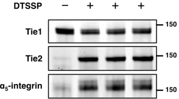
For experiments, cells were plated on plastic plates (for lysate harvesting) or glass-bottom plates (imaging) precoated with 5 μg/cm2 bovine fibronectin (Sigma Aldrich) and 10 μg/cm2 rat tail collagen type I ( Sigma Aldrich). The wells were washed three times with PBST and then incubated with 200 µL of 5% BSA in PBS for 1 h at RT. Cells were washed three times with ice-cold PBS and then incubated with 100 µL of 1 mM DTSSP in PBS (fresh. prepared) for 30 min at RT with gentle rocking.
The cells were then washed twice with PBS and then fixed with 4% paraformaldehyde in PBS for 20 min at room temperature. Cells were rinsed three times with PBS and blocked with the DuoLink blocking buffer (Sigma Aldrich) for 1 h at room temperature. The cells were then run through the Duolink labeling protocol, scaled up to 30 µl reactions per well according to the manufacturer's protocol (DUO92101, Sigma Aldrich).
Cells were visualized using an LSM 710 confocal microscope (Zeiss) with a 40x oil immersion objective and preset filter settings for Texas Red and DAPI. Cells were treated with 5 µg/ml blasticidin for an additional 3 d, and cells from the well treated with the lowest amount of lentivirus that survived selection were expanded and cryopreserved. To validate expression of HTP, treated and untreated EA.hy926 cells were plated on precoated 96-well glass bottom plates and allowed to grow to confluence.
Cells were rinsed three times with complete DMEM and imaged live on an LSM 710 confocal microscope (Zeiss) with a 10× objective. Cells were then plated onto precoated six-well plates at 100,000 cells per well and grown overnight at 37°C with complete DMEM. Cells were then stimulated for 30 min at 37°C with serum-free DMEM containing 500 ng/mL Ang1, Ang2, or Ang4 or no ligand.
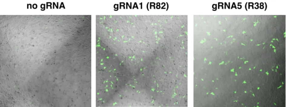
Tie1-gRNA-5F: CACCGCGCAGGTCAGGAAGAAACGC Tie1-gRNA-5R: AAACGCGTTTCTTCCTGACCTGCGC Tie1-gRNA-6F: CACCGTCCGAGCTCCTCCCTGCTC Tie1-gRNA-6R: AAACGAGCAGGGAGGAGCTCGGACRNA-TCTTCGG AAACGACACGCAGGTCAGGAAGAAC Tie1-gRNA-8F: CACCGCCTGACCTGCGTGTCTGGTG Tie1 -gRNA -8R: AAACCACCAGACACGCAGGTCAGGC Ti1-gRNA-9F: CACCGCCTCACCAGACACGCAGGTC Ti1-gRNA-9R: AAACGACCTGCGTGTCTGGTGAGGC. Om gRNAs te toets, is samevloeiende sesput plate van HEK-293T selle getransfekteer met 0.5 μg pCAG-EG(Tie1)FP met 0.5 μg van elk van die gRNA-bevattende pX459 vektore.
Dominant-negative and targeted null mutations in the endothelial receptor tyrosine kinase, tek, reveal a critical role in embryonic vasculogenesis. The receptor tyrosine kinase TIE is required for the integrity and survival of vascular endothelial cells. The orphan receptor Tie1 regulates angiogenesis and vascular remodeling by differentially regulating Tie2 in tip and stalk cells.
Heparin enhances platelet-derived growth factor (PDGF)-BB-induced PDGF-alpha receptor, but not PDGF-beta receptor tyrosine phosphorylation in heparan sulfate-deficient cells. Heparin, heparan sulfate, and dermatan sulfate regulate the formation of insulin-like growth factor-I and insulin-like growth factor-binding protein complexes. Synergistic binding of vascular endothelial growth factor-A and its receptors to heparin selectively modulates complex affinity.
A fluorescent Tie1 reporter allows monitoring of vascular development and endothelial cell isolation from transgenic mouse embryos. Multiple angiopoietin recombinant proteins activate the Tie1 receptor tyrosine kinase and promote its interaction with Tie2. Activation of the orphan endothelial receptor Tie1 modifies Tie2-mediated intracellular signaling and cell survival.
Constitutive association of Tie1 and Tie2 with endothelial integrins is functionally modulated by angiopoietin-1 and fibronectin. One-step generation of mice with reporter and conditional alleles by CRISPR/Cas-mediated genome engineering. Yu, Angiopoietin-1, unlike Angiopoietin-2, is incorporated into the extracellular matrix via the linker peptide region.