with Electron Tunneling Wires
Thesis by Corinna R. Hess
In Partial Fulfillment of the Requirements for the Degree of
Doctor of Philosophy
California Institute of Technology Pasadena, California
2002
(Defended May 30, 2002)
©2002 Corinna R. Hess All Rights Reserved
Aci:lnowledgments
Six years seems like a very long time, at least right now, and two pages much too short, but here goes ...
There are many people to whom I am very grateful for all their help on this project and for all that they have taught me. Dave Dooley gave me the chance to work on this project and has shown great patience despite all the detours we've taken it on.
His group, especially Greg Juda, has sent protein at a moments notice and provided many helpful discussions as well. I've been lucky to have worked with Mike Hill on this project. He has taught me a lot about this "black box" of protein e-chem, and was thankfully always optimistic even when things looked somewhat bleak. The project has also been pushed forward these past few months by the talent and tenacity of a student of his, Ricky Amii. lowe a million thanks to Michele McGuirl, who was enormously helpful in my initial venture into the bio world. She taught me the bulk of what I know about amine oxidase and most importantly has always believed in this project. It seems fitting that my undergraduate advisor, Greg Hillhouse, be here at Caltech as I finish up. I have him to thank for introducing me to research. I wouldn't be here if it weren't for the great experience ofthe 3 and Y2 years I spent in his lab and for his continuing support.
The Gray group has always been made up of many unique personalities who've made the experience of grad school enjoyable, and sometimes very entertaining. I'm very fortunate to have met and worked with many wonderful people; Randy, Mike, Akif, Cissi, have all been great not only to discuss science with, but I've also shared many memorable moments with them and they've been there for periods of high stress as well.
The heart and soul of the Gray group are of course Harry and Jay. I've learned a great deal from Jay and his patience is truly impressive. We've definitely abused his open-door policy. But no matter how busy, I could always plop down on his couch with a look of confusion, and he would take the time to discuss my project with me. He always had a plan B available when things looked grim (actually, I think we've exhausted the first half of the alphabet). And Harry, I couldn't have asked for a better advisor. He always knew when I needed encouragement, when I needed a kick in the ... , and when it had been a particularly frustrating week and I just needed a shot of whiskey.
I am extremely grateful to him for his confidence in this project, his motivation, and for not letting me give up.
All of the ups and downs of these past six years have been shared by my best friends. My appreciation for their friendship cannot be expressed enough. Jenn has been to Brussels and back with me (twice). I'm going to miss all of our adventures (not so much those spring rolls) though I'm sure there are still many ahead. Her sense of humor has always made me laugh, and her generosity is amazing. Natia has also always been there for me and her friendship has been invaluable. Our hour-long discussions about chemistry, life, love, politics, etc. have kept me sane. She has an amazing insight into everything and always had good advice and loads of encouragement.
Finally, I have my family to thank for their unwavering support in all of my efforts. My parents have always encouraged and believed in me. They have celebrated every accomplishment, no matter how small. My brother has always commiserated with me and I've been very luck to have my sister out here these past two years. Their support has meant a lot to me.
Abstract
Amine oxidases (AO) are copper-containing enzymes that catalyze the conversion of amines to aldehydes. The active site is deeply buried within the protein network, a factor which has precluded many investigations into the mechanism. A series of electron tunneling molecular wires has been synthesized, designed to access the cofactors of amine oxidase by means of the substrate channel. These wires are comprised of phenyl-ethynyl units and thus promote rapid rates of electron transfer to the active site. An amine group attached to one end of the oligomer serves as the channel-specific functionality, which binds within the channel. Binding of the oligomers to AO was demonstrated through inhibition and fluorescence quenching studies. A variety of reporter groups has been attached to the chain end opposite the amine group, allowing us to probe the enzyme using a variety of techniques. A Re terminated complex was designed for electron transfer studies with the enzyme. The photophysics of this complex were investigated in the presence of AO. A thiol terminated wire also has been synthesized, which was employed in electrochemical studies of the amine oxidase cofactors. These studies provide evidence for the ability to communicate with the active site using the molecular wires. In addition, these complexes fulfilled our primary goal and enabled us to determine a redox potential of the TPQ cofactor.
Table of Contents
Acknowledgment ---111
Abstract ---V Table of Contents ---VI List of Figures and Tables --- ix
Chapter 1: Introduction --- 1
Amine oxidase background ---2
Thesis overview and objectives --- 8
References --- 11
Chapter 2: Electrochemistry of amine oxidases --- 12
Materials and methods --- 17
Electrochemistry --- 18
Electrode preparations --- 18
Results --- 19
Discussion --- 23
References ---26
Chapter 3: Design and synthesis of molecular wires for amine oxidases ---27
Inhibition studies --- 29
Materials and methods ---29
Results ---32
Synthesis of molecular wires ---32
References --- 37
Chapter 4: Binding of molecular wires to AGAO ---38
Materials and methods --- 40
Results and discussion --- 42
Chapter 5: Photochemical investigations of Re(BzA-1-DEA) with AGAO --- 47
Modeling ofRe(BzA-1-DEA) into the AGAO channel--- 49
Materials and methods --- 51
Results --- 51
Discussion --- 54
Photophysics ofRe(BzA-l-DEA) --- 54
Photophysics ofRe(BzA-l-DEA) with AGAO --- 61
References --- 63
Chapter 6: Electrochemistry of AGAO with DEA-I-PhSH --- 64
Materials and methods --- 67
Electrochemistry --- 68
Electrode preparations --- 68
Modifications of Au electrodes with thiol wires --- 69
Determination of thiol coverage on Au electrodes --- 69
Electrochemistry of DEA-I-PhSAc ---69
Results --- 69
Discussion --- 70
Electrochemistry ofDEA-l-PhSH --- 72
Results ---72
Discussion --- 76
References --- 78
Appendix A: Protein purification and sample preparation --- 79
Protein expression and purification --- 80
AGAO sample preparation ---80
References --- 81
Appendix B: Synthesis and characterization of molecular wires ---82
General --- 83
General procedures for Pd cross-coupling reactions --- 83
Synthesis and purification of oligomers --- 86
Synthesis of Re(BzA -1-DEA) --- 94
References --- 97
List of Figures and Tables
Chapter I
Figure 1.1: Crystal structure of AGAO --- 3
Figure 1.2: Proposed mechanism for TPQ biogenesis --- 6
Figure 1.3: Catalytic cycle of amine oxidase --- 7
Chapter 2 Figure 2.1: Depiction of electrode surfaces for protein electrochemistry --- 14
Figure 2.2: Residues comprising the channel of AGAO --- 16
Figure 2.3: SWV of AGAO on PGE electrodes --- 20
Figure 2.4: SWV of AGAO on (KC)2 modified Au electrodes --- 21
Figure 2.5: SWV of AGAO on (KC)2 modified Au electrodes --- 22
Chapter 3 Table 3.1: Molecules used for inhibition studies of AGAO --- 30
Figure 3.1: Results of inhibition studies of AGAO --- 33
Figure 3.2: Methodology for synthesis of wires --- 34
Figure 3.3: Wires synthesized for communication with the active site AO --- 36
Chapter 4 Figure 4.1: Absorption and emission spectra ofDEA-I-BzA --- 41
Figure 4.2: Time-resolved fluorescence quenching ofDEA-I-BzA by AGAO 43 Figure 4.3: Steady-state fluorescence quenching ofDEA-I-BzA by AGAO -- 44
Figure 4.4: Results of inhibition studies of AGAO with DEA-I-BzA --- 46
Chapter 5
Figure 5.1: Model ofRe(BzA-1-DEA) in the AGAO channel--- 50
Figure 5.2: Absorption spectrum ofRe(BzA-1-DEA) --- 52
Figure 5.3: Emission spectra ofRe(BzA-1-DEA) --- 53
Figure 5.4: Transient absorption spectrum ofRe(BzA-1-DEA) in dioxane --- 55
Figure 5.5: Transient absorption spectra of Re(BzA-1-DEA) --- 56
Figure 5.6: Log lot of transient absorption spectra --- 57
Figure 5.7: Transient absorption spectra of Re(BzA-1-DEA) --- 58
Figure 5.8: Diagram for the excited states ofRe(BzA-1-DEA) --- 60
Chapter 6 Figure 6.1: Depiction of self-assembled protein monolayers on gold --- 66
Figure 6.2: Electrochemistry of AGAO with DEA-1-PhSAc --- 71
Figure 6.3: CVof AGAO on electrodes modified with DEA-I-PhSll/AGAO -- 73
Figure 6.4: OSWV of AGAO on DEA-1-PhSll modified Au electrodes --- 74
Figure 6.5: Difference spectrum for OSWV on DEA-l-PhSll modified Au ---- 75
AppendixB Figure B.1: Synthesis scheme for molecular wire precursors --- 84
Figure B.2: Scheme for synthesis of molecular wires --- 85
Figure B.3: III NMR of iii in CDCh --- 87
Figure B.4: III NMR of vi in dmf-d6 --- 89
Figure B.5: III NMR ofDEA-1-BzA in dmf-d6 --- 90
Figure B.6: III NMR of DE A-I-Ph SAc in CD2Ch --- 92
Figure B.7: III NMR of DE A-I-Ph SAc in CD2Ch --- 93
Figure B.8: IH NMR of Re(BzA-I-DEA) in CD3CN --- 95 Figure B.9: IH NMR of (phen)Re1(COMOH2)]Tf- in dmf-d6 -------96
Chapter 1 Introduction
Amine Oxidase Background
Copper amine oxidases are enzymes that catalyze the conversion of primary amines to aldehydes (Equation 1).
(Eqn.1) Amine oxidases (AOs) are found in virtually all organisms. They have been isolated from bacteria, yeast, fungi, plants and animals. The enzymes encompass a broad range of substrate specificity and serve a diversity of functions in each organism1•2
• In bacteria and yeast, amine oxidase permits growth on amines as the sole source of nitrogen and carbon. In plants, the enzymes are involved in cell wall and lignin formation processes associated with wound healing. They also have been shown to playa role in cell growth through regulation of biogenic amines. The role of amine oxidases in mammals is less well understood. They are believed to be crucial for the regulation of biogenic amines. It has been suggested that the enzymes playa role in cell regulation and cellular signaling processes due to H202 production upon reaction with amines.
Amine oxidase is a homodimer with subunits of molecular mass in the range of 70-95 kDa. Each subunit contains one copper site and one organic cofactor. Crystal structures have been solved for bacterial AOs (E. coli; ECAO and Arthrobacter globiformis; AGAO)3.4, yeast AO (Hansenula polymorpha; HPAOi and plant AO (pea seedling; PSAol (To date, a mammalian AO has not been crystallized.) There are general features common to all amine oxidases. The two subunits together form a mushroom-cap shaped structure (shown in Figure 1.1 for AGAO). Each subunit consists of three domains. Domain 4 is the largest of these and comprises the region for
MW =140 bDa Figure 1.1: Top: Crystal structure of AGAO Bottom: Active site, Cu center and TPQ
Arthrobacter globiformis (AGAO)
i \
../
):.) TPQ382 '\ e H20 e H433 AI e.j:? cu I)
H431 ( jH592intersubunit contact. Domains 2 and 3 are located at the periphery of the enzyme, on the surface of D4. There are two ~ hairpin arms that extend from D4 of each subunit and wrap around the second subunit. Arm II lies along the surface at the top of the molecule, whereas Arm I is located near the bottom and extends into the protein network. Arm I has been suggested to play a role in intersubunit cooperativity. The crystal structures also revealed the presence of a solvent filled channel leading from the protein surface to the active site. This channel is thought to provide the pathway by which substrates access the AO cofactors. The overall volume of the channel varies among AOs, but is ~
20 A deep in most cases. Interestingly, this channel is not present in the structure of ECAO.
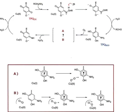
The active site of amine oxidase consists of a copper center and a topaquinone cofactor (TPQ) (Figure 1.1). The copper center is ligated by three histidines and two water molecules in a square pyramidal geometry, and in the resting state of the enzyme contains Cu(II). The nature of the organic cofactor in amine oxidase was not resolved until 1990. Studies by Klinman and coworkers conclusively identified this molecule as the quinone form of 2,4,6-trihydroxyphenylalanine, using labeling studies in combination with mass spectral and NMR techniques? The cofactor was subsequently characterized by resonance Raman as we1l8. The topaquinone is generated by post- translational modification of a tyrosine residue in the precursor protein. This process does not require the presence of chaperones or external cofactors. The precursor enzyme utilizes only molecular oxygen and the protein bound Cu in this reaction. The mechanism for TPQ biogenesis has received considerable attention but is still poorly understood. Of particular interest is the role of the copper center in the six-electron
oxidation of tyrosine, since Cu traditionally acts as a le- oxidant. Based on numerous investigations, a mechanism has been proposed for TPQ biogenesis (Figure 1.2l Tyrosine is believed to initially bind to the Cu site, which then activates the residue for reaction with Oz. This activated intermediate is believed to be either a one-electron oxidized CuI-TyrO complex or one in which charge is delocalized over a Cu"-Tyr- unit, rendering the tyrosine more susceptible to attack by Oz. Formation of this intermediate is believed to be the rate-determining step in the overall reaction. Support for the remaining steps was obtained from resonance Raman and computational studies.
However, none of the postulated intermediates has been directly observed. The degree to which the copper participates in the redox chemistry of the enzyme has yet to be ascertained. The copper site may play primarily a structural role or serve to stabilize the various intermediates. It is uncertain whether the metal itself undergoes a formal change in oxidation state in any of these steps. Copper is required for TPQ biogenesis, nonetheless. Substitution of other metal ions in precursor AO does not result in cofactor formation.
The reaction mechanism of AOs with amines has also been studied extensively and many of the intermediates have been spectroscopically characterizedz,IO. The catalytic cycle is shown in Figure 1.3. In the reductive half-reaction, topaquinone undergoes attack at the C-5 position by the primary amine to initially generate a substrate Schiff base intermediate. This Schiff base complex undergoes proton abstraction by a nearby Asp residue. Subsequent hydrolysis releases the product aldehyde and generates the 2e- reduced aminoquinol (TPQRED). Dioxygen reacts with the aminoquinol in the oxidative half-reaction, producing HzOz, NH3 and the oxidized form
- - - -- -- - - -
Current Opir>ion in Chemical Biology
Figure 1.2: Proposed mechanism for TPQ biogenesis.
Taken from: McGuirl, M.A., Dooley, D.M Curro Gp. Chern. Bio!.
1999,3, 138-144.
Amine Oxidase Mechanism
TPQRED
A)
Hoih _. Hoih
~NH2
0-'~NH2
OH 0/ OH
CD(II) Cu(l)
Figure 1.3: Top: Catalytic cycle of amine oxidase
Bottom: Proposed intermediates for the oxidative half-reaction;
(A) CuI-SQ intermediate, (B) alternative mechanism with 02 binding pocket.
of the cofactor (TPQox).
The mechanism for the oxidative half-reaction has been the subject of considerable debate as well, particularly in regard to the role of the copper center.
Dooley and coworkers obtained EPR evidence for a CuI-semiquinone (CuI-SQ) intermediate (Figure 1.3a), which indicated that Cu was actively involved in the regeneration of TPQoXll. According to their mechanism, the aminoqinol transfers one electron to the copper site to generate CUI-SQ. Dioxygen binds CU(I) and is reduced by one electron to generate a CUll-02·· intermediate, and the second electron is obtained from the semiquinone. Arguments against CUI involvement suggest that formation of the CuI-SQ species is off pathway. Evidence for prebinding of dioxygen to a hydrophobic pocket in the enzyme, not at the copper site, supports this interpretation12• Recent studies in the Klinman lab have demonstrated that the Co(JI) substituted enzyme is also catalytically active at a rate comparable to that of the native enzyme I 3 , confirming that copper is not necessary for enzyme activity. The metal may instead playa structural role or serve to stabilize a superoxide intermediate (Figure 1.3b).
Thesis Overview and Objectives
Our objective was to address some of the mechanistic issues through electrochemical and electron transfer studies of amine oxidase. Prior to our work, the potentials of the amine oxidase cofactors had not been measured. Studies using model complexes of topaquinone indicate that this potential should be ~ -200 mV vs. SCE14.
The protein environment, particularly the interaction with the copper site, and substrate binding are expected to alter the potential of the cofactor compared to the value in
solution. Accurate determination of these values could provide information concerning the mechanisms for the conversion of amines as well as the biogenesis of TPQ. A measurement of the CUIV1 potential and comparison with the aminoquinol potential would shed some light on the nature of the redox equilibrium between the cofactors.
Since a CuI-SQ species has been implicated as a key intermediate in the catalytic cycle, knowing the CUIV1 potential could provide evidence in favor of this mechanism. A Cu(l) intermediate has also been suggested in the initial activation of the Tyr in the precursor enzyme. The potential for oxidation of a metal-bound tyrosine can be estimated as ~ 500 m V vs. NHE. The potential of the copper complex must therefore be greater than this value to generate the tyrosine radical and, if it is in this range, the copper could easily oxidize the aminoquinol as well. It is of interest to compare the potentials of the copper site in the precursor enzyme to that of the fully processed AO.
We have pursued electron transfer studies on amine oxidase in the hope of detecting some of the intermediates throughout TPQ biogenesis. The oxidation of tyrosine is the rate-determining step in the overall mechanism. By performing this initial slow step using an external oxidant in combination with ultrafast spectroscopy, we could potentially detect some of the subsequent intermediates. Substitution of the Cu with Zn or Ni would then allow us to ascertain the role ofthe copper in the mechanism.
One of the major challenges in the investigations of amine oxidases is communicating with the active site, as it is deeply buried within the protein. To meet this challenge, we have employed "molecular wires" designed to access the active site of the enzyme by way of the ~ 20
A
substrate channel. This approach has been used previously in the Gray group to study the mechanisms of cytochrome P45015. Ru-terminated,substrate-linked hydrocarbon chains were used to deliver electrons to the heme site of P450. The intermediates generated by this method could then be detected using ultrafast spectroscopy. Crystal structure studies demonstrated that the substrate-tethered Ru complexes bind tightly in the P450 channel16.
We have designed oligomers, i.e., "wires", consisting ofphenyl-ethynyl bridging units to permit rapid electron transfer to and from the AO active site. These complexes are terminated on one end with amine substituents designed to mimic the natural substrates and bind within the enzyme channel. The nature of the second functional group was varied to permit studies of the enzyme by photophysical and photochemical techniques. Thiol terminated oligomers, which were synthesized for the purpose of electrochemical studies, are the subject of Chapter 5. Metal-terminated complexes for photophysics and photochemistry are treated in Chapter 6. The complexes are all highly fluorescent, such that binding of the oligomers to the enzyme was determined through variations in emission intensities and lifetimes.
The studies described above were carried out primarily on amine oxidase from Arthrobacter globiformis. The crystal structure of AGAO is known, and the substrate charmel is well defined. The oligomers were designed in accordance with the composition and structure of the AGAO channel. The synthesis of these complexes and the results of binding studies as well as electrochemical and photophysical investigations with AGAO are presented in the following chapters.
References
(1) Klinman, J. P.; Mu, D. Annu. Rev. Biochem. 1994,63,299-344.
(2) Halcrow, M.; Phillips, S.; Knowles, P. In Enzyme-catalyzed electron transfer and radical transfer; Holzenberg, Scrutton, Eds.; Plenum Publishers: New York, 2000; Vol.
35, pp 183-231.
(3) Parsons, M. R.; Convery, M. A.; Wilmot, C. M.; Yadav, K. D. S.; Blakeley, V.;
Comer, A. S.; Phillips, S. E. V.; McPherson, M. J.; Knowles, P. F. Structure 1995, 3,
1171-1184.
(4) Wilce, C. J.; Dooley, D. M.; Freeman, H.
c.;
Guss, J. M.; Matsunami, H.;McIntire, W. S.; Ruggiero, C. E.; Tanizawa, K.; Yamaguchi, H. Biochemistry 1997, 36, 16116-16133.
(5) Li, R. B.; Klinman, J. P.; Mathews, F. S. Structure 1998, 6,293-307.
(6) Vignevich, V.; Dooley, D. M.; Guss, J. M.; Harvey, I.; McGuirl, M. A.; Freeman, H. C. J. Mol. BioI. 1993,229,243-245.
(7) Janes, S. M.; Mu, D.; Wemmer, D.; Smith, A. J.; Kaur, S.; Maltby, D.;
Burlingame, A. L.; Klinman, J. P. Science 1990, 981-987.
(8) Brown, D. E.; McGuirl, M. A.; Dooley, D. M.; Janes, S. M.; Mu, D.; K1inman, J.
P. J. BioI. Chern. 1991,266,4049-4051.
(9) Dooley, D. M. J. Bioi. Inorg. Chern. 1999,4, 1-11.
(10) K1inman, J. P. Chern. Rev. 1996,96,2553.
(11) Dooley, D. M.; McGuirl, M. A.; Brown, D. E.; Turowski, P. N.; Mcntire, W. S.;
Knowles, P. F. Nature 1991, 349, 262-264.
(12) Su, Q.; Klinman, J. P. Biochemistry 1998, 37, 12513-12525.
(13) Mills, S. A.; Klinman, J. P. J. Am. Chern. Soc. 2000,122,9897-9904.
(14) Mure, M.; Klinman, J. P. J. Am. Chern. Soc. 1993,115,7117-7127.
(15) Wilker, J. J.; Dmochowski, I. J.; Dawson, J. H.; Winkler, J. R.; Gray, H. B.
Angew. Chern. Int. Ed. 1999, 38, 90-92.
(16) Dunn, A. R.; Dmochowski, I. J.; Bilwes, A. M.; Gray, H. B.; Crane, B. R. Proc.
Natl. Acad. Sci. 2001,98, 12420-12425.
Chapter 2
Electrochemistry of amine oxidases
Many difficulties associated with the electrochemistry of proteins are not encountered with small molecules. Proteins have comparatively slow rates of diffusion to the electrode surface, resulting in diminished currents. The surface characteristics of a bare metal electrode do not afford favorable interactions with the surface environment of a protein. Only a few orientations of a protein may permit electron transfer to the active site. Yet these may not be sites of preferred contact with the electrode surface.
Adsorption phenomena, leading to conformational changes and protein denaturation, tend to be a common problem as well. These obstacles are generally more prominent for enzymes, which usually are larger and often have deeply buried active sites. Several techniques have been developed to circumvent these problems1,2. These methods rely on materials that can provide favorable surfaces for protein-electrode interactions and can be manipulated to accommodate the specific characteristics of a protein. Pyrolitic graphite and modified gold electrodes are two materials that have been successfully employed in the electrochemical investigations of proteins.
Pyrolitic graphite has a crystalline structure much like that of an ideal graphite crystal. This material can be cleaved to generate electrodes of two distinct surface types.
Cleavage along the basal plane generates a hydrophobic surface, whereas the edge plane consists of a surface containing various C-O functionalities (such as carboxyl groups).
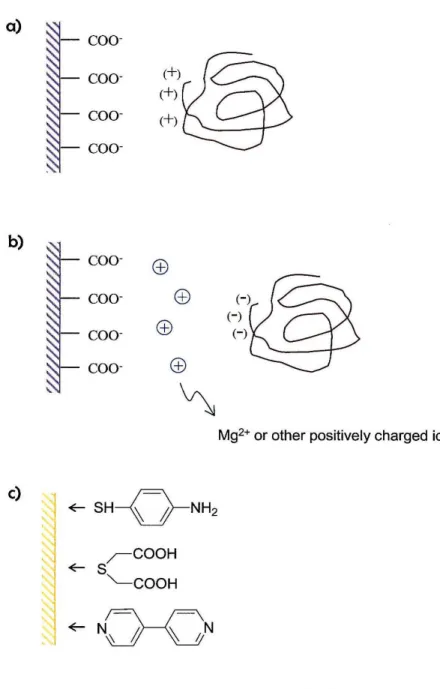
Edge-plane graphite (PGE) electrodes are most commonly used for protein studies. The charged carboxyl groups can participate in electrostatic interactions with residues on the exterior of a protein (Figure 2.la). If the protein to be studied contains negatively
a)
~
b)
c)
COO-
COO-
(+)
coo- (+) coo-
coo- coo- coo-
coo-
C±l
~
SH-o-NH2r -COOH
~S
"-COOH
~NJ eN
(-)
(-) (-)
Mg2+ or other positively charged ions
Figure 2.1: Electrostatic interactions between PGE electrodes and a) positively charged protein surfaces and b) negatively charged protein surfaces. The latter requires the addition of cations to the solution.
c) Commonly used compounds for the modification of gold electrodes.
charged residues at the surface, Mg2+, Cr3+ or other cations can be added to the solution.
This creates a double layer of charge at the PGE-protein interface (Figure 2.1 b).
Gold electrodes can be modified with promoter complexes to create a suitable surface for protein contace. These promoters consist of an X~Y motif. One end of the complex contains the functional group X, which binds to the gold electrode. Thiols, phosphines and pyridyl containing compounds are commonly used with sulfur binding most strongly. The opposing end contains a functional group that interacts with the protein (Y). This group can be varied depending on the properties of the protein to be studied. For example, amino groups can be used for negatively charged proteins, whereas carboxyl containing promoters will favor positively charged proteins. If the active site surroundings are hydrophobic, then the promoter can be designed accordingly. The nature of the linker (~) is variable, but must be oriented in a direction away from the electrode surface.
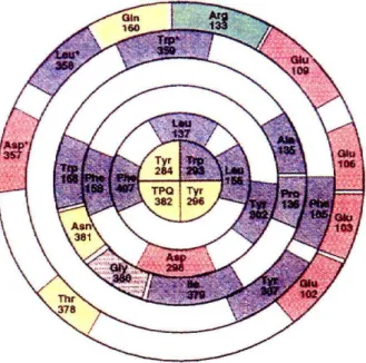
We have used both PGE and modified gold electrodes for electrochemical studies on amine oxidases. We have investigated the redox properties of AOs from Arthrobacter globiformis and bovine plasma. The proteins are negatively charged overall, with pI's of
~ 5 for both AGAO and BPAO. More importantly, the crystal structure of AGAO shows a high concentration of negatively charged residues at the surface of the channel leading to the active site (Figure 2.2). Electrostatic interactions between these residues and protonated amines are thought to be the mechanism by which AO substrates locate this channel and access the active site. The amine-substrates are deprotonated at the channel entrance and are guided to the cofactor site through hydrophobic interactions with residues in the interior of the cavity. Since this channel is the pathway to the active site,
Figure 2.2: Residues comprising the channel of AGAO from the surface to the active site. Residues are color coded. Blue:
hydrophobic, Red: acidic, Green: basic, I., : polar uncharged, Gray: glycines
it was expected to provide the best route for electron transfer to the site as well. Proper orientation of this region at the electrode surface is thus crucial to obtaining electrochemical signals from AO cofactors. The electrostatic charge at the channel entrance was considered in the choice of electrodes and modifiers. In studies with edge- plane graphite electrodes, Mg2
+ ions were added to the protein solution. This creates a cationic layer at the electrode surface, which attracts the negative patch that is associated with the AO channel. For studies using gold electrodes, several amine-terminated modifiers were chosen that could interact with AO in a similar fashion as the substrates.
A pyridyl complex (pyridine 4-aldehyde thiosemicarbazone: PATS-4) and two peptide modifiers (KCTCCA and (KC)2) were used. These complexes bind to the gold surface via sulfur atoms.
Materials and methods
Protein samples: Samples of purified protein were obtained from D. M. Dooley and his group at Montana State University; the general methods for protein purification are described in Appendix A. The concentrations of either AGAO or BP AO used in the electrochemical experiments, ranged from 300 to 500 /lM in 0.1 M potassium phosphate buffer (KPj), pH 7. Protein concentrations were determined as described in Appendix A.
Modifiers: Pyridine 4-aldehydethiosemicarbazone (PATS-4) and the KCTCCA peptide modifier were purchased from Sigma-Aldrich. The (LySCyS)2 peptide was synthesized at the Peptide Synthesis Facility at Caltech. Purity was determined by HPLC and mass
spectral analysis.
A (KPi, pH 7) 0.1 M ionic strength solution was employed in all experiments. When Mg2+ was required, a solution of MgCh (lM: 15-30 ilL) was added to the samples.
Electrochemistry
Apparatus and measurements: Cyclic voltammetry (CV) and Osteryoung square-wave voltammetry (OSWV) were performed on a Bioanalytical Systems (BAS) CV50-W electrochemical analyzer. The electrochemical cell consisted of a small volume (200-300 ilL), three-electrode, two-compartment glass cell. A Standard Calomel Electrode (SCE) served as the reference electrode and Pt wire was the auxiliary electrode. The reference compartment was separated from the working solution by a modified Luggin capillary. All solutions were deoxygenated by flowing Ar over the samples for at least 30 min. In cases where Mg2+ ions were used, a small volume (~
15-30 ilL) of a 1M MgCh solution in water was added to the samples.
Electrode preparations
Edge-plane grapite electrodes: Edge-plane graphite electrodes (PGE) were prepared by encasing a 5 mm diameter cylinder of PGE into a glass tube, sealed with heat-shrink tubing. Electrical contact to the graphite surface was made with a copper wire in the glass tube, and a small amount of mercury. Electrodes were polished with 0.3 Ilm alumina, sonicated for at least 20 min and briefly dried with a heat gun immediately before use.
Modified Au electrodes: Au electrodes were polished with 0.05 !lID alumina and sonicated for ~ 20 min. Electrodes were etched in 1 M sulfuric acid by cycling from -300 mV to 1.5 V (vs. Ag/AgCI). They were subsequently modified with thiol compounds as follows: For PATS-4 modification, electrodes were sonicated in a KPi solution
(containing - 2 mg of the compound) for several minutes and stored in the solution until further use. Electrodes modified with the KCTCCA and (KC)2 peptides were prepared according to procedures described by Hill et al. 4. Au electrodes were placed in a 2 mM solution of the peptide in KPi, and electrochemically modified by cycling the electrode in the solution at reducing potentials (0-800 m V vs. SCE) for - 10 cycles. Electrodes were stored in the peptide solutions until further use.
Results
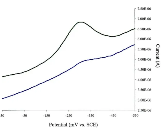
The electrochemistry of AGAO and BP AO was investigated with pyrolitic edge- plane graphite electrodes. Cyclic voltammetry gave no discernible signal. However, redox waves were observed for samples of AGAO by square wave voltammetry. A peak was observed at -0.31 V vs. SCE, which was small in amplitude and poorly defined.
Addition of Mg2+ to the solution enhanced this wave dramatically (Figure 2.3). This was an indication that amine oxidase prefers interactions with positively charged groups. An analogous redox couple was not observed for BP AO, however, even upon addition of Mg ions to the solution.
The redox chemistry of the amme oxidases was also investigated on thiol modified gold electrodes. Electrochemistry usmg both P ATS-4 and the KCTCCA peptide as promoters was unsuccessful. For each enzyme a redox couple was observed with (KC)2 peptide modified electrodes by square wave voltammetry. A broad peak centered around -340 m V (-365 m V) vs. SCE was seen upon reduction of AGAO (BP AO) (Figure 2.4, 2.5), similar to the results with PGE electrodes. Interestingly,
7.50E-06 7.00E-06 6.50E-06 6.00E-06 5.50E-06 n
"
i:l (I):::s 5.00E-06 ...
~
4.50E-06 ~
4.00E-06 3.50E-06 3.00E-06 2.50E-06
50 -50 -150 -250 -350 -450 -550
Potential (m V vs. SCE)
Figure 2_3: Square wave voltammograms of AGAO on PGE electrodes:
(-) bare PGE, no Mg2+; (-) 100
J..LM
Mg2+.[AGAO]
=
500J..LM,
0.1 M KPi , pH 7.E1I2 = -310 mV vs. SCE.
Sweep amplitude: 25 mY; sweep frequency: 5 Hz; potential step: 2 mY.
3.00E-07
2.50E-07
2.00E-07 n
=
::i(1) g
1.50E-07
---
~I.00E-07
---,---~---_,_---,___---~--~---_+ 5.00E-08
50 -50 -150 -250 -350 -450 -550
Potential (mV vs. SCE)
Figure 2.4: Square wave voltammogram of AGAO on (KC)2 modified gold electrode: [AGAO] = 500 J.lM, 0.1 M KPi , pH 7.
ElI2
=
-340 mV vs. SCE.Sweep amplitude: 25 mY; sweep frequency: 5 Hz; potential step: 2 mY.
2.7SE-07
2.2SE-07
n ~
1.7SE-07 ~
1.2SE-07
~-~-~-~--~-~-~--~-~-~--l-7.50E-08
o -so -100 -ISO -200 -2S0 -300 -3S0 -400 -4S0 -SOO
Potential (m V vs. SCE)
Figure 2.5: Square wave voltammogram of BP AO on (KC)2 modified gold electrode: [BPAO] = 500
J..LM,
0.1 M KPi, pH 7.200J..LM
Mg2+ added to solutionEI/2 = -365 mV vs. SCE.
Sweep amplitude: 15 mY; sweep frequency: 5 Hz; potential step: 2 mY.
addition of Mg2+ ions to the BP AO solution improved the signal. In contrast, addition of Mg2+ to AGAO samples gave poor results.
Discussion
The results of the electrochemical experiments on AGAO and BP AO demonstrate the importance of electrostatic interactions between the protein and the electrode surface. As described previously, the surface environment of the AGAO channel consists of several aniqnic aspartic and glutamic side chains. These residues are expected to interact favorably with positively charged functionalities, and this is supported by the results at edge-plane graphite electrodes. Studies using bare PGE, having carboxyl groups at the surface, gave poor results. Addition of Mg2+ ions to the solution led to great improvements in the electrochemistry. The lack of a redox couple for BP AO with PGE electrodes is surprising. This could be due to differences in the surface environment between the two AOs. Although AGAO has a clearly defined acidic patch at the mouth of the channel, this type of patch is not present in all amine oxidases.
Electrostatic interactions may playa large role in controlling the substrate specificity of AGAO. However, different interactions could be responsible for the selectivity of other AOs. The surface residues of BPAO may differ considerably from those of AGAO. As a crystal structure of BPAO is not available, the nature of the channel (if one exists) is not known.
In studies using Au electrodes, the (KC)2 peptide proved to be an effective modifier for both AOs. This is not surprising given that primary amines are the natural substrates for these enzymes. The attraction between the protein and the lysine groups at
the electrode surface is likely to be related to the same interactions that localize amine- substrates to the AO channel. Modification of electrodes with P ATS-4 or the KCTCCA peptide was unproductive for measurements of either protein, despite the fact that these complexes also contain amino groups. Possibly these complexes are too hydrophobic in nature, or are not oriented correctly at the electrode surface. Although the nature of their interaction with AO is unclear, it is safe to say that they do not provide a productive surface environment for protein electrochemistry.
The redox couples seen in the electrochemistry of AGAO and BP AO are likely due to the topaquinone cofactor. Comparison with reported potentials for topaquinone model complexes indicates that the values we have obtained are in the right range. A previous investigation of the electrochemistry of PSAO afforded a value of -300 m V vs.
SCE for a protein-bound topaquinone5, in close agreement with the redox potentials measured for AGAO and BP AO. The voltammograms using PGE and KC modified gold electrodes were unfortunately less than ideal. The redox waves are fairly broad, so information regarding the number of electrons involved or the electrode kinetics could not be extracted.
The poor electrochemical response is presumed to be due to the buried nature of the cofactors. This is a common problem in the direct electrochemistry of enzymes.
Unlike electron transfer proteins, the redox site of an enzyme often lies deep within the protein without a well-defined pathway for electron transfer. The crystal structure of AGAO indicates that the cofactor site lies - 20
A
beneath the protein surface. Thesubstrate channel provides a route to this site. However, the electrochemistry techniques described above, aid only in making contact with the top of the channel at the electrode
surface. Electrons must still be transferred through the solvent interior, and such reactions through water are fairly slow over this distance6• The low rates give poor redox kinetics at the electrode. In light of these factors, we developed a new method that provides direct access to the amine oxidase active site, as described in subsequent chapters.
References
(1) Guo, L.-H.; Hill, H. A. O. In Adv. 1norg. Chem., 1990; Vol. 36, pp 341-375.
(2) Hill, H. A. 0.; Hunt, N. I.; Academic Press, 1993; Vol. 227, pp 501-522.
(3) Frew, J. E.; Hill, H. A. O. Eur. J Biochem. 1988,172,261-269.
(4) Barker, P. D.; Di Gleria, K.; Hill, H. A. 0.; Lowe, V. J. Eur. J Biochem. 1990, 190,171-175.
(5) Sebela, M.; Studnickova, M.; Wimmerova, M. Bioelectrochemistry and Bioenergetics 1996, 41,173-179.
(6) Ponce, A.; Winkler, J. R.; Gray, H. B. JAm. Chem. Soc. 2000,122,8187-8191.
Chapter 3
Design and synthesis of molecular wires for amine oxidases
Our approach to the amme oxidase challenge was to synthesize a senes of
"molecular wires" for the purpose of electrochemical and mechanistic investigations.
These wires were designed to meet very specific requirements. Their structures must be such that they are able to bind tightly within the AO substrate channel, that is, the molecules must be compatible with the depth and volume constraints of the channel and interact favorably with surface and interior residues. For successful electrochemistry, the wires must provide an electron transfer pathway to the active site with rates superior to those through the solvent or the protein backbone. They must also contain a functionality that binds to gold surfaces. The wires can then be used to generate protein mono layers on electrodes.
Phenyl-ethynyl units were selected to comprise the bridge of the wires. The conjugation supplied by these units imparts strong electronic coupling and promotes rapid rates of electron transfer through the molecule. The electron transfer properties of phenyl-ethynyl oligomers on gold surfaces have been interpreted in terms of an experimental rate/distance decay factor
P
of -0.36A·
I I. ThisP
value indicates that electron tunneling rates on the order of 105 S·I will be observed for an oligomer 20A
in length. It follows that high rates can be expected for electron transfer through this type of bridge from the electrode surface to the active site of an amine oxidase.The phenyl-based oligomers are expected to fit inside the AGAO substrate channel, since phenethylamine is the natural substrate for this enzyme. Evidence for the ability of the protein to accommodate fairly large molecules is also given by the use of naphthalene-based inhibitors. The AGAO crystal structure also indicates that the path of
the channel is fairly straight such that flexibility is not required in the molecules. A thiophenol was chosen as the terminal group to bind to the electrode surface. Compared to alternatives such as pyridyl groups, thiols generate the strongest bonds to Au surfaces.
The opposite end of the oligomer serves as the channel-specific group. By definition it must consist of a functional group that can bind specifically to the active site, thereby promoting the insertion of the wire into the protein. It seemed logical to employ an amine group for this purpose, since amines are enzyme substrates. However, we would not like the substrate mimic to engage in reactions with the enzyme, at least not in the first electrochemical experiments. In these experiments, we only want the wire to be a mediator for electron transfer to the redox site, a requirement that suggests the use of tertiary amines. Inhibition studies with a series ofN-containing molecules (Table 3.1) provided information on their ability to bind to AGAO; this, in turn, allowed us to determine which group or groups could function as the channel-specific end of the wire.
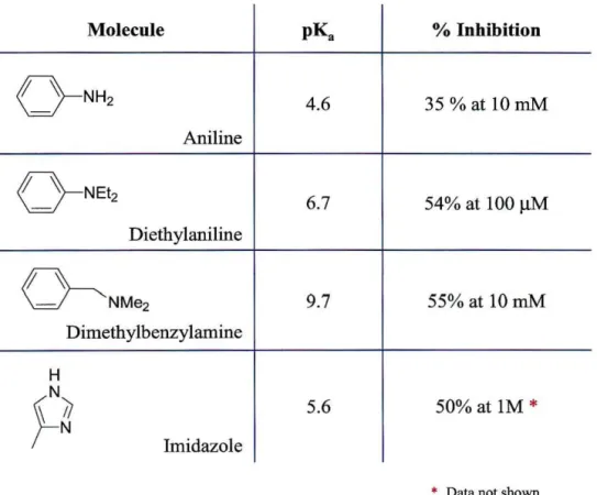
Most of these molecules resemble phenethylamine, and have pKa values in the range 4.5 - 10. With this range of pKa's, we can assess the importance of charge in AO interactions with potential inhibitor wires.
Inhibition studies:
Materials and methods
Inhibition studies were carried out on AGAO using phenethylamine as the substrate. The rate of O2 consumption by the enzyme during the reaction with phenethylamine was used as an assay for substrate inhibition of AGAO by the nitrogen- containing compounds in Table 3.1. The rate of 02 consumption in the presence ofthe
Molecule pK. % Inhibition
o - N H 2 4.6 35 %at IOmM
Aniline
<
} -NEt2 6.7 54% at 100IlM
Diethylaniline
~
- NMe2 9.7 55% at IOmMDimethylbenzylamine
H
~~
N Imidazole 5.6 50% at 1M*
• Data not shown
Table 3.1: Molecules used for inhibition studies of AGAO.
inhibitor compounds was compared to the rates measured in the presence of phenethylamine alone, and a percent inhibition was calculated from the results.
The concentration of AGAO in the assays was 0.17 mg!mL, and the phenethylamine concentration was 100 f.tM. Inhibitor concentrations ranged from 10 f.tM to 10 roM. Aqueous KPi, 0.1 M ionic strength, pH 7 containing 10% DMSO served as the buffer in all experiments. DMSO was used to solubilize the inhibitor compounds, many of which were otherwise insoluble in aqueous solution. All assays were carried out at 25°C in a sealed cuvette, while stirring.
O2 consumption assays were performed usmg a "Microelectrodes" oxygen electrode attached to a voltmeter. The electrode was calibrated by measurement of the voltage at 100%, 21 %, and 0% O2 . For measurements at 100% oxygen, pure O2 was bubbled through a solution ofKPi or KPi with 10% DMSO. Similarly, the 21 % O2 value was measured by bubbling with house air. For the 0% O2 measurement, dithionite was added to react with O2 present in solution. The rate of oxygen consumption by the enzyme was determined by measuring the change in potential due to the amount of O2 over a period of ~ 5-10 min. This was converted to a % O2 based on the above calibration. The % O2 was subsequently converted to a concentration of dioxygen ([02]) using the equation:
where
S
=
(a/22.414) «760-p)/760) (%02/100),S = solubility of O2 in moles! L = [02]
a = absorption coefficient of 02 at 25°C = 0.02831 P = the vapor pressure at 25°C = 23.756.
Results
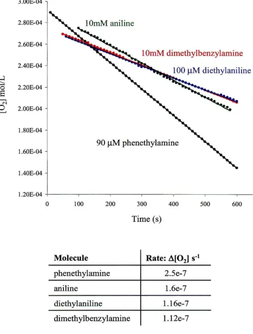
The results of the inhibition studies are shown in Figure 3.1. Aniline proved to be a poor inhibitor, showing only 35% inhibition at concentrations 100-fold greater than that of the substrate. Dimethylbenzylamine showed slightly better inhibition at this concentration. The addition of 10 mM dimethylbenzylamine resulted in a 55% decrease in enzyme turnover rates. However, the best inhibitor proved to be diethylaniline, which impeded enzyme reactivity by more than 50% at a concentration equivalent to that of the substrate. This suggests that the dissociation constant for diethylaniline is comparable to that of phenethylamine (K! = 9 ~M). This result is surprising given that the pKa of diethylaniline is only 6.6, whereas the pKa of dimethylbenzylamine is closer to 10. The charge on the amine has been cited as important for recognition of substrate by the AO channel. In addition, dimethylbenzylamine resembles the AGAO substrate more closely than diethylaniline. However, benzyl amine is also a poor substrate for AGAO (K!
=
36~M). So, it is apparent that factors other than charge play a role in AO substrate specificity. Based on the results of these studies, molecules were synthesized containing diethylaniline as the channel-specific terminal group.
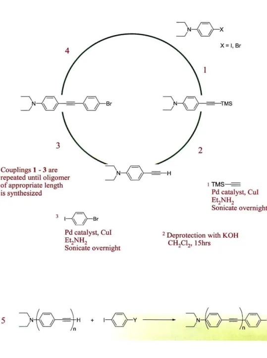
Synthesis of Molecular Wires
Wires were synthesized by means of Pd-catalyzed cross-coupling reactions (Figure 3.2). Beginning with p-iododiethylaniline, the alkyne unit is added by reaction with TMS-acetylene. Deprotection of the terminal alkyne, followed by coupling with p-
iodobromobenzene, affords the bromo-terminated complex. Additional phenyl-alkyne units are added in this manner until an oligomer of the desired length is obtained. The
3.00E-04
2.80E-04
2.60E-04
10mM dimethylbenzylamine
2.40E-04 ...:l
---
'0 2.20E-04
8
~ N
0 ~ 2.00E-04
1.80E-04
90 /-lM phenethylamine
1.60E-04
1.40E-04
1.20E-04
0 100 200 300 400 500 600
Time (s)
Molecule
phenethylamine 2.5e-7
aniline 1.6e-7
diethylaniline 1.16e-7
dimethylbenzylamine 1.12e-7
Figure 3.1: Results of inhbition studies with AGAO using phenethylamine as the substrate. Buffer: aqueous 0.1 M KPj , pH7 containing 10% DMSO.
4
J--Q-==-O---sr
3
4 Couplings 1 -3 are repeated until oligomer of appropriate length is synthesized
5
3 1- o -6r
Pd catalyst, CuI EIzNH2
Sonicate overnight
I
X= I, Sr
lTMS
=
Pd catal yst, CuI EIzNH2
Sonicate overnight
2 Deprotection with KOH CH2CI2, 15hrs
Figure 3.2: Methodology for synthesis of wires by Pd cross-coupling reactions.
The reporter group (Y) is attached to the oligomer end, opposite the channel- specific group in the final step (5).
thiol group is appended to the oligomer in the final coupling of the alkyne terminated oligomer with iodothiophenol. A wire consisting of three phenyl units affords a length of - 24
A.
This length is ideal given the depth of the AGAO channel, and allows the component opposite the amine functionality to protrude from the cavity. This is crucial for the electrochemical wire, since the thiol group must eventually be attached to an electrode surface.The methodology for synthesis of the complexes permits incorporation of an array of functionalities in addition to the thiophenol (Figure 3.3). These "reporter"
groups can be employed for the study of amine oxidases using a variety of techniques. A benzylamine terminated complex was synthesized for fluorescence binding studies. This compound served as the starting material for synthesis of metal terminated wires for electron transfer experiments. Synthetic and characterization procedures are described in full detail in Appendix B.
Wire -Length
22A
22A
29A
3
Figure 3.3: Wires synthesized for communication with the active site of amine oxidases.
References
(1) Creager, S.; Yu, C. J.; Bamdad, C.; O'Connor, S.; MacLean, T.; Lam, E.; Chong, Y.; Olsen, G. T.; Luo, J.; Gozin, M.; Kayyem, J. F. J. Am. Chern. Soc. 1999,121,1059- 1064.
Chapter 4
Binding of molecular wires to AGAO
The inhibiton experiments detailed in Chapter 3 suggest that molecular wires with tertiary amine termini likely will bind to the substrate channel of AGAO. The next step is to verify that oligomers with this functionality actually reside in the AGAO channel and to acquire information regarding the nature of their association with the enzyme. Accordingly, AGAO-inhibition and wire-fluorescence experiments studies were performed with solutions containing both enzyme and wire.
The absorption spectrum of AGAO exhibits a band at 480 nm due to the topaquinone cofactor. This enables fluorescence energy transfer studies with AO, in which the TPQ acts as a quencher for fluorescent probes that emit in this region. The benzylamine terminated complex, DEA-I-BzA (Compound 2, Figure 3.3), was synthesized for the purposes of these studies. Initially, this complex was designed for functionalization with the fluorophore 5-((((2-iodoacetyl)amino)ethyl)amino)napthalene-
I-sulfonic acid (1,5-IAEDANS). Fluorescence due to the dansyl group occurs at 490 nm and thus has significant overlap with TPQ absorption. However, attachment of the fluorophore to the benzyl amine oligomer seemed to prohibit protein binding (data not shown). This was presumed to be due to the flexibility of the dansylated molecule, which allowed "self-stacking" of the napthyl group and the phenyl rings of the oligomer.
The complexes are not very soluble in aqueous solution. Aggregation phenomenon has been observed during studies ofthe phenyl-ethynyl oligomers and is not unlikely for the dansylated compound. Self-stacking would result in a conformation that would render the complex too bulky for insertion into the protein. The phenyl-ethynyl complexes are highly fluorescent even without the addition of other fluorophores. The benzylamine
terminated wire has an emission maximum at ~ 410 nm in aqueous solution (Figure 4.1).
Topaquinone can serve as a quencher of the fluorescence due to DEA-I-BzA as well.
Since the fluorescence energy transfer rate is dependent on the distance between the donor and acceptor ( k u (/r)6 ), we expected to obtain information regarding the proximity of the wire to the TPQ. Fluorescence quenching could thus serve as an indicator ofthe extent of binding within the AGAO channel.
Materials and Methods
Time-resolved fluorescence measurements were performed usmg the third harmonic of a mode-locked Nd:YAG laser
O""x
= 355 nm) for excitation, and a picosecond streak camera (Hamamatsu C5680) for detection. Fluorescence was selected with a long-pass 400 nm cutoff filter. Steady state fluorescence spectra were recorded on a Hitachi F-4500 spectrofluorimeter (I""x = 320 nm); absorption spectra on a Hewlett- Packard 8452 diode array spectrophotometer. Steady state spectra were corrected for the fluorescence due to AGAO upon excitation at 320 nm.Substrate inhibition experiments were performed by measurmg the 02 consumption rate by the enzyme as described in Chapter 3, using 100 11M phenethylamine as the substrate. Concentrations of DEA-I-BzA were 50 11M and 100 11M.
Stock solutions of DEA-I-BzA were prepared in DMSO. Samples containing DEA-I-BzA were prepared in aqueous 0.1 M KPi, pH 7 containing 1% DMSO. Time- resolved fluorescence measurements were made in D20 buffer solutions, to cut down on
Raman scattering from H20.
0.6
0.5
0.4
~
0.3 0.20.1
o
-0.1
250 300 350 400 450 500 550 600
Wavelength (nm)
280
"'ex =
320 run240
Amax =
410 run200
.£ '" <= <1) 160
<=
~
120
80
40
400 450 500 550 600
Wavelength (nm)
Figure 4.1: Absorption spectrum (top) and emission spectrum (bottom) spectra ofDEA-I-BzA in aqueous 0.1 M KPi , 10% DMSO solution.
Results and Discussion
The effect of protein binding on the emission decay rate of DEA-l-BzA was investigated by time-resolved fluorescence spectroscopy. The results are shown in Figure 4.2. The free wire decays in less than 5 ns. Contrary to expectations, the apparent lifetime of emission is not altered in the presence of protein. However, a decrease in the fluorescence intensity is observed. This was the first indication that the complex might be bound to the protein. The intensity change suggests that the fluorescence seen in the presence of AGAO is due to free DEA-l-BzA in solution, whereas fluorescence of the bound wire is completely quenched. This requires that the emissive decay rate due to bound DEA-I-BzA be on the order of5 x 1010•
Fluorescence quenching by AGAO was further investigated by steady state fluorescence experiments (Figure 4.3). The observed intensity of DEA-l-BzA fluorescence correlates with the amount of AGAO present. Addition of one equivalent (two active site equivalents) of AGAO to a 20 /lM solution of DEA-l-BzA results in a 75% decrease in fluorescence intensity compared to the wire only solution. Addition of the second protein equivalent completely quenches the fluorescence. These results provide further evidence for binding of the oligomer to the enzyme.
The fluorescence results do not provide unequivocal proof of binding of DE A-l- BzA within the protein channel. However, given the degree of fluorescence quenching in the presence of AGAO, the complex is presumed to be in close proximity to the TPQ cofactor. Association of the wire with the protein surface should not lead to a perturbation of the emission to this extent. As the rates for the "bound wire" were too high to allow an estimate for the lifetime, kinetics data for DEA-l-BzA binding could
1800
1600
1400
1200 DEA-I-BzA only, 20 11M
.-21000
en 20 J..lM DEA-I-BzA only + 201lM AGAO
..s §
800600 400
200
1.15 1.65 2.15 2.65 3.15 3.65 4.15 4.65
Time (ns)
Figure 4.2: Time-resolved fluorescence quenching of DEA-I-BzA by AGAO, in deuterated aqueous 0.1 M, KPj , pH 7 solution.
"-ex
= 355 nm; 400 nm cutoff filter.400
350 DEA-I-BzA + 2eq. AGAO
300
» 250
.~
~ I!l 200
~ DEA-I-BzA + leq. AGAO
150
100
AGAOonly
50
0
330 350 370 390 410 430 450 470 490
Wavelength (run)
300
250
200
» 150 .~ en
~ s:: 100
~
50
0
-50
3 0 390 410 430 450 470 490
-100 Wavelength (run)
Figure 4.3: Steady-state fluorescence quenching of DEA-I-BzA by AGAO. Top: Uncorrected spectra; Bottom: Spectra corrected for fluorescence due to the protein.
"'ex
= 320 nm. [DEA-I-BzA] = 20 ~ in aqueous 0.1 M KP;, pH 7 containing 1 % DMSO.not be obtained. Substrate inhibition studies of the benzylamine wire were carried out to probe the protein/wire interaction in greater detail. Inhibition of substrate turnover by the complex implies insertion of the complex into the channel, blocking the pathway for access of substrate to the active site. The catalytic rates for reaction of AGAO with phenethylamine were measured in the presence of DEA-I-BzA. A 65% and 75%
reduction in rates was observed in the presence of 50 11M and 100 !lM wire, respectively, compared to the rate for 100 11M phenethylamine alone (Figure 4.4). These results suggest that DEA-I-BzA binding is tighter than that of phenthylamine, and place the dissociation constant in the 10 11M range.
....l
;::"
0
a
~ N
2-
3.00E-04
2.90E-04
2.80E-04
2.70E-04 + lOO /-lM DEA-I-BzA
2.60E-04
2.50E-04
2.40E-04
2.30E-04
2.20E-04 + 50 /-lM DEA-I-BzA
2.IOE-04 100 /-lM PEA only
2.00E-04
0 100 200 300 400 500 600 700
Time (s)
[DEA-I-BzA] Rate: A[02] S-l % inhibition
0 4.4e-7
50mM 1.5e-7 65%
100mM 9.6e-8 78%
Figure 4.4: Results of inhibition studies of AGAO with DEA-I-BzA.
100
f1M
phenethylamine (PEA) was used as the substrate. Studies carried out at 25°C, in aqueous 0.1 M KPj containing 1 % DMSO.ChapterS
Photochemical investigations of Re(BzA-l-DEA) with AGAO
We have synthesized a Re-terminated oligomer (Re(BzA-I-DEA), compound 3, Figure 3.3), with the ultimate goal of performing electron transfer studies on amine oxidases. Since rapid oxidation or reduction of the active site could be triggered by the Re complex, the oligomer seemed suitable for study of the topaquinone biogenesis mechanism. The first, rate-limiting step in formation of the cofactor is activation of a Tyr residue by the Cu site. Slow rates for this step have, thus far, precluded detection of ensuing intermediates and the role of Cu in the reaction is also ill-defined. Cu may be required only for the initial activation of Tyr, rendering it more reactive towards O2• In this case, the Tyr residue could potentially be activated by an external oxidant and formation of TPQ would proceed even in the absence of Cu. Electron transfer studies with Re(BzA-I-DEA), in which Re serves as the one-electron oxidant in a Ni substituted precursor AO, could test this premise. The oxidation of tyrosine occurs at a potential of
~ I V vs. NHE. The ReIIli potential is estimated as ~ 2 V vs. NHE; thus a photooxidized Re complex could easily oxidize this residue. A related Re complex, Rei(phen)(COh(OH2), has been used to label a blue copper protein, P. aeruginosa azurin.
Oxidative flash quench experiments on Re-azurin have led to the generation of tyrosyl radicals!. Rapid electron tunneling rates through the Re-substituted oligomer would permit detection of intermediates formed subsequent to the initial oxidation of tyrosine in precursor AO.

![Figure 2.4: Square wave voltammogram of AGAO on (KC)2 modified gold electrode: [AGAO] = 500 J.lM, 0.1 M KP i , pH 7](https://thumb-ap.123doks.com/thumbv2/123dok/10406879.0/32.774.93.649.193.605/figure-square-wave-voltammogram-agao-modified-electrode-agao.webp)
![Figure 2.5: Square wave voltammogram of BP AO on (KC)2 modified gold electrode: [BPAO] = 500 J..LM, 0.1 M KP i, pH 7.200 J..LM Mg2+ added to solution](https://thumb-ap.123doks.com/thumbv2/123dok/10406879.0/33.774.94.648.156.635/figure-square-voltammogram-modified-electrode-bpao-added-solution.webp)