It is doubtful whether the arrangement shown in the antenna figure is entirely exact. The structure of the fifth leg is largely clear from the figure.
COPEPOD GENUS RIDGEWAYIA — WILSON 147
Cephalic segment, in dorsal view, rounded anteriorly and tapering sharply externally, so that the segment beyond the middle is almost as broad as the second segment; the length, in the midline, slightly greater than that of the other segments combined. Urosome (fig. 1) 4-segmented, the genital segment the longest; the fourth segment is very short, reduced to the width of the caudal rami, with which it is more or less fused medially, but clearly distinct externally.
COPEPOD GENUS RIDGEWAYIA — ^WILSON 149
Aesthetes present on most segments, stouter than setae and of uniform width throughout their length; those of the proximal segments (Fig. 16) more stout than those beyond the middle of the antenna; the longest on segment 10 (reaching to the middle of segment 15).
COPEPOD GENUS RIDGEWAYIA — WILSON 151
The proximal outer part is an unexpanded plate with nine setae, of which the distal six are greatly elongated. Between this group of setae and the basal attachment of the exopod, an extension (epipod?) bearing a single set. Exopod narrowed beyond its middle so as to form two stiff parts, the proximal part bearing five lateral setae; distal somewhat expanded and bears tripartite and three apical setae.
Maxilla (fig. 19) with six clearly developed lobes, two of which belong to the basal division, which is incompletely demarcated from the second. The longest hairs are those of the first lobe and the proximal ones of the second lobe; both naked. Third segment with three plumose setae, between it and the endopod an incompletely separated segment, neither from the endopod nor from the basipod.
COPEPOD GENUS RIDGEWAYIA — WILSON 153
154 PROCEEDINGS OF THE NATIONAL MUSEUM
COPEPOD GENUS RIDGEWAYIA — ^WILSON 155
Apical part of segment 2 is a modified, incompletely separated, rounded toothed flap on the inner margin opposite the placement of the condouter spine; beyond this lobe, the segment is slightly notched anteriorly (the possibility that this tip is at least partially movable is suggested by the different positions found in different dissections; the tip may or may not be interpreted as an incompletely separated third segment). The third segment consists of a shortened segmental part, clearly separated anteriorly from the second segment (Fig. 24), but incompletely separated posteromedially. The segmental part of the anterior surface is deeply incised in the middle with heavy marginal sclerotizations, and partly on its posterior side it forms a base for the attachment of a series of thin, apically and irregularly fragmented membranes (or one folded membrane) and ti*ee. more difficult, extremely flexible processes.
One process, with an irregularly serrated, flared tip, has a broadened base placed in a depression of the posteromedial portion of the segment (Fig. 25). The other process is deeply broadened at its base and attached anteriorly in the segment (Fig. 24), below its attachment. Right condopod elongated-narrow, reaching to the end of the exopod, the tip partly fissured; the front is basally set on with groups of superficial spines (fig. 26).
COPEPOD GENUS RIDGEWAYIA — WILSON 157
Posterior edges of the segments with a complete dorsal fringe, those of segments 1-2 indistinctly serrate, those of segment 3 deeply serrate. Caudal setae look exactly like those of gracilis, except that the two long middle setae are not connected at their base. Antennule longer, reaching to the end of the genital segment; clearly 26-segmented, the proportions of the segments and numerical arrangement are exactly the same as gracilis, except that the last four segments tend to greater elongation, and segment 24 is almost equal to 25 and 26; the long setae of segments and 22 relatively shorter; the superficial spinules of segments 13–22 arranged in single rows extending across the entire distal margin of the segment, with the size of the spinules varying from segment to segment.
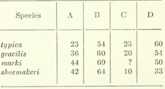
The maxillule is similar to the gracilis except that the first of the two laciniae just distal to the gnathobase has five instead of four setae. Endopod segment 2, first external seta placed below middle of segment, at point about 64 percent of total length; first terminal seta set slightly above middle, at point about 42 percent of total length; spinous process of outer margin short, only about 10 percent of total segment length.
COPEPOD GENUS RIDGEWAYIA — WILSON 159
Antennules extending beyond the metasorae; the left like that of the female, the right (fig. 29) with 24 free segments, four beyond the point of geniculation. The two outer spines of segment 2 are subequal in length, both extending beyond the end of the segment, and with toothed flanges. It is therefore impossible to say with absolute certainty that it does not represent the subadult stage of one or the other of the two species.
There is no indication of alteration of the right antennule, either in the middle or distal portions. Attention is drawn to this to emphasize that the spelling of the generic name is correct. There is also a need to determine the possible presence of the reduced last urosomal segments in both marki and canalis.
COPEPOD GENUS RIDGEWAYIA — WILSON 163 The specific difference noted in the two Tortugas species in the
COPEPOD GENUS RIDGEWAYIA — WILSON 163 The specific difference noted in the two Tortugas species in the. Although this is admittedly an obvious point, it seems appropriate to emphasize the reluctance to include the results of detailed critical examinations of the antennae in published records of any specimens of Ridgewayia or of related genera. Esterly (1911) gave no detail of the female or left male antennae in his account of marki.
Critically considered, it cannot be judged from the text of Gurney's description of the Suez Canal whether or not the segmentation given applies to both antennae or only to the right. If the statement "first antenna with 21 or 22 nodes" refers to both antennae, then segmentation within this group of species varies over a considerable range from 21 to 26 segments. This modification as it occurs in these species of Ridgewayia, though weak, is evidently a specialized joint, giving the distal part of the antennula a flexibility and freedom of movement which is not present in any other part of the appendage.
COPEPOD GENUS RIDGE WAYIA — WILSON 165 comparable to the variously developed geniculations found in many
It therefore appears that geniculation occurs at exactly the same point, although the number of free segments outside the joint is different. There seems no doubt that the modified segments of the middle section, counted as segments 13 and 14, represent two segments (Fig. 30). Comparison of the fixture of the unmodified annulus with that of the modified annulus may not indicate which segments are located in the fused distal region of the right atrial appendage.
Thus, on the basis of comparison of armature, the distal part of the segment preceding geniculation (visible segment 20) is comparable to segment 22 of the un-. These two species of Tortugas are therefore not only unusual among Calanoida in the segmentation of the antennules, but also in the position of the specialized hmge. It is not too surprising to find calanoid copepods with 26-segmented antennae, but the difference in the hmge.
168 PROCEEDINGS OF THE NATIONAL MUSEUM
COPEPOD GENUS RIDGEWAYIA — WILSON 169
170 PROCEEDINGS OF THE NATIONAL MUSEUM
COPEPOD GENUS RIDGEWAYIA — WILSON 171 They are included in table 3 as percentage figures determined in
172 CONTINUATION OF THE NATIONAL MUSEUM vol.los first and second segments are present and strongly developed, and are almost as long as the exopod itself. Its simplicity may be due to reduction or lack of some of the processes and fragmented membranes which complicate the structure in the other species, but it must certainly be correlated with it. Indeed, it closely resembles this portion of the exopod in the Tortugas specimens whenever viewed at relatively low power (fig. 35).
In the case of the latter species, however, the structure of the apical part of the left exopod may be somewhat different. Apparently, the first segments of the basipod are well developed and separated in the Madras specimen. This would include the fusion of the basal segments, the right exopod apex spine, and the loss of the inner parts of the exopod and endopod.
COPEPOD GENUS RIDGEWAYIA — WILSON 173 two species are known in which the setae are present on the right
COPEPOD GENUS RIDGEWAYIA - WILSON 173 two species are known in which the setae are present on the right. Antennula, female and male left, with 26 segments; leg 4, endopod segment 2 with 2 internal setae, segment 3 with 7 setae; leg 5 male, left exopod 2, outer seta longer than its segment, reaching almost the same point as the longest. Antennula, female and male left, with 25 segments; leg 4, endopod 2 with 1 inner seta, segment 3 with 6 setae; leg 5 male, left exopod 2, outer spine truncate, reaching only to elongated apical in middle.
Ridgewayia (as represented by the female of R. typica) was referred to the Calanidae by Thompson and Scott (1903) based on the similarities of the head appendages and legs 1–4. As is now known, the highly modified fifth legs of both sexes and the geniculate ant claw exclude the genus from the Calanidae, but Thompson and Scott are correct in implying that the unreduced, simply modified oral appendages and the first four pairs of legs are essentially be primitive in structure. The segmentation of the body (including the reduced anal segment), the caudalsetae and the large, nonfilamentous rostrum.
COPEPOD GENUS RIDGEWAYIA — ^WILSON 175
Gurney believed that this group represented the most primitive of the Calanoida and was closely related to another group to which only the Calanidae belonged. The more primitive antennal segmentation found in the new Tortugas species emphasizes rather than nullifies its relationship with this group. Too little is currently known about this fact, and Ridgewayia emerges as a highly unique genus exhibiting a combination of primitive features with others with unique or specialized modifications.
Similar taxonomic considerations may apply, with some qualification, to the Pseudocyclopidae, which have been placed by Gurney in an undefined group of 'uncertain positions'. Gurney's view of the Pseudocylopidae may have been somewhat incorrect, to the extent that he appears to be so. As has been noted (M.S. Wilson, 1946), Platycopia is unique among known calanoids and cannot be closely related to any known ones.
COPEPOD GENUS EIDGEWAYIA — WILSON 177 term Calanoida, which, though equivalent to Giesbrecht's term Gjmi-
The distribution of the species of Ridgewayia emphasizes the peculiar faunal relationship between the Indo-West Pacific region and the American tropical Atlantic (West Indian) region (Elanan, 1953; Hyman, 1955). Sewell (1948) listed many species of pelagic copepods in the community and also (1940, p.354) pointed out the similarity of the coastal copepod fauna of the Suez Canal with that of the coasts of India and Ceylon. The coastal copepod fauna of the West Indian region is scarcely known, but Willey (1930) showed that the Bermudan harpaccoid fauna is related to that of the Suez Canal.
Nicho Us (1944) has pointed out the striking similarity between Suez Canal and Bermuda species of Pseudocyclops (P. magnus Esterly, 1911, and P.latensGurney, 1927). The closely related Tortugas and Bermuda species of Ridgewayia highlight the relationship between these two areas of the West Indies region, and, through their demonstrated relationship to species from the Suez Canal and the Indian coast, are another example of coastal animals that zogeographically Connecting Indo-West Pacific and West Indian regions. .
COPEPOD GENUS RIDGEWAYIA — WILSON 179 Hyman, Libbie H