Seven Lubbockia Species
(Copepoda: Cyclopoida) from the Plankton of the Northeast Pacific,
with a Review of the Genus
GAYLE A. HERON and
DAVID M. DAMKAER
SMITHSONIAN CONTRIBUTIONS TO ZOOLOGY • NUMBER 267
Emphasis upon publication as a means of "diffusing knowledge" was expressed by the first Secretary of the Smithsonian. In his formal plan for the Institution, Joseph Henry outlined a program that included the following statement: "It is proposed to publish a series of reports, giving an account of the new discoveries in science, and of the changes made from year to year in all branches of knowledge." This theme of basic research has been adhered to through the years by thousands of titles issued in series publications under the Smithsonian imprint, commencing with Smithsonian Contributions to Knowledge in 1848 and continuing with the following active series:
Smithsonian Contributions to Anthropology Smithsonian Contributions to Astrophysics
Smithsonian Contributions to Botany Smithsonian Contributions to the Earth Sciences Smithsonian Contributions to the Marine Sciences
Smithsonian Contributions to Paleobiology Smithsonian Contributions to Zoology Smithsonian Studies in Air and Space Smithsonian Studies in History and Technology
In these series, the Institution publishes small papers and full-scale monographs that report the research and collections of its various museums and bureaux or of professional colleagues in the world cf science and scholarship. The publications are distributed by mailing lists to libraries, universities, and similar institutions throughout the world.
Papers or monographs submitted for series publication are received by the Smithsonian Institution Press, subject to its own review for format and style, only through departments of the various Smithsonian museums or bureaux, where the manuscripts are given substantive review. Press requirements for manuscript and art preparation are outlined on the inside back cover.
S. Dillon Ripley Secretary
Smithsonian Institution
Seven Lubbockia Species
(Gopepoda: Cyclopoida) from the Plankton of the Northeast Pacific,
with a Review of the Genus
Gayle A. Heron and David M. Damkaer
SMITHSONIAN INSTITUTION PRESS City of Washington
1978
Heron, Gayle A., and David M. Damkaer. Seven Lubbockia Species (Copepoda:
Cyclopoida) from the Plankton of the Northeast Pacific, with a Review of the Genus. Smithsonian Contributions to Zoology, number 267, 36 pages, 22 figures, 2 tables, 1978.—Among the marine plankton cyclopoids, the oncaeids represent a great number and wide variety of relatively little studied species. The family Oncaeidae is defined in this report, and a key is given for its five genera. Of these, the genus Lubbockia is defined in detail, and a key (Figure 2) is given for its 11 species. Relationships within the genus, as well as to other oncaeids, are discussed.
The bulk of the report is based on seven Lubbockia species collected from the plankton of the northeast Pacific Ocean; most of these species are also widely distributed elsewhere. Descriptions, illustrations, and notes on distributions are provided for these seven species, four of which are new. Descriptive notes and illustrations are also given for two other Lubbockia species known from tropical and subtropical areas of the Pacific.
OFFICIAL PUBLICATION DATE is handstamped in a limited number of initial copies and is recorded in the Institution's annual report, Smithsonian Year. SERIES COVER DESIGN: The coral Montastrea cavernosa (Linnaeus).
Library of Congress Cataloging in Publication Data.
Heron, Gayle A.
Seven Lubbockia species (Copepoda: Cyclopoida) from the plankton of the northeast Pacific, with a review of the genus.
(Smithsonian contributions to zoology ; no. 267) Bibliography: p.
1. Lubbockia—Classification. 2. Oncaeidae—Classification. 3. Crustacea—Classification. 4. Crus- tacea—North Pacific Ocean. I. Damkaer, David M., joint author. II. Title. III. Series:
Smithsonian Institution. Smithsonian contributions to zoology ; no. 267.
QL1.S54 no. 267 [QL444.C73] 591'.08s [595'.34J 77-16470
Contents
Page
Introduction 1 Material and Methods 1 Acknowledgments 2
Family ONCAEIDAE Giesbrecht, 1892 3
Key to the Genera of the Family Oncaeidae 3 Genus Lubbockia Claus, 1863 3 Lubbockia carinata, new species 5 Lubbockia forcipula, new species 11 Lubbockia wilsonae Heron and Damkaer, 1969 11 Lubbockia aculeata Giesbrecht, 1891 15 Lubbockia squillimana Claus, 1863 18 Lubbockia minuta Wolfenden, 1905 19 Lubbockia glacialis Sars, 1900 23 Lubbockia species 23 Lubbockia flemingi, new species 25 Lubbockia petersoni, new species 29 Appendix: Tables 33 Literature Cited 35
Seven Lubbockia Species
(Copepoda: Gyclopoida) from the Plankton of the Northeast Pacific,
with a Review of the Genus
Gayle A. Heron and David M. Damkaer
Introduction
Among the Poecilostoma (Copepoda: Cyclo- poida), most attention has been given to the numer- ous families associated with benthic invertebrates (see, for example, Gooding, 1963; Humes and Stock, 1973). The marine plankton poecilostomes, except perhaps for the Corycaeidae, have by comparison been little studied. The "planktonic" poecilostomes (Corycaeidae, Sapphirinidae, and Oncaeidae) are possibly not truly planktonic, but may be associ- ated, at least at times, with larger plankton orga- nisms (Sars, 1918). Sapphirina species, especially, are known to associate as parasites and predators on salps (A. C. Heron, 1973). Oncaea species have been seen to graze on appendicularian houses (Alldredge, 1976). There is no doubt that Lubbockia species would be mechanically able to hold other organisms with their well developed maxillipeds, but their actual food sources are not definitely known. The broad specific differences in maxilliped construc- tion suggest that food sources vary among Lub- bockia species.
At present, 11 named species can be assigned to
Gayle A. Heron and David M. Damkaer, Pacific Marine Environmental Laboratory, Environmental Research Labora- tories, National Oceanic and Atmospheric Administration, Seattle, Washington 98105.
the genus Lubbockia and are included in our key.
Notes on a North Atlantic species, L. extenuata, were provided by G. Boxshall (personal communica- tion), but the published report (Boxshall, 1977) was not available before the present paper went to press. Neither L. extenuata nor L. brevis, originally described from off Ireland, were found in the northeast Pacific, and they are not described in this report. A description of both sexes of L. brevis, and also of L. glacialis, is included in a report of Arctic Oncaeidae to be published soon (Heron and English, in prep.). Lubbockia squillimdna and L.
aculeata, from tropical and subtropical areas, also were not found in our northeast Pacific samples, but descriptive notes are given here for these species as well as for the seven species (including four which are new) that are known for the northeast Pacific. Only the male of L. forcipula remains un- known.
Most Lubbockia species have a wide distribution, and of those species found in the northeast Pacific, we have also studied specimens from other areas: the Antarctic (L. carinata, L. flemingi, L. forcipula, and L. wilsonae; Tables 1, 2), the Atlantic (L. minuta;
Tables 1, 2), and the Arctic oceans (L. glacialis;
Heron and English, in prep.). Lubbockia petersoni has been found only in the northeast Pacific.
MATERIAL AND METHODS.—Sources of the sped-
mens examined during this study are listed in Table 1 and the species are itemized in Table 2.
Those specimens that were studied in detail are enumerated under "Material Studied" in each species presentation.
Type specimens or reference specimens of all species described in this report have been deposited in the National Museum of Natural History, Smith- sonian Institution, Washington, D. C, where they bear the catalog numbers of the former United States National Museum (USNM).
The samples from NOAA Surveyor were collected using a 60 cm diameter closing ringnet of 211 /tin mesh, hauled vertically through discrete layers. The Brown Bear and Thompson samples from Mr.
Willis K. Peterson were collected with a specially designed plankton net, mesh aperture 110 jan, which accompanied a deep water-bottle cast. Two Clarke-Bumpus nets were sewed together with the open cod end of the shorter one reduced to about 5 cm diameter and extended into the mouth of the other; a small collecting bucket was attached to the end of the outer net. A steel ring was sewed into the mouth of the common opening, and fastened by a short bridle to the hydrographic wire. Another short bridle extended from the bucket, and this bridle was fastened to the wire beneath the net.
Thus the sampler was held parallel to the hydro- graphic wire and it sampled only as it was re- trieved. The net was attached beneath the deepest water-bottle on a hydrographic cast (1800 m or more). The maximum depth of the net sample was estimated from the deepest thermometric depth.
The constricted cod end of the inner net and the relatively great length of the entire device limited the escape of plankton when an ascending haul was stopped to remove water bottles.
The other Thompson samples were collected us- ing a multiple net trawl described by Frost and McCrone (1974).
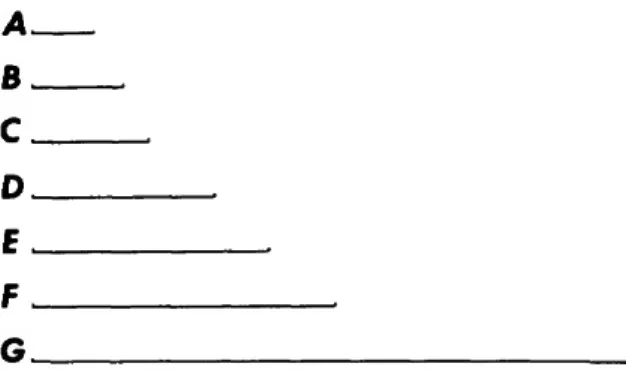
Figures were drawn with the aid of a Wild M20 drawing tube. In the figure legends, the capital letter following the explanation of each figure com- ponent refers to the particular scale to which the component was drawn. These scales are illustrated in Figure 1. Some Lubbockia species have a large number of spinules, sensilla, and gland vents (Fahrenbach, 1962), the latter associated with in- ternal tubules. Only such external features that are conspicious have been illustrated. Swimming legs
A
B C D EF G
FIGURE 1.—Scales used in drawing figure components (each scale = 0.1 mm).
are illustrated in anterior view.
Terminology used in describing the armature of swimming legs is as follows: setae are represented by arabic numerals, spines by roman numerals;
Si = inner border of segments, Se = outer border, St = terminal border. In describing the armature of antenna 1, arabic numerals used alone represent the number of setae. For use of "uropod" and "pediger,"
see Bowman (1971, 1976).
ACKNOWLEDGMENTS.—We are indebted to and thank the following persons for loans of specimens or samples: Dr. Thomas E. Bowman, National Museum of Natural History (NMNH), Smithsonian Institution, Washington, D. C, the depository of specimens bearing catalog numbers of the former United States National Museum (USNM); Dr.
Frank D. Ferrari, Smithsonian Oceanographic Sort- ing Center (SOSC), Washington, D. C ; and Dr.
Bruce W. Frost, Mr. Willis K. Peterson, and Mr.
David S. Thoreson, Department of Oceanography, University of Washington (UW), Seattle, Washing- ton.
We are grateful to Dr. Takasi Tokioka, Seto Marine Biological Laboratory, for providing the rare Marukawa (1927) publication, and also to Dr.
Shigeko Ooishi, Faculty of Fisheries, Mie Univer- sity, for translating the relevant text.
We also appreciate the information kindly given by Drs. T. Bjornberg, Universidade de Sao Paulo, Brasil, and G. Boxshall, British Museum (Natural History), concerning Lubbockia species studied by them.
This study was supported in part by the Alaska Outer Continental Shelf Environmental Assessment Program, sponsored by the National Oceanic and
Atmospheric Administration (NOAA) and the U.S.
Department of Interior, Bureau of Land Manage- ment (BLM). This publication is Contribution Number 950, Department of Oceanography, Uni- versity of Washington WB-10, Seattle, Washington.
Family ONCAEIDAE Giesbrecht, 1892 Prosome and urosome divisions well defined, the latter generally slender. Prosome, and urosome of female, 5-segmented; urosome of male 6-segmented.
Prosome elongate to elongate-oval, or cyclopiform.
Antenna 1 short, with reduced number of segments.
Antenna 2 3-segmented, subprehensile to prehensile.
Labrum medially incised. Mandible complex, with 3-5 subterminal elements. Maxilla 1 small, bilobed.
Maxilliped a well developed claw in both sexes. Legs 1-4, exopods and endopods essentially 3-segmented.
Leg 5 a single free segment or represented by 1-3 setae.
Sars revised the family Oncaeidae and questioned the inclusion of the genus Pachos (Claus, 1863). On the basis of a female and a male specimen in the genus Pachos that occurred in sample TA 45 (Table 1), it is apparent that the antenna 1 and the oral
appendages of this genus differ from any of the other genera Sars included in the family Oncaeidae:
Conaea, Lubbockia, Oncaea, and Pseudolubbockia.
As a result of having examined specimens from each of these genera and from the genus Epi- calymma, recently described as an addition to the Oncaeidae (G. A. Heron, 1977), we affirm Sars' supposition and exclude Pachos from the Oncaeidae.
Humes and Stock (1973) stated that Oncaeidae are rather closely related to the sabelliphilid and lichomolgid poecilostomes, but that several impor- tant features establish their distinctions. It is likely that Humes and Stock considered only the genus Oncaea when they included among these principal differing features a nonprehensile antenna 2 and the form of the mandible. In our opinion, antenna 2 of Oncaeidae is at least subprehensile, grading through Oncaea species toward a prehensile type, found also in Epicalymma and Conaea species, while in Pseudolubbockia and Lubbockia species, especially in females, it is definitely prehensile. The form of the mandible in Lubbockia species also approaches the lichomolgoid type. We believe, therefore, that Oncaeidae are even more closely related to the Lichomolgoidea than has previously been suggested.
Key to the Genera of the Family Oncaeidae
1. Legs 1-4 with inner coxal seta 2 Legs 1—4 without inner coxal seta 3 2. Mandible with narrow serrate tip; leg 5 a free segment armed with 4 elements
Pseudolubbockia Sars, 1909 Mandible terminating in a lash or lashed element; leg 5 absent (1 species) or a free segment armed with 2 elements Lubbockia Claus, 1863 3. Uropod with conspicuous expansion on dorsal surface, surrounding insertion of dorsal seta .... 4 Uropod without expansion on dorsal surface Oncaea Philippi, 1843 4. Legs 1-3 third exopod segment with II, III, III outer spines ....Epicalymma G. A. Heron, 1977 Legs 1-3 third exopod segment with III, II, II outer spines Conaea Giesbrecht, 1891
Genus Lubbockia Claus, 1863
Prosome elongate to elongate-oval; urosome slender. Antenna 1 7-segmented, with 1 or more incomplete sutures on female, the third segment with 2 spines; at least the 3 terminal segments al- ways fused on male. Antenna 2 with elongate third segment; female armature formula: 0, 1, 3 (sub- apical) f 3 (terminal) f 3 or 4 terminal claws.
Mandible blade with scalelike denticles on median edge and terminating in a lash; 1 inner and 2
dorsal elements. Maxilla 1 a bilobed appendage, lobes with 2, 3 or 3, 3 elements. Maxilla 2 with a short setule and 3 (1 bifurcate) or 4 ornamented elements. Oral appendages of male degenerate in some species, maxilliped usually dimorphic.
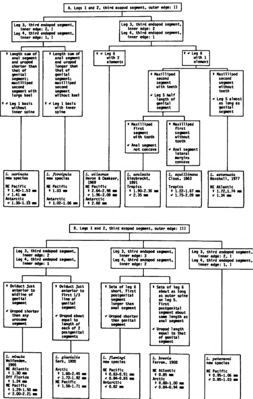
Variable armature characteristics, as noted under species descriptions, are: (1) inner edge of leg 1 basis, (2) outer edge of third exopod segment in legs 1 and 2, and (3) inner edge of third endopod segment in legs 3 and 4. Such characteristics appear to reflect natural groups in the Oncaea-Epicalym-
[ A . Legs 1 and 2, third exopod segment, outer edge: II |
Leg 3, third endopod segment.
Inner edge: 2, I Leg 4, third endopod segment.
Inner edge: 1, I
Leg 3, third endopod segment, Inner edge: 2
Leg 4, third endopod segment, Inner edge: 1
» Length sum of anal segment and uropod shorter than that of genital segment;
maxilliped second segment with large keel s " Leg 1 basis without Inner spine
« Length sum of anal segment and uropod longer than that of genital segment;
maxilliped second segment without keel
* Leg 1 basis with Inner spine
Leg 6 with 1 element
« Maxilliped second segment with teeth i Leg 5 half
length of genital segment
« Maxilliped second segment without teeth
•f Leg S almost as long as genital segment
s Maxilliped f i r s t segment with tooth
* Anal segment not concave
L. oarinata new species NE Pacific
« 1.40-1.53
* 1.41 mm Antarctic
" 1.30-1.33 i
I. foraipula new species NE Pacific
s 1.03 im Antarctic
« 1.02-1.06 i
L. wileonae Heron i Damkaer,
1969 NE Pacific
» 2.64-2.98 ran
<f 1.96-2.08 ran Antarctic
* 2.60 Ma
L. aculeata Giesbrecht,
1891 Tropics
1 1.90-2.36 i 2.35 mi
» Maxilliped first segment without tooth
* Anal segment lateral margins concave
L. aquillimana Claus, 1863 Tropics
8 1.22-1.67 mn
* 1.75-2.09 mn
L. extenuata Boxshail. 1977 NE Atlantic
• 1.72,1.74 im i 1.34 mm
B. Legs 1 and 2, third exopod segment, outer edge: III
Leg 3, third endopod segment.
Inner edge: 2
Leg 4, third endopod segment, Inner edge: 1
« Oviduct just anterior to midline of genital segment
" Uropod shorter than any urosome segment
L. minuta Wolfenden,
1905 NE Atlantic
» 1.30 mm Off Florida
» 1.24 m NE Pacific
« 1.29-1.50 i
* 2.00-2.21
» Oviduct just anterior to f i r s t 1/3 line of genital segment ' Uropod about
equal to length of each of 2 postgenital segments
L. glaaialie Sars, 1900 Arctic
' 1.60-2.45 m
" 1.72-1.92 ran NE Pacific
» 1.58-1.71 ma
Leg 3, t h i r d endopod segment, Inner edge: 3
Leg 4 , t h i r d endopod segment.
Inner edge: 2
Leg 3, third endopod segment, Inner edge: 2, I Leg 4, third endopod segment,
Inner edge: 1, I
» Seta of leg 6 short. First postgenital segment longer than anal segment
* Uropod shorter than genital segment
L. flemingi new species NE Pacific
« 0.83-0.91
* 0.84-0.85 Antarctic
* 0.82 mi
s Seta of leg 6 about as long as outer spine on leg 5.
First postgenital segment about same length as anal segment
* Uropod length equal to that of genital segment
L.
Fa NE s
brevie -ran, 1908
Atlantic 0.85 mi Arctic
s a
0.88-1.00 m 0.84-0.94 mm
L. petersoni new species NE Pacific
» 0.95-1.06
* 0.85-1.03
FIGURE 2.—Diagrammatic key to the species of the genus Lubbockia.
ma-Conaea complex but probably no such groups exist within Lubbockia species. The characteristics enumerated above are among those used to prepare a diagrammatic key to the species of Lubbockia (Figure 2). The generic pattern of armature is as follows, with a numeral representing a constant value and an asterisk indicating a variable count.
All species, except Lubbockia flemingi, with hya- line papilla partially covering vent on outer edge of leg 1 coxa. Lubbockia, as well as Oncaea, species usually have small or conspicuous conical projec- tions between 2 terminal spines on the endopods of legs 2 and 3, and sometimes leg 4. These terminal projections appear to support a vent, which is
Coxa Basis Endopod Exopod
Leg 1 2 3 4
Si 1 1 1 1
Si
• 0 0 0
Se 1 1 1 1
1 Si 1 1 1 1
2 Si
1 2 2 2
Si 4 3
•
• 3 St I II II II
Se I 1 I I
1 Se I I I I
Si 1 1 1 1 2
Se I I I I
Si 4 5 5 5
3 St I I I I
Se
•
• II II
present even when the projection is lacking. Often a vent is also on the inner distal corner of the basis of all swimming legs.
Leg 5 a free segment bearing 2 terminal elements, or leg 5 absent and represented by 1 lateral seta.
Lubbockia is an interesting and complex genus, far more variable than many other polytypic poe- cilostome genera. Specific differences include slight variations in body shape, but major differences in maxilliped and leg 5 structure, and armature and relative length of endopods and exopods of legs 1-4.
The individual Lubbockia species show various relationships to other oncaeid genera, depending on the morphological features compared. The relation- ship of Lubbockia to Pseudolubbockia is seen to be close when the comparison is based on L. carinata, L. jorcipula, and L. petersoni.
With considerable foresight, Claus (1863) named Lubbockia "for the deserving English investigator"
John Lubbock, afterward Sir John Lubbock, Lord Avebury (1834-1913). It is difficult to select a single area of endeavor to characterize Lubbock, for he became preeminent in many fields and was the author of a great number of scientific and popular books. His earliest published papers dealt with Copepoda (1853-1863), including specimens collected by Darwin on H.M.S. Beagle. Although much of Lubbock's efforts were in taxonomy, he felt strongly that these investigations were not ends in themselves, but only instruments for the understanding of animal behavior. Lubbock was the first to experiment on color vision in insects, and he did extensive research on social-insect be-
havior. By profession a banker, Lubbock instigated major alterations in English banking practices. He was a Member of Parliament, where he introduced far sighted legislation having a present day cast.
Among these proposals were: Open Spaces Act, Ancient Monuments Acts, and Wild Birds Pro- tection Act. Lubbock was the first to correctly at- tribute Stonehenge to the Bronze Age; his arch- eology texts, in which he coined the terms "paleo- lithic" and "neolithic," were standard for a gen- eration. Lubbock's father, Sir John Lubbock, was a physical astronomer, dealing especially with tidal theory; both men were vice-presidents of the Royal Society. Young Lubbock was a friend, neighbor, and finally pallbearer of Charles Darwin, who said that he relied on the opinions of three men only—
Hooker, Huxley, and Lubbock. Lubbock's name is also perpetuated in some genera of insects, and in some copepod species. There are several biogra- phies of Lubbock, including a recent one by Pum- phrey (1959).
Lubbockia carinata, new species
FIGURES 3-6
MATERIAL STUDIED (Tables 1, 2).—NE PACIFIC: 4 females, 1.40-1.53 mm, NP 4 et al. (including: holotype, 1.44 mm, NP 4, USNM 168487; 1 paratype, 1.41 mm, NP 12, USNM 168489); 1 male, allotype, 1.41 mm, NP 7, USNM 168488;
1 juvenile, stage IV, 1.16 mm, NP 32. ANTARCTIC OCEAN:
2 males, 1.30-1.33 mm, AA 41, 42.
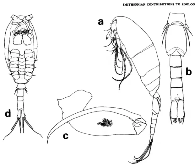
FEMALE.—Average length 1.47 mm. Prosome
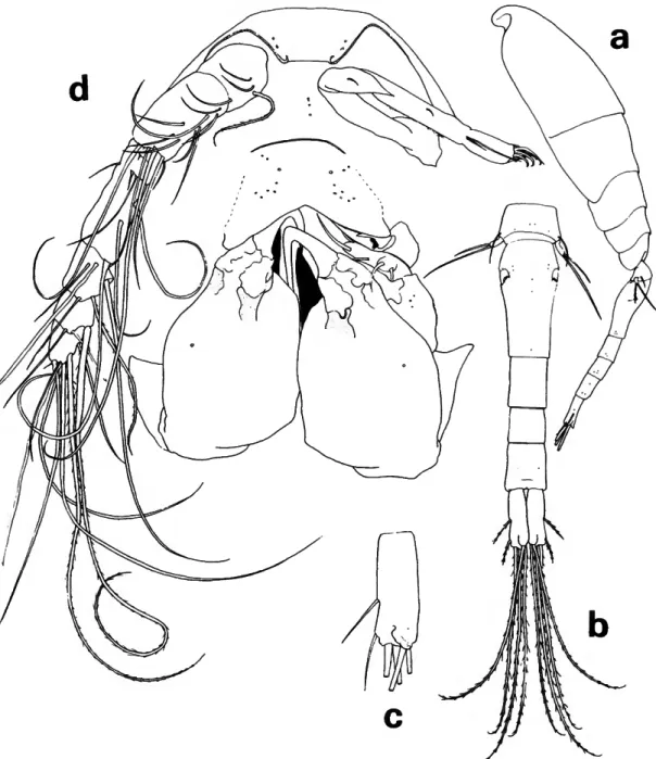
FIGURE 3.—Lubbockia carinata, new species, female: a, lateral (A); b, urosome, dorsal (C); c, uropod, dorsal {E); d, cephalosome, ventral (maxilliped keel darkened) (£).
FIGURE 4.—Lubbockia carinata, new species, female: a, labrum, ventral (F); b, mandible, right (G); c, maxilla 1, left (G); d, maxilla 2, right (G); e, maxilliped, right (E); /, leg 1 (E).
(Figure 3a) about 1.6 times length of urosome, with rounded posterior corners.
Pediger 5 (Figure 36) with faint transverse ridge dorsoposteriorly. Genital segment with areas of ex- ternal genital apparatus on anteriolateral third of segment, each area with 3 setules; genital segment and each postgenital segment with delicate spinules on posteroventral margin. Uropod approximately half the length of genital segment, with short spi- nules on ventral surface; dorsal, innermost, and outer long terminal setae nearly as long as inner long terminal seta; rounded laminal flap (Figure 3c) extending over base of dorsal seta.
Rostrum (Figure 3d) with thickened posteroven- tral margin, truncate as shown in ventral view.
Antenna 1 (Figure 3d) 7-segmented, with rudi- mentary articulation between segments 3-4 and 5-6. Armature formula: 4, 13 + 1 spinule, 1 + 2 spines, 4, 4 + 1 esthete, 2 + 1 esthete, 7 + 1 esthete.
Antenna 2 (Figure 3d) 3-segmented, with incom- plete suture at distal third of terminal segment.
Second segment with 1 seta. Third segment with inner setules and 3 subapical setae; armed termi- nally with 3 setae and 4 claws, all short.
Labrum (Figure 4a) with free margin divided into 2 deltoid posteroventral lobes; a number of conspicuous gland vents on outer surface and rows of setules inserted on under surface.
Mandible (Figure 46) with 2 outer elements orna- mented with rows of setules; mandible blade with row of graduated, scalelike denticles on the median edge; apical lash slender and long; concave edge of base with a hirsute element and rows of long setules supported by 2 riblike processes.
Maxilla 1 (Figure Ac) flat, bilobed; 3 barbed ele- ments on outer lobe and 1 barbed and 1 setose element on inner lobe.
Maxilla 2 (Figure 4d) 2-segmented. First segment with expanded base and outer row of setules. Sec- ond segment outer margin with a hyaline setule and setose, stout seta; terminally bearing 2 setulose elements; a constriction and sclerotization suggest- ing segmentation near the anterior base but poste- rior base fused; curved setulose element on inner margin.
Maxilliped (Figure 4e) apparently 4-segmented;
complicated sclerotized pattern in area of segment 3. Second segment with a remarkable anterior keel- like process, curving posteriorly beside an inner pocket which serves as a sheath for the terminal
claw; segment 2 with short outer spinules.
Leg 1 (Figure 4/) without inner spine on basis, third exopod segment with 2 short outer spines.
Leg 2 (Figure 5a) third exopod segment with 2 short outer spines. Leg 3 (Figure 56) inner edge of third endopod segment with 2 setae and 1 spine.
Leg 4 (Figure 5c) inner edge of third endopod seg- ment with 1 seta and 1 spine. Legs 2-4 endopods terminate with a small vented conical projection between spines.
Leg 5 (Figure 36) with short free segment; 2 terminal elements, the longer tapering in a way which suggests there is a fine serrate, hyaline flange;
length less than twice that of the outer. Seta on body near leg.
Leg 6 probably represented by 3 short, hyaline setules in area of external genital apparatus.
MALE.—Average length 1.35 mm. Prosome (Fig- ure 6a) about 1.7 times the length of urosome.
Urosome (Figure 66) with the 4 postgenital seg- ments bearing spinules on posteroventral margins.
Uropodal setae with relative lengths as in female.
Rostral area and mouthparts, except antenna 1 and maxilliped, as in female.
Antenna 1 with segments corresponding to termi- nal 3 of female, fused in male; armature similar to corresponding segments of female.
Maxilliped (Figure 6c) with segment 2 and claw elongate compared to female; 2 short segments proximal to claw.
Swimming legs and leg 5 resemble those of female.
Leg 6 (Figure 66) probably represented by poster- oventral pointed flap on genital segment.
STAGE IV.—Length 1.16 mm, with 4 segments and uropod in urosome; leg 5 small and segmented (Figure 6d). Maxilliped with keel-like process well developed.
ETYMOLOGY.—The specific name carinata, from the Latin carinatus (keeled) refers to the keel-like process on the maxilliped of the female.
REMARKS.—Lubbockia carinata superficially re- sembles L. forcipula, but differs from it by the rela- tively shorter uropods, distinctively keeled female maxilliped and by the lack of an inner spine on leg 1 basis. Lubbockia carinata, L. glacialis, and L. minutus are the only Lubbockia species without a spine on leg 1 basis.
Lubbockia carinata also resembles L. forcipula
FIGURE 5.—Lubbockia carinata, new species, female: a, leg 2 (E); b, leg 3 (E); c, leg 4 (E).
in having a seta on body near leg 5. Lubbockia petersoni has such a seta but lacks leg 5. These are the only known Lubbockia species with this seta, which is probably a remnant of leg 5 basis. A simi- larly placed seta is characteristic of the other genera of Oncaeidae: Pseudolubbockia, Oncaea, Epica- lymma, and Conaea.
Lubbockia carinata and L. forcipula have arma- ture of female antenna 1 resembling that of Pseudo-
lubbockia dilatata Sars, 1909, except for having 2 additional setae and 1 spinule on segment 2. These 3 Oncaeidae species also differ from all other Lub- bockia females by having an additional seta on seg- ments 3 and 5, and 7 setae and 1 esthete on the terminal segment instead of the usual 5 setae and 2 esthetes.
We have restudied Pseudolubbockia dilatata for comparison with Lubbockia species and have modi-
FIGURE 6.—Lubbockia carinata, new species, male: a, lateral (A); b, urosome, ventral (C); c, maxilliped, left (£). Stage IV: d, ventral (maxilliped keel darkened; antenna 1, except basal segment, and swimming leg rami omitted) (A).
fied our diagnosis concerning details of P. dilatata antenna 1. Antenna 1, 7-segmented, with an in- complete suture on inner surface between 5-6.
Armature formula: 4, 11, 1 + 2 spines, 4, 4 + 1 esthete, 2 + 1 esthete, 7 + 1 esthete. The esthetes are relatively short, narrow, and each is associated with a short seta, on proximal third of segment 5 and distal margins of segments 6 and 7. The short seta on the posterior margin of segment 5 is an error in figure 2, Heron and Damkaer, 1969.
The maxillipeds of several species of caligoid copepods have been described with a prominent second segment keel or similar process associated with the tip of the terminal claw, as in L. carinata:
Dysgamus atlanticus Steenstrup and Lutken, 1861 (pi. 4: fig. 8), Dysgamus pacificus Wilson, 1950 (pi.
6: fig. 55), and Lewis (1968, fig. 8d) for Dissonus heronensis Kabata, 1966. Lewis (pers. comm.) has stated that the legends for his figure %d,e were in- advertently transposed and that figure Sd is the maxilliped of female D. heronensis.
The long, resilient uropodal setae of L. carinata and L. forcipula resemble the uropodal setae in Conaea and Epicalymma species, but differ from those of Oncaea, Pseudolubbockia, and the other Lubbockia species.
DISTRIBUTION.—Northeast Pacific and Antarctic Ocean (Tables 1, 2).
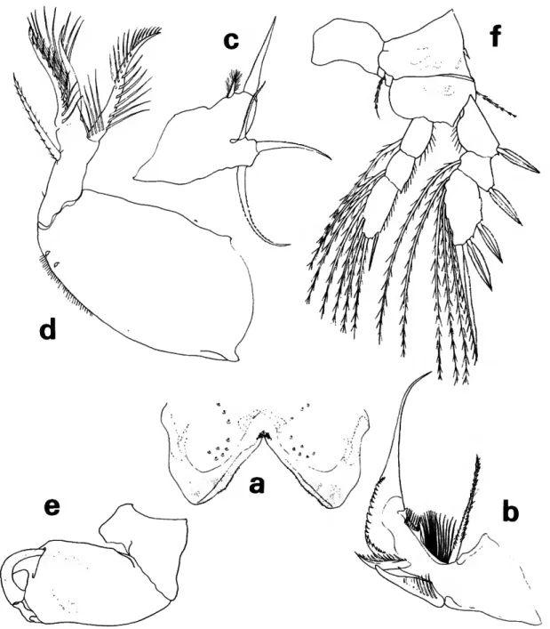
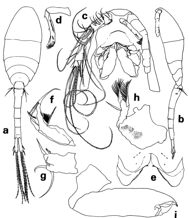
FIGURE 7.—Lubbockia forcipula, new species, female: a, dorsal (C); b, lateral (C); c, cephalosome, ventral (E); d, antenna 2, right (E); e, labrum, ventral (G); /, mandible, left (G); g, maxilla 1, right (G); h, maxilla 2, right (G); i, maxilliped, right (G).
Lubbockia forcipula, new species
FIGURES 7-8
MATERIAL STUDIED (Tables 1, 2).—NE PACIFIC: 1 female, holotype, 1.03 mm, NP 11, USNM 168492. ANTARCTIC OCEAN: 2 females, 1.08, 1.12 mm, A A 42.
FEMALE.—Length about 1.08 mm. Prosome (Fig- ure 7a,b) 1.2 times the length of urosome.
Pediger 5 with transverse sclerotized ridge dorso-
posteriorly. Genital segment with areas of external genital apparatus on anteriolateral third of seg- ment; each area with a setule. Genital segment and each postgenital segment with delicate minute spi- nules on posteroventral margin. Uropod about z/3
length of genital segment, posteriorly dilated over base of dorsal seta; dorsal, innermost, and outer long terminal setae nearly as long as inner long terminal seta.
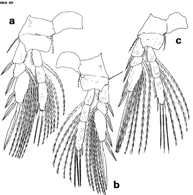
FIGURE 8.—Lubbockia forcipula, new species, female: a, leg 1 (E); b, leg 2 (E); c, leg 3 (E);
d, leg 4 (E).
Rostrum with thickened posteroventral margin slightly depressed and indented as shown in ventral view of cephalosome (Figure 7c).
Antenna 1 (Figure 7c) and antenna 2 (Figure lc,d) with armature similar in number to that of L. carinata.
Labrum (Figure le) with free margin divided into 2 posteroventral lobes, each with a subapical cluster of setules; complex pattern of layers of tis- sues with hyaline setules inserted on under surfaces.
Mandible (Figure 7/), maxilla 1 (Figure 7g), and maxilla 2 (Figure Ik) with armature similar in number to that of L. carinata.
Maxilliped (Figure 7t) 4-segmented, cheliform.
Second segment beaklike, projecting beyond and somewhat enclosing the tip of short terminal claw, the complex forming a short, strong pincer.
Legs 1-4 (Figure Sa-d) with armature similar in number to that of L. carinata except for the addi- tion of an inner spine on leg 1 basis.
Leg 5 (Figure la) similar in general form to that of L. carinata, including seta on body near leg.
Leg 6 probably represented by short, hyaline setule in area of external genital apparatus.
MALE.—Unknown.
ETYMOLOGY.—The specific name, from the Latin forcipula (nippers, tongs), refers to the appearance of the maxilliped in the female.
REMARKS.—The truncate shape of the L. for- cipula rostrum resembles L. carinata, but the de- pressed and indented posterior margin is similar to that of Pseudolubbockia dilatata.
Of the known species of Lubbockia, only L. for- cipula, L. carinata, and L. petersoni have an inner spine on legs 3 and 4 third endopod segment; L.
carinata and L. forcipula differ from L. petersoni by having 2 (rather than 3) outer spines on legs 1 and 2 third exopod segment. Lubbockia forcipula and L. carinata also differ from other Lubbockia species by having 13 setae (rather than 7) on antenna 1 second segment.
The unusual maxilliped of L. forcipula, having a short, hooded terminal claw, separates it from L.
carinata, which it otherwise resembles. The maxilli- ped of L. forcipula resembles that of Phyllotherus cornutus (Milne-Edwards, 1840), a caligoid copepod parasitic on sharks (see Cressey, 1967, fig. 130).
DISTRIBUTION.—Northeast Pacific and Antarctic Ocean (Tables 1, 2).
Lubbockia wilsonae Heron and Damkaer, 1969
FIGURE 9
Lubbockia aculeata.—Vervoon, 1951:151; 1957:148. [Not L.
aculeata Giesbrecht, 1891.]
Lubbockia wilsonae Heron and Damkaer, 1969:9-15, figs. 11- 19.
The original description of Lubbockia wilsonae was based on 2 females (2.70, 2.75 mm) from the Gulf of Alaska. Additional specimens of both sexes from the northeast Pacific have made it possible to describe the male and amend the original descrip- tion of the female.
MATERIAL STUDIED (Tables 1, 2)—NE PACIFIC; Prince William Sound, Alaska: 3 females, 2.80-2.98 mm, NP 1. Near the Canadian weatherstrip sta P: 6 females, 2.64-2.88 mm, NP 38, 39; 3 males, 1.96-2.08 mm, NP 38, USNM 168503, 168504; 8 juveniles, stage V, NP 38, 39; 2 juveniles, stage IV, both 1.48 mm, NP 39; 2 juveniles, stage III, 0.97, 1.06 mm, NP 7, 4 (frequency of sex and stage categories by depth for samples NP 38, 39 shown in accompanying tabulation).
ANTARCTIC OCEAN: 3 males, 2.42-2.44 mm, AA 42.
6 Aug 7 Aug
m 165-110 220-165 275-220 330-275 385-330 440-385
$ 1
1 1
$ 1
2 V
1 3 1
$
2
1 V
1 2
IV
1 1
FEMALE.—Rostrum (Figure 9a) rounded poste- riorly. Antenna 1 in all Lubbockia species appears to be fundamentally 7-segmented; segment 3 often with incomplete or no suture separating 4. Female Lubbockia specimens have been described as having from 4 or 5 to 7 segments in antenna 1; some su- tures are incomplete on inner surface, while articu- lation is usually more distinct on outer surface.
Lubbockia wilsonae female antenna 1 is 7-seg- mented (rather than 6) with segments 3-4 partially or completely fused. Armature should be corrected from original description to 4, 6 + 1 esthete + 1 spinule, 2 spines, 4, 3 + 1 esthete, 2 + 1 esthete, 5 + 2 esthetes (1 seta, about equal to combined length of segments 5-7, in place of 2 short setae shown on figure 14 of Heron and Damkaer, 1969).
Maxilla 1 (Figure 96) bilobed; inner lobe with 2 elements and short hyaline setules; 3 elements on larger lobe.
MALE.—Length about 2.01 mm (NE Pacific) to
FIGURE 9.—Lubbockia wihonae Heron and Damkaer, female: a, rostrum, ventral (E); b, maxilla 1, right (F). Male: c, lateral (A); d, dorsal (A); e, first 3 urosomal segments, ventral (D); /, rostrum, ventral (E); g, antenna 1 distal esthete bases x, y, and z (G); h, antenna 2, left (E);
i, maxilliped, right (E). Stage V, female: ;, urosome, ventral (A). Stage V, male: k, urosome, ventral (A). Stage III: /, ventral (antenna 1, except basal segment, and swimming leg rami omitted) (A).
2.43 mm (Antarctic). Body (Figure 9c,d) slender, with prosome to urosome ratio and relative lengths of urosomal segments similar to female. Prosome with conspicuous sensilla, gland vents, and minute spinules. Urosomal segments covered with minute spinules, somewhat larger on most ventral surfaces;
posteroventral margins with spinule patterns similar to those of female. Genital segment (Figure 9e) with conspicuous spinules on ventral surface. Relative lengths of uropodal setae as in female.
Rostrum (Figure 9/) more tapered postero- ventrally than in female.
Antenna 1 distinctly segmented only between segments 4 and 5 and outer surface of 1 and 2. Arm- ature similar in number to that of female except for additions of esthete on segment 2 and 2 esthetes on segment 4. All esthetes, except 1 each on segments corresponding to segments 2 and 7 (labeled "x" in Figure 9g) longer and of a different structure than those of female. Terminal esthete (labeled "y" in Figure 9g) with broad base and wide setalike rib supporting entire length; nearly reaches urosome segment 3. The 5 other esthetes extend from a short base (labeled "z" in Figure 9g) and also have a sup- porting rib.
Antenna 2 (Figure 9h) third segment sexually di- morphic; 3 terminal claws of female modified to 3 short setae, and 1 seta modified to long lash; 2 clus- ters of long setules on inner surface.
Labrum with hood extending to cover vertex be- tween the 2 posteroventral lobes.
Mandible, maxilla 1, and maxilla 2 degenerate, variable between specimens and right and left.
Maxilliped (Figure 9i) with outer surfaces of seg- ments 1 and 2 rugose; second segment with inner row of setules; cluster of long, delicate setules on inner distal surface; terminal claw undulant.
Swimming legs and leg 5 as in female.
Leg 6 probably represented by posteroventral flap on genital segment, bearing 2 elements.
STAGE V.—Female (Figure 9;") 1.98 mm, male (Fig- ure 9k) 1.75 mm, with 5 segments and uropod in urosome; leg 5 well developed. Male with a faint sclerotized ventral line on genital segment in area of adult anterior margin of posteroventral flap.
STAGE IV.—Length 1.48 mm, with 4 segments and uropods in urosome; leg 5 small with 2 spines; inner spine reaching beyond posterior edge of next seg- ment.
STAGE III.—Two specimens, 0.97, 1.06 mm, with
5 segments in the prosome and 3 segments and uropods in urosome (Figure 91); leg 5 represented by 1 spine. Antenna 2 with 6 elements similar to that in adult female. Maxilliped with relative size sim- ilar to that in adult female, 2 small triangular spines on second segment and small hyaline spinules on terminal claw.
REMARKS.—The 3 L. wilsonae males from the Antarctic differ from the northeast Pacific speci- mens by their larger size and greater number of spinules on the urosome. All other morphological characters appear to be similar.
Among the Lubbockia species with 2 outer spines on legs 1 and 2 third exopod segment, only L. wil- sonae has 2 elements representing leg 6.
DISTRIBUTION.—Northeast Pacific and Antarctic Ocean (Tables 1, 2; Heron and Damkaer, 1969).
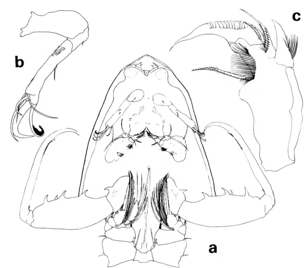
Lubbockia aculeata Giesbrecht, 1891
FIGURES 10-11
Lubbockia aculeata Giesbrecht, 1891:477; 1892:606-611, pi. 48:
figs. 3 , 9 , 1 1 , 13,16,20.
Giesbrecht described Lubbockia aculeata from specimens collected in the tropical eastern Pacific Ocean (female, 2.30 mm; male, 2.35 mm).
Our specimens agree with the characters which Giesbrecht outlined for the species. Additional species in the genus now require more detailed comparisons, so we include figures of several fea- tures not emphasized by Giesbrecht.
MATERIAL STUDIED (Tables 1, 2).—ATLANTIC OCEAN, off Bahamas: 2 females, 1.90, 2 1 3 mm, TA 44. Near Florida: 2 females, 2 1 0 , 2 3 6 mm, TA 45. PACIFIC OCEAN, near Philippine Islands: 1 female, 2.05 mm, T P 43.
FEMALE.—Average length about 2.09 mm. Body and all appendages with unusually thick exoskele- ton. Rostrum with complex pattern of sclerotiza- tion and pointed posteroventral margin as shown in ventral view of cephalosome (Figure 10a).
Antenna 1 as in L. wilsonae.
Antenna 2 (Figure 106) and mandible {Figure 10c) with armature similar in number to that of L. carinata.
Maxilliped similar to L. squillimana and L. wil- sonae, with large dentiform processes on second segment, but L. aculeata with 1 large dentiform process on first segment (Figure lla). First segment
FIGURE 10.—Lubbockia aculeata Giesbrecht, female: a, cephalosome and somite of leg 1, ventral (C); b, antenna 2, left (E); c, mandible, left (G).
may have small spinules in addition to dentiform process (Figure 10a).
Leg 1 (Figure 116) with inner spine on basis, third exopod segment with 2 short outer spines.
Leg 2 (Figure lie) third exopod segment with 2 short outer spines. Leg 3 (Figure lid) inner edge of third endopod segment with 2 setae. Leg 4 (Fig- ure 1 le) inner edge of third endopod segment with 1 seta. Legs 2-4 endopods terminate with a vent between spines.
REMARKS.—Fryer (1957:14) stated that the swim- ming legs of certain freshwater cyclopoids are al- ways directed forward while feeding, but that the
legs play no part in the actual feeding mechanism.
When legs are directed either posteriorly or an- teriorly, the usual cases in preserved specimens, the direction of the inner spine on the basis is not clearly apparent (see spine darkened on Figure 10a).
However, legs of some specimens were preserved in intermediate positions, and the direction of this spine was obvious (see spine darkened on Figure lla); it appears that this spine would be useful in holding food. Three of the 11 named Lubbockia species (4 of 12 including stages IV and V of "Lub- bockia species") lack this spine: L. carinata, L.
glacialis, and L. minuta.
17
FIGURE 11.—Lubbockia aculeata Giesbrecht, female: a, maxilliped basal segment and leg 1, ventral (C); b, leg 1 (D); c, leg 2 (D); d, leg 3 (D); e, leg 4 (D).
DISTRIBUTION.—Lubbockia aculeata has a definite tropical distribution, although a few specimens have been collected just outside tropical waters. Records and summaries are from the Pacific Ocean (Tables 1, 2; Giesbrecht, 1891; Farran, 1936; Bjornberg, 1973); Indian Ocean and Indo-Pacific (Sewell, 1947); Atlantic Ocean (Tables 1, 2; Cleve, 1904;
Farran, 1926; Owre and Foyo, 1967; Vives, 1972);
and the Mediterranean Sea (Razouls, 1974).
Lubbockia squillimana Claus, 1863
Figure 12
Lubbockia squillimana Claus, 1863:164, 165, pi. 25: figs. 1-5.—
Giesbrecht, 1892:606-611, pi. 4: fig. 6, pi. 48: figs. 1, 2, 4-8 17-19,21.
Lubbockia minuta.— Marukawa, 1927:1237, fig. 2384. [Not L.
minuta Wolfenden, 1905.]
Lubbockia marukawai Mori, 1937:122, 123, pi. 67: figs. 10-13.
Specimens that we studied from different areas varied as little as did ones from within the same sample, and all specimens agreed with Giesbrecht's (1892) excellent description. We have supplemented this description of L. squillimana, because of the additional Lubbockia species that have been added since Giesbrecht's study.
MATERIAL STUDIED (Tables 1, 2)—MEDITERRANEAN SEA, Gulf of Naples: 20 females, 1.29-1.67 mm, M 47. USNM
FIGURE 12.—Lubbockia squillimana Claus, female: a, rostrum, ventral (E); b, rostrum, lateral (E);
c, labrum, ventral (G); d, maxilla 2, right (G); e, leg 2 (E); /, leg 3 (E).
168505; 3 males, 1.98-2.09 mm, M 47, USNM 168505. Western Mediterranean: 3 females, 1.33-1.48 mm, M 46. ATLANTIC OCEAN, near Florida: 82 females, 1.22-144 mm, TA 45;
3 males, 1.75-1.86 mm, TA 45, USNM 168506. Off Bahamas:
1 damaged male, TA 44.
FEMALE.—Body and all appendages with un- usually thick exoskeleton, with complex sclerotized pattern in some areas (Figure I2a,b).
Antenna 1 as in L. wilsonae.
Labrum (Figure 12c) with free margin divided into 2 posteroventral lobes, each margin with a row of processes on under surface; short medial hood over apex.
Maxilla 2 (Figure 12d) with armature similar in number to that of L. carinata.
Legs 1-4 armature similar to that of L. aculeata;
legs 2 and 3 (Figure I2e,f) and leg 4 endopods ter- minate with a vent between spines.
DISTRIBUTION.—Lubbockia squillimana, like L.
aculeata, has a definite tropical distribution, al- though a few specimens have been collected just outside tropical waters. Principal records and sum- maries are from the Pacific Ocean (Giesbrecht, 1891;
Farran, 1936; Mori, 1937, 1942; Wilson, 1950); In- dian Ocean and Indo-Pacific (Scott, 1909; Krishna- swamy, 1953); Atlantic Ocean (Giesbrecht, 1892;
Marques, 1958; Owre and Foyo, 1967; Vives, 1972);
and the Mediterranean Sea (Razouls, 1974).
Lubbockia minuta Wolfenden, 1905
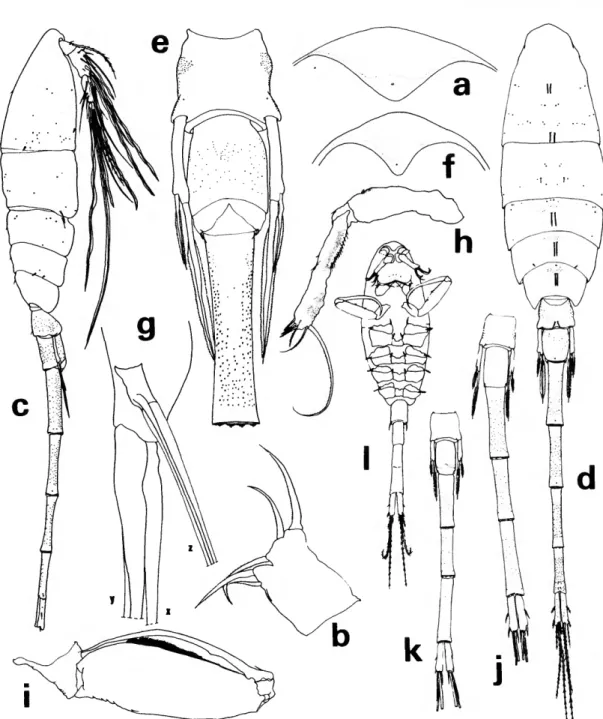
FIGURES 13-15 Lubbockia minuta Wolfenden, 1905:15, 16.
Lubbockia glacialis.—Heron and Damkaer, 1969:16-18, figs.
20, 21.—Boxshall, 1977:118, fig. 8.—Deevey and Brooks, 1977:
287. [Not L. glacialis Sars, 1900.]
Wolfenden collected 1 female (1.30 mm) off the west coast of Ireland, which he described without illustrations. The copepod collection of Richard Norris Wolfenden, M.D. (1856-1923) was recently made available to the authors by his heirs. While a number of copepod type specimens have been found in this collection, we have unfortunately not located the Lubbockia minuta. Some L. squillimana were found (see above), and also an empty slide labeled
"Lubbockia." The Wolfenden Collection will be cataloged and deposited in NMNH.
MATERIAL STUDIED (Tables 1, 2).—NE PACIFIC: 32 females, 1.29-150 mm, NP 2 et al., USNM 168508-168512; 2 males, 2.00, 2.03 mm, NP 9, 14, USNM 168507; 5 juveniles, stage V,
1.03-1.10 mm, NP 9 et al.; 8 juveniles, stage IV, 0.89-0.97 mm, NP 9 et al. ATLANTIC OCEAN, near Florida: 1 female, 1.24 mm, TA 45.
FEMALE.—Average length about 1.41 mm. Pro- some of equal or slightly greater length than urosome (Figure 13a). Urosome (Figure 136,c) with delicate hyaline spinules on posteroventral margin of postgenital segments. Genital segment length twice that of uropod; area of external genital ap- paratus located just anterior to midlength of seg- ment, each area with a setule. Second postgenital segment usually about y4 length of genital segment, occasionally as long as genital segment
Rostrum rounded posteriorly as shown in ventral view of cephalosome (Figure 13d).
Antenna 1 (Figure 13a,d) 7-segmented with vari- able rudimentary articulations between segments 3^1 and 5-6. Armature formula: 4, 7 + 1 spinule, 2 spines, 4, 3 + 1 esthete, 2 + 1 esthete, 5 + 2 esthetes; esthetes elongate.
Antenna 2 (Figure 13d) with 1 seta on second segment. Third segment with inner setules and 3 subapical setae; armed terminally with 4 short claws and 3 short setae, 1 nude, 1 setose, and 1 lashlike.
Labrum (Figure 13e) with free margin divided into 2 rounded posteroventral lobes, each margin with a row of short setules on under surface.
Mandible (Figure 13/) with stout, setulose outer element; mandible blade with row of size-graduated scalelike denticles and lashlike element adjacent to blade; concave inner edge of mandible base with a setulose element and row of long setules.
Maxilla 1 (Figure 13g) flat, bilobed; 3 elements on each lobe.
Maxilla 2 (Figure 13A) with expanded first seg- ment. Second segment outer margin with a hyaline setule and stout setulose element; terminates in element adjacent to setulose seta; curved setulose element on inner margin.
Maxilliped (Figure 13i) 4-segmented. Second seg- ment with inner cluster of short, delicate hyaline setules. Terminal segment a sharply pointed claw.
Leg 1 (Figure 14a) without inner spine on basis, third exopod segment with 3 short outer spines.
Leg 2 (Figure 146) third exopod segment with 3 short outer spines. Leg 3 (Figure 14c) inner edge of third endopod segment with 2 setae. Leg 4 (Fig- ure 15a) inner edge of third endopod segment with 1 seta. Legs 2-4 endopods terminate with a small vented projection between spines.
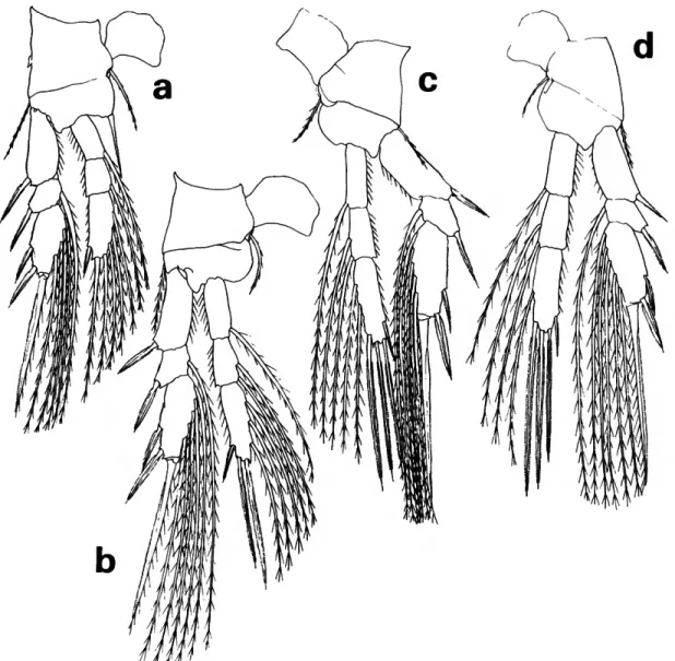
FIGURE 13.—Lubbockia minuta Wolfenden, female: a, lateral (A); b, urosome, dorsal (C); c, postgenital segments and uropods, ventral (C); d, cephalosome, ventral (E); e, labrum, ventral (G); /, mandible, right (G); g, maxilla 1, left (G); h, maxilla 2, left (G); i, maxilliped, right (E).
FIGURE 14.—Lubbockia minuta Wolfenden, female: a, leg 1 (E); b, leg 2 (E); c, leg 3 (E).
Leg 5 (Figure 156) with slender free segment not extending to area of external genital apparatus; 2 terminal spines with narrow, delicate hyaline flange.
Leg 6 probably represented by small, hyaline setule in area of external genital apparatus.
MALE.—Urosome about 1.3 times length of pro- some (Figure 15c). Solophenyl blue 2RL stain (English and Heron, 1976) revealed minute hyaline setules on ventral posterior margins of 3 postgenital segments. Third postgenital segment shows sexual dimorphism in being i/g longer than preceding seg- ment rather than only slightly longer as in female.
Rostrum as in female.
Antenna 1 with a single distinct articulation be- tween segments 2, 3 of female; a partial line sepa-
rates segments 1, 2; armature similar to correspond- ing segments of female, except for addition of 1 esthete on segment 2 and 2 esthetes on segment 4.
These additional esthetes and esthete on segment 6 elongate, supported by setalike rib; a terminal esthete reaches fifth urosomal segment.
Antenna 2 third segment with the 4 terminal claws of the female modified into 4 setae.
Mandible, maxilla 1, and maxilla 2 degenerate.
Maxilliped 3-segmented, without short segment proximal to claw of female.
Swimming legs as in female.
Leg 5 with relative length of spines differing slightly from female.
Leg 6 probably represented by posteroventral flap
FIGURE 15.—Lubbockia minuta Wolfenden, female: a, leg 4 (E); b, last prosomal segment and first 2 urosomal segments, lateral (E). Male: c, ventral (A). Stage V: d, dorsal (B). Stage IV: e, dorsal (B).
on genital segment, bearing a small pointed poste- rior process.
STAGE V.—Average length of 5 specimens 1.06 mm (1.03-1.10 mm). These specimens resemble L. glaci- alis, but are slightly smaller and more slender, and have shorter uropods in relation to the preceding (anal) segment and urosome (Figure \5d).
STAGE IV.—Average length of 8 specimens 0.95 mm (0.89-0.97 mm). The same criteria used to separate stage V were used to distinguish this stage (Figure \5e).
REMARKS.—A single damaged male of Lubbockia minuta was reported earlier as L. glacialis from the Gulf of Alaska (Heron and Damkaer, 1969); the size and swimming legs of that male resembled those of female L. glacialis (male unknown at that time).
Since the females of L. minuta have now been recog- nized with males in northeast Pacific samples, it is believed that these males are sexually dimorphic males of L. minuta. Lubbockia minuta closely re- sembles L. glacialis, including the sexually di- morphic characters. A forthcoming study of Arctic Ocean Oncaeidae includes the description of the male L. glacialis, 1.72-1.92 mm x = 1.82 mm).
Lubbockia minuta has a shorter leg 5 and more distal female genital operculum than L. glacialis.
The inner spine of L. glacialis male leg 5 reaches the middle of the second postgenital segment.
The female specimen of L. minuta found near Florida is slightly smaller (1.24 mm) than the small- est northeast Pacific female (1.29 mm), but appears to be similar except for more distinct segmentation between segments 3, 4 on antenna 1.
DISTRIBUTION.—NE Atlantic (Wolfenden, 1905) and tropical Atlantic Ocean (Tables 1, 2); NE Pacific Ocean (Tables 1, 2).
Lubbockia glacialis Sars, 1900
FIGURE 16
Lubbockia glacialis Sars, 1900:114-118, pi. 33: figs. 1-15.
MATERIAL STUDIED (Tables 1, 2).—NE PACIFIC: 6 females, 1.58-1.71 mm (x = 1.63 mm), NP 6 et al., USNM 168513- 168515; 1 juvenile, stage V, 1.20 mm, NP 4; 1 juvenile, stage IV, 1.03 mm, NP 27.
REMARKS.—The specimens from the northeast Pacific enumerated above were so much smaller than those which Sars described (2.45 mm) from the Norwegian North Polar Expedition that it took a
considerable amount of study to conclude that these are the same species. Seventy-five L. glacialis females from the Arctic Ocean (which are not listed in Tables 1, 2 but are to be discussed in a forth- coming report, Heron and English, in prep.), measured 1.60-1.92 mm (x = 1.72 mm).
The relative lengths of the 2 postgenital seg- ments of L. glacialis females are slightly variable.
The second postgenital segment of the northeast Pacific specimens is slightly longer than the first postgenital segment, while these 2 segments are about the same length in Arctic specimens. The prosome appeared larger in relation to the urosome on Arctic specimens, but this could be an effect of their diet. The intestinal structures of all the Arctic females and juveniles appear to be bulging with a material which is conspicuously darker than the exoskeleton, even in unstained specimens. The ex- panded prosome, variable in extent and occasionally asymmetrical, may be an effect of the gut content.
The Pacific specimens resemble the Arctic speci- mens in all other characters.
Lubbockia glacialis females resemble L. minuta in many characters, but differ in having the external genital apparatus on the anterior third of the geni- tal segment, and in having a longer uropod in rela- tion to the anal segment (Figure \6a,b).
STAGE V.—One specimen, 1.20 mm, NP 4, differed from the very similar L. minuta by the slightly larger size and longer uropodal length relative to the preceding segment (Figure 16c).
STAGE IV.—One specimen, 1.03 mm, NP 27, was separated from the similar specimens of L. minuta on the same criteria as those used for stage V (Figure 16d).
DISTRIBUTION.—Arctic Ocean (Sars, 1900; Grice, 1962; Johnson, 1963; Minoda, 1967; Heron and English, in prep.) and the North Pacific Ocean (Tables 1, 2; Minoda, 1971). Hardy and Gunther (1935) reported L. glacialis from the Antarctic, but with some doubt.
Lubbockia species
FIGURE 17
STAGE V.—Seven specimens, 1.37-1.44 mm, (x = 1.40 mm) from the northeast Pacific (Tables 1, 2).
Body proportions (Figure 17a) and appendages are similar to those in L. minuta and L. glacialis. Areas
FIGURE 16.—Lubbockia glacialis Sars, female: a, urosome, dorsal (C); b, last prosomal segment and first 2 urosomal segments, lateral (£). Stage V: c, dorsal (B). Stage IV: d, dorsal (B).
of dense refractile points on fourth and fifth uroso- mal segments separate this species from the others.
These specimens were larger than the juveniles of L. minuta and L. glacialis from the northeast Paci-
fic, but were approximately the same size as L.
glacialis juveniles from the Arctic Ocean (Heron and English, in prep.).
STAGE IV.—A specimen (1.14 mm) (Table 2) was
FIGURE 17.—Lubbockia species, stage V: a, dorsal (B). Stage IV: b, dorsal (B).
compared with similar size specimens of Arctic L.
glacialis, and each urosomal segment of this unde- scribed species appeared more robust and wider in relation to the length (Figure 176).
Lubbockia flemingi, new species
FIGURES 18-20
MATERIAL STUDIED {Tables 1, 2).—NE PACIFIC: 6 females, 0.83-0.91 mm, NP 7 et al., {including: holotype, 0.83 mm, NP 7, USNM 168493; 2 paratypes, 0.84 mm, NP 13, 17, USNM 168495, 168496); 2 males, 0.84, 0.85 mm, NP 16, 26 {including: allotype, 0.84 mm, NP 16, USNM 168494).
ANTARCTIC OCEAN: 1 male, 0.82 mm, AA 42.
FEMALE.—Average length about 0.85 mm. Pro- some (Figure 18a,b) about 1.7 times length of urosome. Pediger 5 with slight transverse dorsopos- terior sclerotized ridge. Genital segment with areas of external genital apparatus on anterior third of dorsolateral surface, each area with a setule. Genital segment and each postgenital segment with fine spinules on posteroventral margins. Uropod about 3 times as long as wide; outermost proximal seta about midsegment.
Rostrum with thickened posteroventral margin, deltoid in ventral view as shown in figure of orien- tation of oral appendages (Figure 18c).
Antenna 1 with armature and segmentation simi- lar in number to that of L. minuta.
Antenna 2 (Figure 18c) with 1 seta on second segment. Third segment with inner setules and 3 subapical setae; armed terminally with 4 short claws and 3 setae.
Labrum (Figure 18d) with free margin divided into 2 rounded posteroventral lobes, each margin with row of hyaline setules and small dentiform processes on under surface; complex pattern of layers of tissue over- and underlying sclerotized areas.
Mandible (Figure 18e) with 2 outer elements, both with rows of setules; mandible blade with row of size-graduated scalelike denticles on median edge, and apical lash; concave edge of mandible base with a setulose element and rows of long setules.
Maxilla 1 (Figure 18/) flat, bilobed; 3 elements on each lobe.
Maxilla 2 (Figure 19a) first segment expanded.
Second segment with a hyaline setule and stout setulose element; terminal bifurcate, setulose ele- ment; curved setulose element on inner margin.
FIGURE 18.—Lubbockia flemingi, new species, female: a, lateral (C): b, dorsal (C); c, cephalo- sorae, ventral (E); d, labrum, ventral {G); e, mandible, right (G); /, maxilla 1, right (G).
Maxilliped (Figure 196) 4-segmented. Second seg- ment with a row and cluster of setules on inner sur- face (Figure 18c). Terminal segment a long claw.
Leg 1 (Figure 19c) with inner spine on basis, third
exopod segment with 3 short outer spines. Leg 2 (Figure 19<2) third exopod segment with 3 short outer spines. Leg 3 (Figure 19e) inner edge of third endopod segment with 3 setae. Leg 4 (Figure 19/)
FIGURE 19.—Lubbockia flemingi, new species, female: a, maxilla 2, right (G); b, maxilliped, left (E); c, leg 1 (E); d, leg 2 (E); <r, leg 3 (E); /, leg 4 (E).
inner edge of third endopod segment with 2 setae.
Legs 2-4 endopods with a small vented projection between terminal spines. Leg 1 coxa lacks an outer papilla, but a vent is present.
Leg 5 (Figure 186) with free segment about i/s
length of genital segment; 2 terminal spines with serrate, hyaline flange; outer spine slightly more than 14 length of inner, which reaches beyond pos-