AND DISEASE
Thesis by Scott A. Detmer
In Partial Fulfillment of the Requirements for the Degree of
Doctor of Philosophy
California institute of Technology Pasadena, California
2007
(Defended March 1, 2007)
Scott Detmer All Rights Reserved
Acknowledgements
First, I thank my advisor, David Chan. David’s ideas are usually very creative or logical, often both, and I have enjoyed being part of his lab. I will try to approach experimental design and analysis with the level of acuity and rigor that David has brought to our discussions. Also, to my committee members: Pamela Bjorkman, Ray Deshaies, and Scott Fraser—thank you for pragmatism in goal setting, practical experimental advice, and encouragement.
Erik Griffin, Hsiuchen Chen, Takumi Koshiba, Yan Zhang, Priscilla Tee, Zhiyin Song, Tara Suntoke, Toby Rosen, Lloyd Lytle, Tadato Ban, and Sungmin Park-Lee, you have all made the Chan lab a great place to work. I am also grateful to Christine Vande Velde from Don Cleveland’s lab at UCSD for her skills and enthusiasm relating to mitochondria and motor neurons.
I have been lucky to have really incredible classmates and friends at Caltech—Stijn Cassenaer, Dan Gold, Davin Malasarn, Erik Griffin, Dave Buchbinder—it has been grand. Also, to the many people in biology who have always been very warm and helpful, thank you—especially to Liz Ayala, Diane Solis, Gwen Williams, Marta Murphy, and Carole Worra.
My family has been supportive throughout my schooling, even after they stopped asking when I would be done. Mom and Dad, Dave and Debbie, Dan and Jesse, thank you for the encouragement and diversions over the last thirty years. And more recently, Kris and Steve, Eleanor and Stan, Roselle and Joe, thank you for the same.
Finally, to Robin. You have absolutely been the highlight of grad school and marrying you has been the best thing to come of it. Your support, patience, love, advice, and occasional needling—especially at the end—have been all that I needed. Many adventures to come, not the least of which begins in May…
Abstract
We have investigated the role of mitofusin proteins in mitochondrial fusion and Charcot-Marie-Tooth disease Type 2A (CMT2A). Mitofusins (Mfn1 and Mfn2) are required for mammalian mitochondrial fusion. In structure-function analysis, we have identified loss-of-function mutations in mitofusin GTPase and heptad-repeat domains that disrupt homotypic and heterotypic domain interactions. Mutations in Mfn2 cause CMT2A, a progressive peripheral neuropathy. We have functionally characterized Mfn2 disease mutations and find that wild-type Mfn1, but not Mfn2, can efficiently complement nonfunctional CMT2A alleles to restore mitochondrial fusion. This finding demonstrates the importance of Mfn1-Mfn2 heterooligomers and suggests that Mfn1 expression is important in determining the cell-type specificity of CMT2A. To study the consequences of an Mfn2 CMT2A allele in vivo, we generated transgenic mice that express Mfn2 T105M in motor neurons. These animals demonstrate gait impairments due to distal muscle loss, axonopathy and altered mitochondrial morphology and distribution in motor neurons. In a second approach, we have generated CMT2A knock- in mice by replacing the endogenous genomic Mfn2 with Mfn2 alleles L76P or R94Q.
Preliminary characterizations suggest that heterozygous animals have no disease symptoms, but homozygous Mfn2 R94Q animals are severely affected. Together, these mouse models provide means to assess the pathology of Mfn2 CMT2A alleles and the role of mitochondrial dynamics in vivo.
Table of Contents
Acknowledgements iii
Abstract v
Table of Contents vi
List of Figures and Tables x
Chapter 1: New Functions for Mitochondrial Dynamics 1
Introduction 2
Mitochondria as dynamic organelles 3
Control of mitochondrial shape by fusion and fission 3 Control of mitochondrial distribution by active transport 4
Dynamic internal structure 5
Mechanisms of mitochondrial dynamics 6
Mediators of fusion 6
Mediators of fission 9
Other regulators of morphology 10
Proteins required for mitochondrial transport 10 Proteins mediating inner membrane morphology 11 Biological functions of mitochondrial dynamics 12
Protection of respiratory function 13
Essential developmental functions 13
Mitochondrial distribution and recruitment in neurons 14
Involvement in human disease 16
OPA1 and ADOA 16
Mfn2 and CMT2A 17
GDAP1 and CMT4A 19
Regulation of apoptosis 20
Lymphocyte chemotaxis 22
Perspectives 22
References 24
Figures 39
Thesis overview 45
Chapter 2: Multiple Independent Modes of Mitofusin Domain
Interactions are Required for Mitochondrial Fusion 48
Abstract 49
Introduction 49
Results 51
Discussion 60
Experimental procedures 63
References 66
Figures 69
Chapter 3: Complementation Between Mouse Mfn1 and Mfn2 Protects
Mitochondrial Fusion Defects Caused by CMT2A Disease Mutations 79
Abstract 80
Introduction 80
Results 81
Discussion 86
Materials and methods 88
References 88
Supplementary figures 90
Chapter 4: Mfn2 Disease Allele Causes Gait Defects and Axonopathy
in Transgenic Mouse Model of CMT2A 95
Introduction 96
Results 98
Discussion 105
Experimental procedures 107
References 110
Figures 114
Chapter 5: Generation of Mitofusin 2 Knock-In Mouse Models of
Charcot-Marie-Tooth Disease Type 2A 125
Introduction 126
Results and discussion 127
References 136
Table and figures 139
Chapter 6: Future Directions 143
References 149
List of Figures and Tables
Chapter 1:
Figure 1-1. Mitochondria as dynamic organelles 41 Figure 1-2. Mitochondrial fusion and fission determine morphology 42
Figure 1-3. Mitochondrial fusion 43
Figure 1-4. Mitochondrial fission 44
Chapter 2:
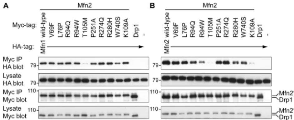
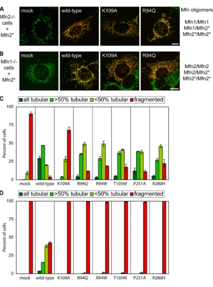
Figure 2-1. Structure-function analysis of Mfn1 72 Figure 2-2. Structure-function analysis of Mfn2 73 Figure 2-3. Co-immunoprecipitation of full-length Mfn1 mutants 74 Figure 2-4. Mitofusin N-N and N-C domain interactions 75 Figure 2-5. Characterization of Mfn1 N-C interaction 76 Figure 2-6. Characterization of non-HR N-C interaction 77 Figure 2-7. Mfn2 CMT2A disease mutations disrupt N-C interaction 78 Chapter 3:
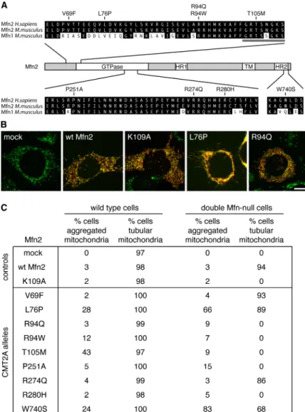
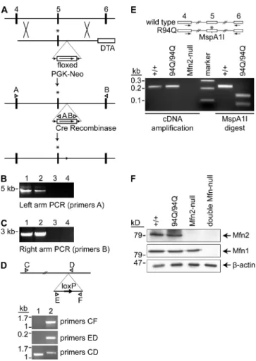
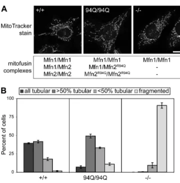
Figure 3-1. Functional analysis of Mfn2 CMT2A alleles 81 Figure 3-2. Lack of mitochondrial fusion activity in many CMT2A alleles 82 Figure 3-3. Construction of MEFs containing homozygous Mfn2R94Q
Knockin mutations 83
Figure 3-4. Tubular mitochondria in Mfn2R94Q—Mfn2R94Q cells 84 Figure 3-5. Physical association of mutant Mfn2 with wild-type Mfn1
and Mfn2 84
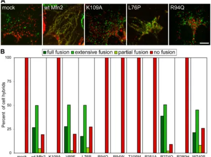
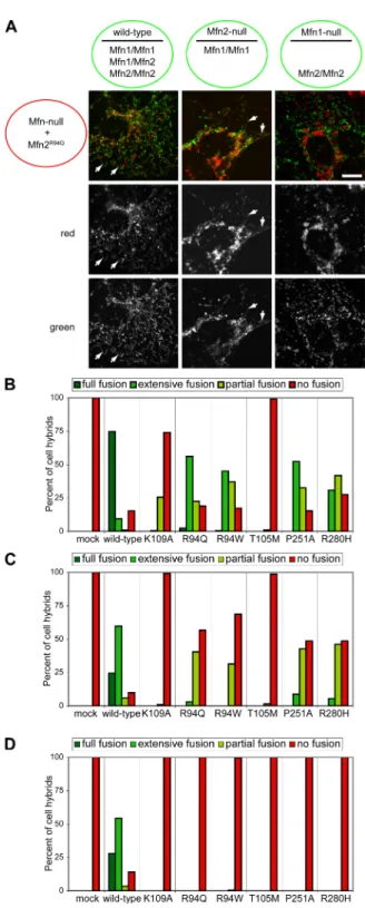
Figure 3-6. Mfn1 but not Mfn2 complements Mfn2 CMT2A alleles 85 Figure 3-7. Mfn1 complements Mfn2 CMT2A mutants in trans 86 Figure 3-8. Mfn1 complements mutant Mfn2 to preserve mitochondrial
fusion in most CMT2A cells 87
Supplementary figure 3-1. Mitochondrial profiles in MEFs expressing
Mfn2 CMT2A alleles 92
Supplementary figure 3-2. Mitochondrial aggregation in wild-type cells
highly overexpressing Mfn2 CMT2A alleles 93
Supplementary figure 3-3. Quantification of retroviral expression levels 94 Chapter 4:
Figure 4-1. Generation of Mfn2 T105M transgenic mice 118
Figure 4-2. Tg1/Tg1 mouse phenotype 119
Figure 4-3. Transgene expression in motor neurons 120
Figure 4-4. Hindlimb muscle mass 121
Figure 4-5. Loss of motor neurons in Tg1/Tg1 animals 122 Figure 4-6. Altered mitochondrial distribution in Tg1/Tg1 motor neurons 123 Supplementary figure 4-1. Transgene allelic series for tail phenotype 124 Chapter 5:
Table 5-1. Summary of genotype/phenotype for knock-in animals 140 Figure 5-1. 94Q/94Q animals have runted phenotype 141 Figure 5-2. Mitochondrial morphologies in CMT2A MEFs 142
Chapter 1
New functions for mitochondrial dynamics
Scott A. Detmer and David C. Chan
This chapter has been submitted to Nature Reviews Molecular Cell Biology.
Introduction
Seminal electron microscopy studies in the 1950s revealed the double-membranes and cristae that are hallmarks of mitochondria. These studies of fixed samples led to the canonical view of mitochondria as bean-shaped organelles, although little was known about their versatile and dynamic nature. It is now clear that mitochondria have drastically different morphologies depending on the cell type. Even within the same cell, mitochondria can take on a range of morphologies, from small spheres or short rods to long tubules. In fibroblasts, for example, mitochondria visualized with fluorescent proteins or specific dyes typically form tubules with diameters of approximately 0.5 microns, but their lengths can range from one to ten or more microns.
Even more remarkably, when mitochondria are tracked in living cells, they behave as dynamic organelles that move constantly and undergo structural transitions.
Mitochondrial tubules move with their long axis aligned along cytoskeletal tracks.
Moreover, individual mitochondria can encounter each other during these movements and undergo fusion. Fusion results in merging of the double membranes, resulting in both lipid and content mixing. Conversely, an individual mitochondrion can divide by fission to yield two or more shorter mitochondria.
What are the molecular mechanisms underlying these unusual behaviors, and do they have consequences for mitochondrial function and cell physiology? In this review, we discuss the dynamic nature of mitochondria and summarize the mechanisms that drive mitochondrial fusion and fission. In addition, we discuss recent insights into how these processes affect the function of mitochondria. Fusion and fission control not only the
shape of mitochondria, but also the functional capacity of the mitochondrial population, including its respiratory activity. Moreover, mitochondrial dynamics plays a key role in mammalian development, neuronal function and several neurodegenerative diseases, and apoptosis.
Mitochondria as dynamic organelles
Control of mitochondrial shape by fusion and fission
It is very clear that an important function of mitochondrial fusion and fission is to control the shape, size, and number of mitochondria (Fig. 1a). At steady-state, the frequencies of fusion and fission events are balanced (Nunnari et al., 1997) to maintain the overall morphology of the mitochondrial population. When this balance is experimentally perturbed, dramatic transitions in mitochondrial shape can occur. Genetic studies in yeast and mammals indicate that cells with a high fusion-to-fission ratio have fewer mitochondria that are longer and more highly interconnected (Fig. 2) (Bleazard et al., 1999; Chen et al., 2003; Sesaki and Jensen, 1999; Smirnova et al., 2001).
Conversely, cells with a low fusion-to-fission ratio have numerous mitochondria that are small spheres or short rods, often referred to as "fragmented mitochondria." Such changes in mitochondrial dynamics are used in vivo to developmentally control mitochondrial morphology, as during Drosophila spermatogenesis, when many mitochondria synchronously fuse to form the Nebenkern structure that is required for sperm motility (Hales and Fuller, 1997).
Control of mitochondrial distribution by active mitochondrial transport
Mitochondrial transport is required to distribute mitochondria throughout the cell (Fig. 1b). In most cells, mitochondria are highly motile and travel along cytoskeletal tracks. Mitochondrial transport depends on the actin cytoskeleton in budding yeast (Fehrenbacher et al., 2004) and on both actin and microtubules in mammalian cells (Hollenbeck and Saxton, 2005; Ligon and Steward, 2000b; Morris and Hollenbeck, 1995). Depending of the cellular context, these transport processes serve to ensure proper inheritance of mitochondria or to recruit mitochondria to active regions of the cell.
For example, in budding yeast, mitochondria are transported into and retained in the developing bud to ensure mitochondrial inheritance to the daughter cell (Fehrenbacher et al., 2004). In neurons, mitochondria are specifically recruited to active growth cones where demand for ATP is presumed to be great (Morris and Hollenbeck, 1993).
Rates of neuronal mitochondria transport have been reported between 0.4 micron/min (Li et al., 2004) and 0.1-1 micron/sec (Miller and Sheetz, 2004; Morris and Hollenbeck, 1995; Pilling et al., 2006). Mitochondria move in a saltatory manner, with pauses often followed by reversal of direction. At a given time, 50-75% of mitochondria are stationary (Bereiter-Hahn and Voth, 1994; Chang et al., 2006; Ligon and Steward, 2000a; Miller and Sheetz, 2004; Morris and Hollenbeck, 1995; Pilling et al., 2006).
These pauses and reversals of direction may reflect attachment and detachment of cytoskeletal motors. Although these movements can appear chaotic, several lines of evidence from neuronal studies suggest that mitochondrial transport is not random. First,
neuronal mitochondria most often pause at sites lacking other mitochondria, resulting in a well-spaced axonal mitochondrial distribution (Miller and Sheetz, 2004). Second, nearly 90% of mitochondria with high membrane potential move in the anterograde direction, whereas 80% of mitochondria with low potential move in the retrograde direction (Miller and Sheetz, 2004). These results suggest that active mitochondria are recruited to distal regions with high-energy requirements, whereas impaired mitochondria are returned to the cell soma for destruction or repair. Third, mitochondria accumulate at both pre- and post-synaptic sites in an activity-dependent manner (Chang et al., 2006; Li et al., 2004).
Finally, mitochondria, but not other organelles, accumulate at axonal sites of with high local nerve growth factor concentration, perhaps related to mitochondrial collection at active growth cones (Chada and Hollenbeck, 2004; Morris and Hollenbeck, 1993).
Dynamic internal structure
In addition to changes in the overall shape of mitochondria, the internal structures of mitochondria are also dynamic. Three-dimensional tomography of cryo-preserved samples (Mannella et al., 1994; Perkins et al., 1997) has provided new views of mitochondrial internal structure and its plasticity. The inner membrane can be divided into distinct regions: the inner boundary membrane, the cristae membrane, and the cristae junctions (Fig. 1c). The inner boundary membranes are the regions where inner membrane is in close proximity to the outer membrane. These regions are likely important for protein import and may be the sites of coupled outer and inner membrane
fusion. The cristae junctions are narrow "neck" regions that separate the inner boundary membrane from the involuted cristae membrane.
The various regions of the mitochondrial inner membrane are not only morphologically distinct, they appear to constitute separate functional domains of the inner membrane. Proteins involved in translocation of proteins through the inner membrane, such as the TIM23 complex, are enriched in the inner boundary membrane, whereas proteins involved in oxidative phosphorylation are enriched in the cristae membranes (Gilkerson et al., 2003; Vogel et al., 2006; Wurm and Jakobs, 2006). In addition, the structure of mitochondrial membranes is linked to the metabolic state of mitochondria (Fig. 1c). “Orthodox” cristae morphology, with narrow cristae and few cristae junctions per cristae compartment, is found in low ATP conditions. “Condensed”
morphology, with larger cristae having several junctions per cristae, is found in high ATP conditions (Mannella, 2006). The conversion between these states likely involves inner membrane fusion and fission (Mannella, 2006). Taken together, these observations suggest that inner membrane morphology is dynamically related to bioenergetics, although the causal relationship remains unclear.
Mechanisms of mitochondrial dynamics
Mediators of fusion
An important inroad into the molecular analysis of mitochondrial morphology came with the discovery in 1997 of the Drosophila fusion factor fuzzy onions (Fzo), a
mitochondrial outer membrane GTPase required for the fusion of mitochondria during spermatogenesis (Hales and Fuller, 1997). The yeast ortholog, Fzo1, was found to have a conserved role in mitochondrial fusion (Hermann et al., 1998), and yeast genetics provided the tools to identify additional modulators of mitochondrial fusion and fission (Okamoto and Shaw, 2005; Shaw and Nunnari, 2002). Therefore, the core machineries mediating mitochondrial fusion and fission are most fully understood in yeast. Several of these components have functionally conserved mammalian homologs. More comprehensive discussions of the molecular mechanisms of mitochondria fusion and fission have been presented in recent reviews (Chan, 2006; Griffin et al., 2006).
In yeast, the core mitochondrial fusion machinery consists of two GTPases, Fzo1p and Mgm1p (Fig. 3). Fzo1 is located on the mitochondrial outer membrane and is essential for fusion of the outer membranes (Hermann et al., 1998; Meeusen et al., 2004).
The mammalian orthologs of Fzo1 are the mitofusins Mfn1 and Mfn2. These two related proteins form homo-oligomeric and hetero-oligomeric complexes that are functional for fusion (Chen et al., 2003). Mitofusins are required on adjacent mitochondria during the fusion process, implying that they form complexes in trans between apposing mitochondria (Koshiba et al., 2004; Meeusen et al., 2004). A heptad repeat region of Mfn1 has been shown to form an anti-parallel coiled coil that is likely involved in tethering mitochondria during fusion (Koshiba et al., 2004). Mgm1 is a dynamin-related protein that is essential for fusion of the mitochondrial inner membranes, a function consistent with its localization to the intermembrane space and association of the inner membrane (Meeusen et al., 2006). The mammalian ortholog OPA1 is also essential for mitochondrial fusion (Chen et al., 2005; Cipolat et al., 2004). In yeast, the outer
membrane protein Ugo1 physically links Fzo1 and Mgm1, but no mammalian ortholog has been discovered.
The membrane potential across the mitochondrial inner membrane, maintained by the electron transport chain, is essential for mitochondrial fusion (Legros et al., 2002;
Meeusen et al., 2004). Ionophores that dissipate the mitochondrial membrane potential cause mitochondrial fragmentation due to an inhibition of mitochondrial fusion (Legros et al., 2002; Malka et al., 2005). In an in vitro fusion assay, both the proton and electrical gradient are important components of the requirement for membrane potential (Meeusen et al., 2004). The link between membrane potential and fusion remains to be resolved, but one factor appears the dependence of OPA1 post-translational processing on the membrane potential (Ishihara et al., 2006).
Recent work has also identified mitochondrial lipids as important factors in fusion. Mitochondrial morphology screens in yeast identified members of the ergosterol synthesis pathway as being required for normal mitochondrial morphology (Altmann and Westermann, 2005; Dimmer et al., 2002). Recently, MitoPLD has been identified as important for mitochondrial fusion (Choi et al., 2006). This mitochondrial outer membrane enzyme hydrolyzes cardiolipin to generate phosphatidic acid. Interestingly, erosterol has been linked to yeast vacuole fusion (Fratti et al., 2004), and phosphatidic acid is thought to play a role in generating membrane curvature required for SNARE- mediated fusion (Vitale et al., 2001). Thus, specific lipids may play similar roles in distinct types of membrane fusion.
Mediators of fission
The opposing process, mitochondrial fission, requires the recruitment of a dynamin-related protein (Dnm1 in yeast and Drp1 in mammals) from the cytosol to mitochondria (Fig. 4). Both Dnm1 and Drp1 assemble into punctate spots on mitochondrial tubules, and a subset of these complexes lead to a productive fission event (Bleazard et al., 1999; Sesaki and Jensen, 1999; Smirnova et al., 2001). By analogy with classical dynamin in endocytosis, Dnm1 and Drp1 are thought to assemble into rings and spirals that encircle and constrict the mitochondrial tubule during fission (Shaw and Nunnari, 2002). Consistent with this model, purified Dnm1 indeed can form helical rings and spirals in vitro, with dimensions similar to those of constricted mitochondria (Ingerman et al., 2005). Moreover, Dnm1 assembly is required for fission activity (Bhar et al., 2006).
Recruitment of Dnm1 to yeast mitochondrial fission sites involves three other components. It is dependent on Fis1, a mitochondrial integral outer membrane protein that is essential for fission (Fekkes et al., 2000; Mozdy et al., 2000; Tieu and Nunnari, 2000). Fis1 binds indirectly to Dnm1 through either one of two molecular adaptors, Mdv1 and Caf4 (Fig. 4b) (Griffin et al., 2005). Either Mdv1 or Caf4 are sufficient to allow Fis1-dependent recruitment of Dnm1, although Mdv1 has a more important role in mediating fission. Fis1 in mammals is also essential for mitochondrial fission (Lee et al., 2004), but no orthologs of Mdv1 and Caf4 are currently known. Both Fis1 and Drp1 are also required for fission of peroxisomes (Koch et al., 2003; Koch et al., 2005).
Other regulators of morphology
In addition to these core fusion and fission components, other genes can affect mitochondrial morphology. For example, genes such as Mmm1, Mdm10, and Mdm12 are required to maintain yeast mitochondria in a tubular shape (Okamoto and Shaw, 2005). Additional genes have been identified through visual screens for aberrant mitochondrial morphology in large-scale collections of mutant yeast (Altmann and Westermann, 2005; Dimmer et al., 2002). These screens suggest that several cellular pathways influence mitochondrial morphology and inheritance, including ergosterol biosynthesis, mitochondrial protein import, actin dynamics, vesicular fusion, and ubiquitin-mediated protein degradation. Another fruitful approach has been the identification of proteins that physically associate with the core components, such as Mfn2, Mgm1, and Drp1 (Eura et al., 2006; Hajek et al., 2006; Herlan et al., 2003;
McQuibban et al., 2003; Nakamura et al., 2006)}.
Proteins required for mitochondrial transport
Kinesin and dynein microtubule motors are known to be required for anterograde and retrograde mitochondrial transport, respectively (Hollenbeck and Saxton, 2005).
Recent work has clarified the linkage between mitochondria and the molecular motors.
Screens for essential genes in drosophila identified Milton and dMiro, both of which are required for anterograde mitochondrial transport in neurons (Guo et al., 2005; Stowers et al., 2002). Milton interacts directly with kinesin and associates indirectly with
mitochondria. dMiro is a mitochondrial outer membrane protein that interacts directly with Milton. Thus, dMiro links Milton and kinesin to mitochondria (Glater et al., 2006).
dMiro contains both GTPase and EF hand domains which are likely important in regulating mitochondrial transport.
Proteins mediating inner membrane morphology
Studies of mitochondrial inner membrane structure are complicated by the intimate link, discussed above, between mitochondrial bioenergetics and cristae structure.
Nevertheless, several proteins have been shown to have a specific role in control of cristae structure. In addition to their roles in mitochondrial fusion, Mgm1 and OPA1 are important for cristae structure. Loss of Mgm1 in yeast or knock-down of OPA1 in mammalian cells results in disorganized inner membrane structures (Amutha et al., 2004;
Frezza et al., 2006; Olichon et al., 2003; Sesaki et al., 2003). In both cases, Mgm1 or OPA1 homo-oligomeric interactions are involved (Frezza et al., 2006; Meeusen et al., 2006). The role of OPA1 in cristae structure appears separable from its role in mitochondrial fusion (Frezza et al., 2006).
Mitochondrial F1FoATP synthase, a rotary enzyme embedded in the inner membrane that couples proton pumping to ATP synthesis, is essential for normal cristae structure (Paumard et al., 2002). This role in inner membrane structure involves a dimeric form of ATP synthase containing the additional subunits e and g. As visualized by electron microscopy, the ATP synthase dimer has a dimeric interface with a sharp angle that could distort the local lipid membrane. This distortion may contribute to the
high membrane curvature that characterizes cristae tubules (Dudkina et al., 2005;
Minauro-Sanmiguel et al., 2005). Mgm1 is required for oligomerization of ATP synthase, providing a link between these two modulators of cristae structure (Amutha et al., 2004).
Additional proteins modulate inner membrane dynamics. In yeast, Mdm33 is required for normal mitochondrial morphology, and its over-expression leads to septation and vesiculation of the inner membranes (Messerschmitt et al., 2003). Because of these phenotypes, Mdm33 has been suggested to play a role in inner membrane fission.
Depletion of Mitofilin by RNAi in mammalian cells results in formation of complex sworls of inner membrane (John et al., 2005). Depletion of Mmm1p, Mdm31 and Mdm32, yeast proteins implicated in mtDNA maintenance, also cause aberrant cristae morphologies (Dimmer et al., 2005; Hobbs et al., 2001). A challenge for the future will be determining whether these proteins have direct or indirect effects on cristae morphology.
Biological functions of mitochondrial dynamics
Mitochondrial fusion and fission were first studied because of their important role in regulation of mitochondrial morphology. In addition to this function, it is now clear that these processes have additional roles in maintaining the health of the mitochondrial population, with consequences for development, disease, and apoptosis.
Protection of respiratory function
Cells with abnormalities in mitochondrial fusion have severe mitochondrial dysfunction in addition to mitochondrial fragmentation. Mouse fibroblasts lacking either Mfn1 or Mfn2 show mild heterogeneity in mitochondrial membrane potential (Chen et al., 2003), although respiratory activity is intact. Cells lacking both mitofusins, however, show great heterogeneity in mitochondrial shape and membrane potential and reduced respiratory capacity (Chen et al., 2005). Cells lacking OPA1 show similar defects, with an even greater reduction in respiratory capacity. Because other cells with fragmented mitochondria can have normal mitochondrial function, these observations show that fusion is beneficial to the mitochondrial population apart from its effect on mitochondrial shape. Fusion and fission allow the mitochondrial population to form a dynamic network capable of genetic and biochemical complementation. Indeed, functional complementation between mutant mitochondrial DNA genomes has been observed (D'Aurelio et al., 2004; Ono et al., 2001)
Essential developmental functions
Perturbations in mitochondrial dynamics result in specific developmental defects.
Mice with loss of either mitofusins die in mid-gestation, with Mfn2-null mice showing a specific defect in placental development (Chen et al., 2003). Mice lacking OPA1 also die in early embryogenesis (Alavi et al., 2007).
Mitochondrial fission is also an essential process. Worms deficient in mitochondrial division die before reaching adulthood (Labrousse et al., 1999). An infant with a dominant-negative Drp1 allele has been reported. This patient died at about one month of age and had a wide spectrum of abnormalities, including reduced head growth, increased lactic acid, and optic atrophy. Fibroblasts from this patient showed elongation of both mitochondria and peroxisomes (Waterham et al., 2007).
Mitochondrial distribution and recruitment in neurons
Mitochondrial dynamics is probably a ubiquitous phenomenon that is important for all cells. However, neurons appear to be particularly dependent on proper control of mitochondrial dynamics. Abundant mitochondria have been noted at neuronal synapses since the 1950s (Palay, 1956). More recent time-lapse imaging has confirmed enrichment and retention of mitochondria at both pre- and post-synaptic sites (Chang et al., 2006). Due to their extreme length (the longest are greater than half a body length), neurons rely heavily on active transport to recruit organelles, including mitochondria, to nerve termini. Fibroblasts lacking the anterograde mitochondrial motor Kif5b have perinuclearly aggregated mitochondria but no reported functional defects (Tanaka et al., 1998). In contrast, disruption of anterograde mitochondrial transport in neurons results in defective synaptic transmission (Guo et al., 2005; Stowers et al., 2002; Verstreken et al., 2005).
Fly neurons lacking Milton, Miro, or Drp1 show loss of synaptic mitochondria and are defective for synaptic transmission (Guo et al., 2005; Stowers et al., 2002;
Verstreken et al., 2005). Miro-deficient larvae had altered neuromuscular junctions with smaller and more abundant pre-synaptic boutons compared to wild-type larvae, indicating synaptic overgrowth in the absence of productive synapse formation (Guo et al., 2005).
Both mutant Miro and Drp1 synapses were found to have elevated resting Ca2+ levels but normal Ca2+ dynamics upon moderate stimulation, with more severe defects appearing during sustained stimulation (Guo et al., 2005; Verstreken et al., 2005). Both studies suggest that mitochondria are critically important for Ca2+ homeostasis only during sustained and intense stimulation. The more important defect in Drp1 mutants appears to be decreased synaptic levels of ATP and subsequent immobilization of the reserve vesicle pool. Forward-filling of synapses with ATP partially rescued synaptic transmission in Drp1 mutant neuromuscular junctions (Verstreken et al., 2005).
Mitochondria are also required post-synaptically in dendrites of hippocampal neurons. Mitochondrial localization at dendritic spines (potential post-synaptic sites) increased following repetitive stimulation (Li et al., 2004). Expression of mutant Drp1 caused elongation of mitochondria and a decrease in both the abundance of dendritic mitochondria and the density of dendritic spines. Conversely, over-expression of wild- type Drp1 decreased mean mitochondrial length and increased both the density of dendritic mitochondria and spines (Li et al., 2004). Dendritic spine density was also increased by creatine, which stimulates mitochondrial activity.
Involvement in human disease
OPA1 and ADOA
Heterozygous mutations in OPA1 cause autosomal dominant optic atrophy (ADOA). This disease is the most common heritable form of optic neuropathy and is due to degeneration of retinal ganglion cells, whose axons form the optic nerve (Delettre et al., 2000). Over 100 mutations in OPA1 have been reported, with the majority of the mutations occurring in the GTPase domain(Ferre et al., 2005). Roughly half of the pathogenic alleles contain nonsense mutations predicted to encode a truncated protein. A few nonsense mutations abolish nearly the entire coding sequence(Delettre et al., 2000), suggesting that haploinsufficiency of OPA1 can cause ADOA. It remains possible that other less severe truncations may have dominant-negative activity.
Fibroblasts knocked-down for OPA1 have fragmented mitochondria, defects in respiration, aberrant cristae structure, and increased susceptibility to apoptosis (Chen et al., 2005; Cipolat et al., 2004; Griparic et al., 2004; Olichon et al., 2003). In the disease state, the pathophysiology remains to be clarified. Monocytes from a patient with a C- terminal truncation in OPA1 had aggregated mitochondria (Delettre et al., 2000), and skin fibroblasts from a patient with a GTPase missense mutation in OPA1 had fragmented mitochondria and increased sensitivity to apoptosis (Olichon et al., 2006). In addition, OPA1 mutations have been associated with reduced ATP production and mtDNA content (Kim et al., 2005; Lodi et al., 2004). It is unclear why mutations in OPA1, which is broadly expressed, have such cell-type specific defects.
A mouse model of ADOA has been constructed with an OPA1 gene containing a splice site mutation that causes processed transcripts to lack exon 10 (Alavi et al., 2007).
Although this mutation would be expected to produce an internally truncated protein, the mutant mice do not produce any detectable protein from the mutant OPA1 gene.
Heterozygous mice show several features of ADOA, including progressive decline in retinal ganglion cell numbers and loss of axons in the optic nerve. These results further support haploinsufficiency of OPA1as a mechanism for ADOA. Interestingly, mice homozygous for the OPA1 mutation die at mid-gestation (Alavi et al., 2007), consistent with an essential requirement for mitochondrial fusion during embryonic development (Chen et al., 2003).
Mfn2 and CMT2A
Charcot-Marie-Tooth (CMT) disease, one of the most common hereditary neuropathies, is caused by mutations in at least 30 different genes (Zuchner and Vance, 2006). Affected individuals have progressive distal motor and sensory impairments beginning in the feet and hands due to loss of function of long neurons. Depending on the type of CMT, these diseases can be caused by a primary defect in the Schwann cells that myelinate peripheral nerves or in the neurons themselves (Zuchner and Vance, 2006). CMT2A is an axonopathy caused by the latter defect, and has been associated with over 40 mutations in Mfn2. Nearly all of these disease alleles contain missense mutations or short, in-frame deletions (Zuchner et al., 2004). Most mutations cluster in or near the GTPase domain but some also occur in each of the heptad repeat domains of
Mfn2. In addition to loss of peripheral nerve function, a subset of CMT2A patients also have optic atrophy, suggesting a similar mechanistic and clinical outcome for disruption of mitochondrial dynamics with mutation of OPA1 and Mfn2 (Chung et al., 2006;
Zuchner et al., 2006).
Because of difficulties in studying patient nerve tissue, the pathogenic mechanisms leading to peripheral nerve degeneration in CMT2A are not well understood.
Only one study has examined ultrastructural defects in mitochondria from nerves of CMT2A patients. Mitochondria in the sural nerve of two patients show structural aberrations in the mitochondrial outer and inner membranes, along with swelling that is suggestive of mitochondrial dysfunction (Verhoeven et al., 2006). Aggregation of mitochondria is also observed. Interestingly, over-expression of Mfn2 CMT2A alleles (Baloh et al., 2007; Detmer and Chan, 2007) causes mitochondrial aggregation and subsequent mitochondrial transport defects in neurons (Baloh et al., 2007). In fibroblasts, the mitochondrial aggregation phenotype is dependent on significant over-expression of the CMT2A alleles (Detmer and Chan, 2007), and therefore its relevance to disease pathogenesis remains to be clarified.
Several perplexing issues remain to be resolved concerning the molecular genetics of CMT2A. How does mutation of one copy of Mfn2 lead to disease? Why are long peripheral neurons selectively affected, given that Mfn2 is a broadly expressed protein?
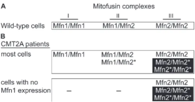
Clues to these issues have come from analysis of CMT2A alleles in mice (Detmer and Chan, 2007). Many CMT2A alleles of Mfn2 are nonfunctional for fusion when expressed alone. However, the fusion activity of these nonfunctional alleles can be efficiently complemented by wild-type Mfn1 but not Mfn2. Because of this
complementation, in CMT2A patients, cells that express Mfn1 are protected from gross loss of fusion activity. In contrast, cells with little or no Mfn1 expression would suffer a greater relative loss of fusion activity. In part, these properties of the CMT2A alleles may underlie the selective loss of sensory and motor neurons. Consistent with this model, Mfn2 appears to be more highly expressed in central and peripheral nervous tissue than Mfn1 (S. A. Detmer and D. C. Chan, unpublished observations). Even within the peripheral nerves, it appears that mitochondrial fusion defects are partial, because only the longest nerves are affected. Most likely, the extreme dimensions of long peripheral nerves make them most vulnerable to changes in mitochondrial dynamics. Hopefully, the molecular basis of this effect can be dissected in animal models.
GDAP1 and CMT4A
Another form of CMT is associated with defects in mitochondrial dynamics.
GDAP1 (ganglioside-induced differentiation-associated protein 1) is mutated in CMT4A, one of the few recessive forms of CMT disease. CMT4A contains both demyelinating and axonal features, and consistent with this mixed clinical presentation, GDAP1 is expressed in both Schwann cells and neurons (Niemann et al., 2005). GDAP1 is an integral outer membrane protein that likely affects mitochondrial division (Niemann et al., 2005; Pedrola et al., 2005). Disease alleles either fail to localize to mitochondria or are defective in stimulating mitochondrial division when over-expressed (Niemann et al., 2005). If GDAP1 disease alleles disrupt normal mitochondrial fission, they may cause mitochondrial distribution defects similar to those induced by Drp1 mutations discussed
above (Li et al., 2004; Verstreken et al., 2005). Again, it will be important determine whether patient samples substantiate these ideas.
Regulation of apoptosis
A regulated switch in mitochondrial morphology appears to be important for some forms of apoptosis. In apoptosis mediated by Bcl-2 family members, the outer membrane of mitochondria are permeabilized to release contents of the intermembrane space, such as cytochrome c, into the cytoplasm. Cytochrome c subsequently activates a cascade of caspases that propagate and execute the apoptotic program. This mitochondrial outer membrane permeabilization is an early step in apoptosis and is temporally associated with mitochondrial fragmentation (Desagher and Martinou, 2000;
Frank et al., 2001). In principle, therefore, genes controlling mitochondrial morphology can potentially impact progression of apoptosis.
The first functional link between mitochondrial fission and apoptosis was the observation that inhibition of fission can partially block apoptosis (Frank et al., 2001).
Over-expression of dominant-negative Drp1 prevents mitochondrial fragmentation and reduces membrane depolarization, cytochrome c release, and cell death in response to apoptotic stimuli (Frank et al., 2001). Similarly, RNAi-mediated down-regulation of Fis1 and Drp1 inhibits both fragmentation and apoptosis (Lee et al., 2004). These results suggest that mitochondrial fission is an important component of mitochondrial permeabilization and progression of apoptosis. In addition, over-expression of rat mitofusin protects against apoptosis, whereas RNAi down-regulation of mitofusin leads
to fragmentation of mitochondria and increased susceptibility to apoptosis (Sugioka et al., 2004). Mitochondrial fusion is inhibited during apoptosis following Bax activation (Karbowski et al., 2004). Interestingly, Bax, which localizes to discrete puncta on mitochondria upon induction of apoptosis, colocalize with both Drp1 and Mfn2 (Karbowski et al., 2002). The functional nature of these interactions is not understood but suggests that the fission and fusion machinery may be modified in early apoptosis.
Surprisingly, the apoptotic proteins Bax and Bak appear to have a role in normal mitochondrial morphology. Bax and Bak double knock-out cells have fragmented mitochondria due to a fusion defect (Karbowski et al., 2006). In addition, Bax and Mfn2 interact in a yeast-2-hybrid analysis, and Bax may be involved in Mfn2 distribution on the mitochondrial outer membrane (Karbowski et al., 2006).
In spite of these compelling observations, the causal relationship of mitochondrial fission to apoptosis remains to be definitively resolved. Perplexingly, depending on the biological system, mitochondrial fragmentation has been reported to occur before cytochrome c release in some cases, but after in other cases (Arnoult, 2007). In some experimental systems, inhibition of mitochondrial fission by knock-down of Fis1 or Drp1 does not inhibit apoptosis, even though mitochondrial fragmentation is efficiently inhibited (Parone et al., 2006).
In addition to outer membrane permeabilization, remodeling of the cristae membranes is required for rapid and efficient release of cytochrome c (Goldstein et al., 2000; Scorrano et al., 2002). The reason for this requirement is that only a minority (~15%) of cytochrome c resides in the intermembrane space, with the bulk found in cristae compartments (Scorrano et al., 2002). OPA1 appears to be a regulator of cristae
junctions and therefore cytochrome c release (Cipolat et al., 2006; Frezza et al., 2006).
Over-expression of OPA1 blocks cytochrome c release following induction of apoptosis by maintaining narrow cristae junctions (Frezza et al., 2006).
Lymphocyte chemotaxis
Mitochondrial dynamics appears to be important for proper mitochondrial redistribution in lymphocytes during chemotaxis (Campello et al., 2006). Mitochondria redistribute to the trailing edge in lymphocyte cell lines migrating in response to chemical attractants. Modulation of mitochondrial fusion or fission effected both mitochondrial redistribution and cell migration. For example, fragmentation of mitochondria enhanced both redistribution and migration whereas conditions that promoted fusion, and subsequently larger mitochondria, impaired both redistribution and migration. These observations suggest that mitochondrial dynamics is important for recruiting mitochondria to local cellular areas that have the greatest energy requirements.
Perspectives
The study of mitochondrial dynamics has undergone great advances in the last few years. It is now clear that, in addition to control of organelle morphology, mitochondrial dynamics is important for the functional state of mitochondria. As a result, these processes play crucial roles in mammalian development, apoptosis, and disease. As our knowledge of mitochondrial dynamics increases, we can expect to learn about its
involvement in other processes. The link between defects in mitochondrial fusion and neurodegenerative disease is particularly intriguing. In future studies, the pathophysiological mechanisms underlying such neurodegenerative diseases will hopefully be dissected in appropriate animal models.
References
Alavi, M.V., S. Bette, S. Schimpf, F. Schuettauf, U. Schraermeyer, H.F. Wehrl, L.
Ruttiger, S.C. Beck, F. Tonagel, B.J. Pichler, M. Knipper, T. Peters, J. Laufs, and B. Wissinger. 2007. A splice site mutation in the murine Opa1 gene features pathology of autosomal dominant optic atrophy. Brain.
Altmann, K., and B. Westermann. 2005. Role of essential genes in mitochondrial morphogenesis in Saccharomyces cerevisiae. Mol Biol Cell. 16:5410-7.
Amutha, B., D.M. Gordon, Y. Gu, and D. Pain. 2004. A novel role of Mgm1p, a dynamin-related GTPase, in ATP synthase assembly and cristae formation/maintenance. Biochem J. 381:19-23.
Arnoult, D. 2007. Mitochondrial fragmentation in apoptosis. Trends Cell Biol. 17:6-12.
Baloh, R.H., R.E. Schmidt, A. Pestronk, and J. Milbrandt. 2007. Altered axonal mitochondrial transport in the pathogenesis of Charcot-Marie-Tooth disease from mitofusin 2 mutations. J Neurosci. 27:422-30.
Bereiter-Hahn, J., and M. Voth. 1994. Dynamics of mitochondria in living cells: shape changes, dislocations, fusion, and fission of mitochondria. Microsc Res Tech.
27:198-219.
Bhar, D., M.A. Karren, M. Babst, and J.M. Shaw. 2006. Dimeric Dnm1-G385D interacts with Mdv1 on mitochondria and can be stimulated to assemble into fission complexes containing Mdv1 and Fis1. J Biol Chem. 281:17312-20.
Bleazard, W., J.M. McCaffery, E.J. King, S. Bale, A. Mozdy, Q. Tieu, J. Nunnari, and J.M. Shaw. 1999. The dynamin-related GTPase Dnm1 regulates mitochondrial fission in yeast. Nat Cell Biol. 1:298-304.
Campello, S., R.A. Lacalle, M. Bettella, S. Manes, L. Scorrano, and A. Viola. 2006.
Orchestration of lymphocyte chemotaxis by mitochondrial dynamics. J Exp Med.
Chada, S.R., and P.J. Hollenbeck. 2004. Nerve growth factor signaling regulates motility and docking of axonal mitochondria. Curr Biol. 14:1272-6.
Chan, D.C. 2006. Mitochondrial fusion and fission in mammals. Annu Rev Cell Dev Biol.
22:79-99.
Chang, D.T., A.S. Honick, and I.J. Reynolds. 2006. Mitochondrial trafficking to synapses in cultured primary cortical neurons. J Neurosci. 26:7035-45.
Chen, H., A. Chomyn, and D.C. Chan. 2005. Disruption of fusion results in mitochondrial heterogeneity and dysfunction. J Biol Chem. 280:26185-92.
Chen, H., S.A. Detmer, A.J. Ewald, E.E. Griffin, S.E. Fraser, and D.C. Chan. 2003.
Mitofusins Mfn1 and Mfn2 coordinately regulate mitochondrial fusion and are essential for embryonic development. J Cell Biol. 160:189-200.
Choi, S.Y., P. Huang, G.M. Jenkins, D.C. Chan, J. Schiller, and M.A. Frohman. 2006. A common lipid links Mfn-mediated mitochondrial fusion and SNARE-regulated exocytosis. Nat Cell Biol. 8:1255-62.
Chung, K.W., S.B. Kim, K.D. Park, K.G. Choi, J.H. Lee, H.W. Eun, J.S. Suh, J.H.
Hwang, W.K. Kim, B.C. Seo, S.H. Kim, I.H. Son, S.M. Kim, I.N. Sunwoo, and B.O. Choi. 2006. Early onset severe and late-onset mild Charcot-Marie-Tooth disease with mitofusin 2 (MFN2) mutations. Brain. 129:2103-18.
Cipolat, S., O. Martins de Brito, B. Dal Zilio, and L. Scorrano. 2004. OPA1 requires mitofusin 1 to promote mitochondrial fusion. Proc Natl Acad Sci U S A.
101:15927-32.
Cipolat, S., T. Rudka, D. Hartmann, V. Costa, L. Serneels, K. Craessaerts, K. Metzger, C.
Frezza, W. Annaert, L. D'Adamio, C. Derks, T. Dejaegere, L. Pellegrini, R.
D'Hooge, L. Scorrano, and B. De Strooper. 2006. Mitochondrial rhomboid PARL regulates cytochrome c release during apoptosis via OPA1-dependent cristae remodeling. Cell. 126:163-75.
D'Aurelio, M., C.D. Gajewski, M.T. Lin, W.M. Mauck, L.Z. Shao, G. Lenaz, C.T.
Moraes, and G. Manfredi. 2004. Heterologous mitochondrial DNA recombination in human cells. Hum Mol Genet. 13:3171-9.
Delettre, C., G. Lenaers, J.M. Griffoin, N. Gigarel, C. Lorenzo, P. Belenguer, L.
Pelloquin, J. Grosgeorge, C. Turc-Carel, E. Perret, C. Astarie-Dequeker, L.
Lasquellec, B. Arnaud, B. Ducommun, J. Kaplan, and C.P. Hamel. 2000. Nuclear gene OPA1, encoding a mitochondrial dynamin-related protein, is mutated in dominant optic atrophy. Nat Genet. 26:207-10.
Desagher, S., and J.C. Martinou. 2000. Mitochondria as the central control point of apoptosis. Trends Cell Biol. 10:369-77.
Detmer, S.A., and D.C. Chan. 2007. Complementation between mouse Mfn1 and Mfn2 protects mitochondrial fusion defects caused by CMT2A disease mutations. J Cell Biol. 176:405-14.
Dimmer, K.S., S. Fritz, F. Fuchs, M. Messerschmitt, N. Weinbach, W. Neupert, and B.
Westermann. 2002. Genetic basis of mitochondrial function and morphology in Saccharomyces cerevisiae. Mol Biol Cell. 13:847-53.
Dimmer, K.S., S. Jakobs, F. Vogel, K. Altmann, and B. Westermann. 2005. Mdm31 and Mdm32 are inner membrane proteins required for maintenance of mitochondrial shape and stability of mitochondrial DNA nucleoids in yeast. J Cell Biol.
168:103-15.
Dudkina, N.V., J. Heinemeyer, W. Keegstra, E.J. Boekema, and H.P. Braun. 2005.
Structure of dimeric ATP synthase from mitochondria: an angular association of monomers induces the strong curvature of the inner membrane. FEBS Lett.
579:5769-72.
Eura, Y., N. Ishihara, T. Oka, and K. Mihara. 2006. Identification of a novel protein that regulates mitochondrial fusion by modulating mitofusin (Mfn) protein function. J Cell Sci. 119:4913-25.
Fehrenbacher, K.L., H.C. Yang, A.C. Gay, T.M. Huckaba, and L.A. Pon. 2004. Live cell imaging of mitochondrial movement along actin cables in budding yeast. Curr Biol. 14:1996-2004.
Fekkes, P., K.A. Shepard, and M.P. Yaffe. 2000. Gag3p, an outer membrane protein required for fission of mitochondrial tubules. J Cell Biol. 151:333-40.
Ferre, M., P. Amati-Bonneau, Y. Tourmen, Y. Malthiery, and P. Reynier. 2005. eOPA1:
an online database for OPA1 mutations. Hum Mutat. 25:423-8.
Frank, S., B. Gaume, E.S. Bergmann-Leitner, W.W. Leitner, E.G. Robert, F. Catez, C.L.
Smith, and R.J. Youle. 2001. The role of dynamin-related protein 1, a mediator of mitochondrial fission, in apoptosis. Dev Cell. 1:515-25.
Fratti, R.A., Y. Jun, A.J. Merz, N. Margolis, and W. Wickner. 2004. Interdependent assembly of specific regulatory lipids and membrane fusion proteins into the vertex ring domain of docked vacuoles. J Cell Biol. 167:1087-98.
Frezza, C., S. Cipolat, O. Martins de Brito, M. Micaroni, G.V. Beznoussenko, T. Rudka, D. Bartoli, R.S. Polishuck, N.N. Danial, B. De Strooper, and L. Scorrano. 2006.
OPA1 controls apoptotic cristae remodeling independently from mitochondrial fusion. Cell. 126:177-89.
Gilkerson, R.W., J.M. Selker, and R.A. Capaldi. 2003. The cristal membrane of mitochondria is the principal site of oxidative phosphorylation. FEBS Lett.
546:355-8.
Glater, E.E., L.J. Megeath, R.S. Stowers, and T.L. Schwarz. 2006. Axonal transport of mitochondria requires milton to recruit kinesin heavy chain and is light chain independent. J Cell Biol. 173:545-57.
Goldstein, J.C., N.J. Waterhouse, P. Juin, G.I. Evan, and D.R. Green. 2000. The coordinate release of cytochrome c during apoptosis is rapid, complete and kinetically invariant. Nat Cell Biol. 2:156-62.
Griffin, E.E., S.A. Detmer, and D.C. Chan. 2006. Molecular mechanism of mitochondrial membrane fusion. Biochim Biophys Acta. 1763:482-9.
Griffin, E.E., J. Graumann, and D.C. Chan. 2005. The WD40 protein Caf4p is a component of the mitochondrial fission machinery and recruits Dnm1p to mitochondria. J Cell Biol. 170:237-48.
Griparic, L., N.N. van der Wel, I.J. Orozco, P.J. Peters, and A.M. van der Bliek. 2004.
Loss of the intermembrane space protein Mgm1/OPA1 induces swelling and localized constrictions along the lengths of mitochondria. J Biol Chem.
279:18792-8.
Guo, X., G.T. Macleod, A. Wellington, F. Hu, S. Panchumarthi, M. Schoenfield, L.
Marin, M.P. Charlton, H.L. Atwood, and K.E. Zinsmaier. 2005. The GTPase dMiro is required for axonal transport of mitochondria to Drosophila synapses.
Neuron. 47:379-93.
Hajek, P., A. Chomyn, and G. Attardi. 2006. Identification of a novel mitochondrial complex containing mitofusin 2 and stomatin-like protein 2. J Biol Chem.
Hales, K.G., and M.T. Fuller. 1997. Developmentally regulated mitochondrial fusion mediated by a conserved, novel, predicted GTPase. Cell. 90:121-9.
Herlan, M., F. Vogel, C. Bornhovd, W. Neupert, and A.S. Reichert. 2003. Processing of Mgm1 by the rhomboid-type protease Pcp1 is required for maintenance of mitochondrial morphology and of mitochondrial DNA. J Biol Chem. 278:27781- 8.
Hermann, G.J., J.W. Thatcher, J.P. Mills, K.G. Hales, M.T. Fuller, J. Nunnari, and J.M.
Shaw. 1998. Mitochondrial fusion in yeast requires the transmembrane GTPase Fzo1p. J Cell Biol. 143:359-73.
Hobbs, A.E., M. Srinivasan, J.M. McCaffery, and R.E. Jensen. 2001. Mmm1p, a mitochondrial outer membrane protein, is connected to mitochondrial DNA (mtDNA) nucleoids and required for mtDNA stability. J Cell Biol. 152:401-10.
Hollenbeck, P.J., and W.M. Saxton. 2005. The axonal transport of mitochondria. J Cell Sci. 118:5411-9.
Ingerman, E., E.M. Perkins, M. Marino, J.A. Mears, J.M. McCaffery, J.E. Hinshaw, and J. Nunnari. 2005. Dnm1 forms spirals that are structurally tailored to fit mitochondria. J Cell Biol. 170:1021-7.
Ishihara, N., Y. Fujita, T. Oka, and K. Mihara. 2006. Regulation of mitochondrial morphology through proteolytic cleavage of OPA1. Embo J. 25:2966-77.
Ishihara, N., A. Jofuku, Y. Eura, and K. Mihara. 2003. Regulation of mitochondrial morphology by membrane potential, and DRP1-dependent division and FZO1- dependent fusion reaction in mammalian cells. Biochem Biophys Res Commun.
301:891-8.
John, G.B., Y. Shang, L. Li, C. Renken, C.A. Mannella, J.M. Selker, L. Rangell, M.J.
Bennett, and J. Zha. 2005. The mitochondrial inner membrane protein mitofilin controls cristae morphology. Mol Biol Cell. 16:1543-54.
Karbowski, M., D. Arnoult, H. Chen, D.C. Chan, C.L. Smith, and R.J. Youle. 2004.
Quantitation of mitochondrial dynamics by photolabeling of individual organelles shows that mitochondrial fusion is blocked during the Bax activation phase of apoptosis. J Cell Biol. 164:493-9.
Karbowski, M., Y.J. Lee, B. Gaume, S.Y. Jeong, S. Frank, A. Nechushtan, A. Santel, M.
Fuller, C.L. Smith, and R.J. Youle. 2002. Spatial and temporal association of Bax
with mitochondrial fission sites, Drp1, and Mfn2 during apoptosis. J Cell Biol.
159:931-8.
Karbowski, M., K.L. Norris, M.M. Cleland, S.Y. Jeong, and R.J. Youle. 2006. Role of Bax and Bak in mitochondrial morphogenesis. Nature. 443:658-62.
Kim, J.Y., J.M. Hwang, H.S. Ko, M.W. Seong, B.J. Park, and S.S. Park. 2005.
Mitochondrial DNA content is decreased in autosomal dominant optic atrophy.
Neurology. 64:966-72.
Koch, A., M. Thiemann, M. Grabenbauer, Y. Yoon, M.A. McNiven, and M. Schrader.
2003. Dynamin-like protein 1 is involved in peroxisomal fission. J Biol Chem.
278:8597-605.
Koch, A., Y. Yoon, N.A. Bonekamp, M.A. McNiven, and M. Schrader. 2005. A role for Fis1 in both mitochondrial and peroxisomal fission in mammalian cells. Mol Biol Cell. 16:5077-86.
Koshiba, T., S.A. Detmer, J.T. Kaiser, H. Chen, J.M. McCaffery, and D.C. Chan. 2004.
Structural basis of mitochondrial tethering by mitofusin complexes. Science.
305:858-62.
Labrousse, A.M., M.D. Zappaterra, D.A. Rube, and A.M. van der Bliek. 1999. C. elegans dynamin-related protein DRP-1 controls severing of the mitochondrial outer membrane. Mol Cell. 4:815-26.
Lee, Y.J., S.Y. Jeong, M. Karbowski, C.L. Smith, and R.J. Youle. 2004. Roles of the mammalian mitochondrial fission and fusion mediators Fis1, Drp1, and Opa1 in apoptosis. Mol Biol Cell. 15:5001-11.
Legros, F., A. Lombes, P. Frachon, and M. Rojo. 2002. Mitochondrial fusion in human cells is efficient, requires the inner membrane potential, and is mediated by mitofusins. Mol Biol Cell. 13:4343-54.
Legros, F., F. Malka, P. Frachon, A. Lombes, and M. Rojo. 2004. Organization and dynamics of human mitochondrial DNA. J Cell Sci. 117:2653-62.
Li, Z., K. Okamoto, Y. Hayashi, and M. Sheng. 2004. The importance of dendritic mitochondria in the morphogenesis and plasticity of spines and synapses. Cell.
119:873-87.
Ligon, L.A., and O. Steward. 2000a. Movement of mitochondria in the axons and dendrites of cultured hippocampal neurons. J Comp Neurol. 427:340-50.
Ligon, L.A., and O. Steward. 2000b. Role of microtubules and actin filaments in the movement of mitochondria in the axons and dendrites of cultured hippocampal neurons. J Comp Neurol. 427:351-61.
Lodi, R., C. Tonon, M.L. Valentino, S. Iotti, V. Clementi, E. Malucelli, P. Barboni, L.
Longanesi, S. Schimpf, B. Wissinger, A. Baruzzi, B. Barbiroli, and V. Carelli.
2004. Deficit of in vivo mitochondrial ATP production in OPA1-related dominant optic atrophy. Ann Neurol. 56:719-23.
Malka, F., O. Guillery, C. Cifuentes-Diaz, E. Guillou, P. Belenguer, A. Lombes, and M.
Rojo. 2005. Separate fusion of outer and inner mitochondrial membranes. EMBO Rep. 6:853-9.
Mannella, C.A. 2006. Structure and dynamics of the mitochondrial inner membrane cristae. Biochim Biophys Acta. 1763:542-8.
Mannella, C.A., M. Marko, P. Penczek, D. Barnard, and J. Frank. 1994. The internal compartmentation of rat-liver mitochondria: tomographic study using the high- voltage transmission electron microscope. Microsc Res Tech. 27:278-83.
Mattenberger, Y., D.I. James, and J.C. Martinou. 2003. Fusion of mitochondria in mammalian cells is dependent on the mitochondrial inner membrane potential and independent of microtubules or actin. FEBS Lett. 538:53-9.
McQuibban, G.A., S. Saurya, and M. Freeman. 2003. Mitochondrial membrane remodelling regulated by a conserved rhomboid protease. Nature. 423:537-41.
Meeusen, S., R. DeVay, J. Block, A. Cassidy-Stone, S. Wayson, J.M. McCaffery, and J.
Nunnari. 2006. Mitochondrial inner-membrane fusion and crista maintenance requires the dynamin-related GTPase Mgm1. Cell. 127:383-95.
Meeusen, S., J.M. McCaffery, and J. Nunnari. 2004. Mitochondrial fusion intermediates revealed in vitro. Science. 305:1747-52.
Messerschmitt, M., S. Jakobs, F. Vogel, S. Fritz, K.S. Dimmer, W. Neupert, and B.
Westermann. 2003. The inner membrane protein Mdm33 controls mitochondrial morphology in yeast. J Cell Biol. 160:553-64.
Miller, K.E., and M.P. Sheetz. 2004. Axonal mitochondrial transport and potential are correlated. J Cell Sci. 117:2791-804.
Minauro-Sanmiguel, F., S. Wilkens, and J.J. Garcia. 2005. Structure of dimeric mitochondrial ATP synthase: novel F0 bridging features and the structural basis of mitochondrial cristae biogenesis. Proc Natl Acad Sci U S A. 102:12356-8.
Morris, R.L., and P.J. Hollenbeck. 1993. The regulation of bidirectional mitochondrial transport is coordinated with axonal outgrowth. J Cell Sci. 104 ( Pt 3):917-27.
Morris, R.L., and P.J. Hollenbeck. 1995. Axonal transport of mitochondria along microtubules and F-actin in living vertebrate neurons. J Cell Biol. 131:1315-26.
Mozdy, A.D., J.M. McCaffery, and J.M. Shaw. 2000. Dnm1p GTPase-mediated mitochondrial fission is a multi-step process requiring the novel integral membrane component Fis1p. J Cell Biol. 151:367-80.
Nakamura, N., Y. Kimura, M. Tokuda, S. Honda, and S. Hirose. 2006. MARCH-V is a novel mitofusin 2- and Drp1-binding protein able to change mitochondrial morphology. EMBO Rep. 7:1019-22.
Niemann, A., M. Ruegg, V. La Padula, A. Schenone, and U. Suter. 2005. Ganglioside- induced differentiation associated protein 1 is a regulator of the mitochondrial network: new implications for Charcot-Marie-Tooth disease. J Cell Biol.
170:1067-78.
Nunnari, J., W.F. Marshall, A. Straight, A. Murray, J.W. Sedat, and P. Walter. 1997.
Mitochondrial transmission during mating in Saccharomyces cerevisiae is determined by mitochondrial fusion and fission and the intramitochondrial segregation of mitochondrial DNA. Mol Biol Cell. 8:1233-42.
Okamoto, K., and J.M. Shaw. 2005. Mitochondrial morphology and dynamics in yeast and multicellular eukaryotes. Annu Rev Genet. 39:503-36.
Olichon, A., L. Baricault, N. Gas, E. Guillou, A. Valette, P. Belenguer, and G. Lenaers.
2003. Loss of OPA1 perturbates the mitochondrial inner membrane structure and integrity, leading to cytochrome c release and apoptosis. J Biol Chem. 278:7743- 6.
Olichon, A., T. Landes, L. Arnaune-Pelloquin, L.J. Emorine, V. Mils, A. Guichet, C.
Delettre, C. Hamel, P. Amati-Bonneau, D. Bonneau, P. Reynier, G. Lenaers, and P. Belenguer. 2006. Effects of OPA1 mutations on mitochondrial morphology and apoptosis: Relevance to ADOA pathogenesis. J Cell Physiol.
Ono, T., K. Isobe, K. Nakada, and J.I. Hayashi. 2001. Human cells are protected from mitochondrial dysfunction by complementation of DNA products in fused mitochondria. Nat Genet. 28:272-5.
Palay, S.L. 1956. Synapses in the central nervous system. J Biophys Biochem Cytol.
2:193-202.
Parone, P.A., D.I. James, S. Da Cruz, Y. Mattenberger, O. Donze, F. Barja, and J.C.
Martinou. 2006. Inhibiting the mitochondrial fission machinery does not prevent Bax/Bak-dependent apoptosis. Mol Cell Biol. 26:7397-408.
Paumard, P., J. Vaillier, B. Coulary, J. Schaeffer, V. Soubannier, D.M. Mueller, D.
Brethes, J.P. di Rago, and J. Velours. 2002. The ATP synthase is involved in generating mitochondrial cristae morphology. Embo J. 21:221-30.
Pedrola, L., A. Espert, X. Wu, R. Claramunt, M.E. Shy, and F. Palau. 2005. GDAP1, the protein causing Charcot-Marie-Tooth disease type 4A, is expressed in neurons and is associated with mitochondria. Hum Mol Genet. 14:1087-94.
Perkins, G., C. Renken, M.E. Martone, S.J. Young, M. Ellisman, and T. Frey. 1997.
Electron tomography of neuronal mitochondria: three-dimensional structure and organization of cristae and membrane contacts. J Struct Biol. 119:260-72.
Pilling, A.D., D. Horiuchi, C.M. Lively, and W.M. Saxton. 2006. Kinesin-1 and Dynein are the primary motors for fast transport of mitochondria in Drosophila motor axons. Mol Biol Cell. 17:2057-68.
Scorrano, L., M. Ashiya, K. Buttle, S. Weiler, S.A. Oakes, C.A. Mannella, and S.J.
Korsmeyer. 2002. A distinct pathway remodels mitochondrial cristae and mobilizes cytochrome c during apoptosis. Dev Cell. 2:55-67.
Sesaki, H., and R.E. Jensen. 1999. Division versus fusion: Dnm1p and Fzo1p antagonistically regulate mitochondrial shape. J Cell Biol. 147:699-706.
Sesaki, H., S.M. Southard, M.P. Yaffe, and R.E. Jensen. 2003. Mgm1p, a dynamin- related GTPase, is essential for fusion of the mitochondrial outer membrane. Mol Biol Cell. 14:2342-56.
Shaw, J.M., and J. Nunnari. 2002. Mitochondrial dynamics and division in budding yeast.
Trends Cell Biol. 12:178-84.
Smirnova, E., L. Griparic, D.L. Shurland, and A.M. van der Bliek. 2001. Dynamin- related protein Drp1 is required for mitochondrial division in mammalian cells.
Mol Biol Cell. 12:2245-56.
Stowers, R.S., L.J. Megeath, J. Gorska-Andrzejak, I.A. Meinertzhagen, and T.L.
Schwarz. 2002. Axonal transport of mitochondria to synapses depends on milton, a novel Drosophila protein. Neuron. 36:1063-77.
Sugioka, R., S. Shimizu, and Y. Tsujimoto. 2004. Fzo1, a protein involved in mitochondrial fusion, inhibits apoptosis. J Biol Chem. 279:52726-34.
Tanaka, Y., Y. Kanai, Y. Okada, S. Nonaka, S. Takeda, A. Harada, and N. Hirokawa.
1998. Targeted disruption of mouse conventional kinesin heavy chain, kif5B, results in abnormal perinuclear clustering of mitochondria. Cell. 93:1147-58.
Tieu, Q., and J. Nunnari. 2000. Mdv1p is a WD repeat protein that interacts with the dynamin-related GTPase, Dnm1p, to trigger mitochondrial division. J Cell Biol.
151:353-66.
Verhoeven, K., K.G. Claeys, S. Zuchner, J.M. Schroder, J. Weis, C. Ceuterick, A.
Jordanova, E. Nelis, E. De Vriendt, M. Van Hul, P. Seeman, R. Mazanec, G.M.
Saifi, K. Szigeti, P. Mancias, I.J. Butler, A. Kochanski, B. Ryniewicz, J. De Bleecker, P. Van den Bergh, C. Verellen, R. Van Coster, N. Goemans, M. Auer- Grumbach, W. Robberecht, V. Milic Rasic, Y. Nevo, I. Tournev, V.
Guergueltcheva, F. Roelens, P. Vieregge, P. Vinci, M.T. Moreno, H.J. Christen, M.E. Shy, J.R. Lupski, J.M. Vance, P. De Jonghe, and V. Timmerman. 2006.
MFN2 mutation distribution and genotype/phenotype correlation in Charcot- Marie-Tooth type 2. Brain. 129:2093-102.
Verstreken, P., C.V. Ly, K.J. Venken, T.W. Koh, Y. Zhou, and H.J. Bellen. 2005.
Synaptic mitochondria are critical for mobilization of reserve pool vesicles at Drosophila neuromuscular junctions. Neuron. 47:365-78.
Vitale, N., A.S. Caumont, S. Chasserot-Golaz, G. Du, S. Wu, V.A. Sciorra, A.J. Morris, M.A. Frohman, and M.F. Bader. 2001. Phospholipase D1: a key factor for the exocytotic machinery in neuroendocrine cells. Embo J. 20:2424-34.
Vogel, F., C. Bornhovd, W. Neupert, and A.S. Reichert. 2006. Dynamic subcompartmentalization of the mitochondrial inner membrane. J Cell Biol.
175:237-47.
Waterham, H., J. Koster, C. van Roermund, P. Mooyer, R. Wanders, and J. Leonard.
2007. A Lethal Defect of Mitochondrial and Peroxisomal Fission. New England Journal of Medicine:in press.
Wurm, C.A., and S. Jakobs. 2006. Differential protein distributions define two sub- compartments of the mitochondrial inner membrane in yeast. FEBS Lett.
580:5628-34.
Zuchner, S., P. De Jonghe, A. Jordanova, K.G. Claeys, V. Guergueltcheva, S.
Cherninkova, S.R. Hamilton, G. Van Stavern, K.M. Krajewski, J. Stajich, I.
Tournev, K. Verhoeven, C.T. Langerhorst, M. de Visser, F. Baas, T. Bird, V.
Timmerman, M. Shy, and J.M. Vance. 2006. Axonal neuropathy with optic atrophy is caused by mutations in mitofusin 2. Ann Neurol. 59:276-81.
Zuchner, S., I.V. Mersiyanova, M. Muglia, N. Bissar-Tadmouri, J. Rochelle, E.L. Dadali, M. Zappia, E. Nelis, A. Patitucci, J. Senderek, Y. Parman, O. Evgrafov, P.D.
Jonghe, Y. Takahashi, S. Tsuji, M.A. Pericak-Vance, A. Quattrone, E. Battaloglu, A.V. Polyakov, V. Timmerman, J.M. Schroder, and J.M. Vance. 2004. Mutations in the mitochondrial GTPase mitofusin 2 cause Charcot-Marie-Tooth neuropathy type 2A. Nat Genet. 36:449-51.
Zuchner, S., and J.M. Vance. 2006. Mechanisms of disease: a molecular genetic update on hereditary axonal neuropathies. Nat Clin Pract Neurol. 2:45-53.
Figure Legends
Figure 1-1. Mitochondria as dynamic organelles. a. Mitochondrial fusion and fission
control mitochondrial number and size. When mitochondria fuse, two mitochondria become a single larger mitochondrion with continuous outer and inner membranes.
Conversely, single mitochondrion can divide to form two distinct mitochondria by fission. b. Mitochondria are distributed throughout the cytoplasm by active transport along microtubules and actin in mammalian systems. Distinct molecular motors transport mitochondria in anterograde or retrograde directions. c. Inner membrane dynamics.
Cristae are dynamic and can have a variety of different shapes, often in response to the bioenergetic state of the cell. OM, outer membrane; IM, inner membrane; IMS, inner membrane space; IBM, inner boundary membrane; CM, cristae membrane; CJ, cristae junction.
Figure 1-2. Mitochondrial fusion and fission determine morphology. Mitochondrial
morphology is determined by the relative rates of mitochondrial fusion and fission. In wild type cells (center), mitochondria form tubules of variable length. In the absence of mitochondrial fusion (left), unopposed fission results in fully fragmented mitochondria.
Conversely, decreased fission relative to fusion (right) cause elongated and highly interconnected mitochondria. Drp1 K38A, dominant-negative form of Drp1; Mfn-null, cells lacking Mfn1 and Mfn2; scale bar, 10 µm.