THE REGULATION OF SATIETY IN DROSOPHILA
Thesis by
Anne Christina Hergarden
In Partial Fulfillment of the Requirements for the degree of
Doctor of Philosophy
CALIFORNIA INSTITUTE OF TECHNOLOGY
Pasadena, California 2011
(Defended January 20, 2011)
2011
Anne Christina Hergarden All Rights Reserved
ACKNOWLEDGEMENTS
I am grateful to my Mom and Dad who are responsible for where I am today. They provided me with the best education available, and I believe that the Montessori schools I attended built a foundation of learning that was invaluable. In addition, I am truly lucky to have parents who have supported my life decisions and adventures without judgment or criticism.
I am thankful for the family vacations that instilled in me an interest in Biology, which was partially inspired by my uncle Rü, who was always pointing out and explaining interesting facts about the flora and fauna on our many hikes. I thank my brother for the many afternoons spent collecting whatever pest happened to be overpopulating my mom’s garden. My first memories were of observing and playing with doodle bugs and my mistaken attempt to befriend a wasp. I am lucky to be able to find a job continuing to observe and play with insects.
I am indebted to the many teachers and mentors that believed in me during various stages of my education and encouraged me in my endeavors. I am especially thankful for the efforts of Dr. Coe and Mrs. Shields in my education and Dr. Thompson for sparking my interest in Biology. I thank Janet Swaffar for giving me confidence. I thank Wulfila Gronenberg for introducing me to insect neurobiology. Ardem Patapoutian was
tremendously supportive and gave me an invaluable opportunity as a technician in his lab, which I am grateful for. He and Andrea Peier were crucial in starting my true training as a scientist.
I thank my thesis advisor, David Anderson, for demanding high standards, for his enthusiasm in engaging me in scientific banter, for teaching me to think for myself, and for finding endless teaching opportunities. From the day I walked into the lab, I was impressed that he listened to everyone’s opinions and allowed logic to reign.
I thank my committee members, Kai Zinn, Paul Sternberg, and David Prober, for their time, comments, and helpful advice.
I thank Timothy Tayler for allowing me to collaborate with him in generating and characterizing a collection of neuropeptide-Gal4 transgenic flies and for giving great feedback and advice. I thank Tim Lebestky and Greg Suh, who taught me fly pushing and
the past and present Anderson lab members who have been kind and helpful. I need to especially thank Gabriele Mosconi, lab mom to all, and who was the heart of the lab.
I am indebted to Holly Beale for coaching me, holding me accountable, and giving advice and support while I was writing my thesis. I thank her and Alice Robie, Ashley Wright, Hidehiko Inagaki, and Allen Wong for giving me feedback on various drafts and for helpful advice and discussions.
Caltech has been a true coming home for me, finding intelligent, inquisitive, and interesting individuals everywhere I look. I was lucky to have found lasting friendships with my classmates: Ashley Wright, Alice Robie, Stephen Smith, Adeline Seah, Surelys Galano, and Jennie Green. I will always remember and be grateful for VJAM, a small journal club in which Jasper Simon, Vivek Jayaraman, Maria Papadopoulou, and Anusha Narayan each brought unique perspectives to our common interest of invertebrate
behavioral biology. I am grateful for my lab rotations with Michael Dickinson and Gilles Laurent, which not only introduced me to some fascinating research, but also to many fascinating people that I have the pleasure to call my friends. I am lucky to have received mentorship from the late Seymour Benzer, founder of the molecular genetics of behavior in Drosophila. I am also thankful to the Benzer lab members, especially Gil Carvalho, Bill Ja, and Bader Al-Anzi for helpful comments and discussions.
I am humbled by and grateful for the amount of support and encouragement I’ve received from my friends and colleagues throughout the years. I thank Mary Laura Lind, who kept me sane with runs and backpacking trips, and with her open door and open fridge policy. I owe thanks to Kenji Sasaki, for the Hot Pockets, the BB gun for stress relief, for doing the dishes, and providing a supportive shoulder during my many crises. I am grateful for Amber Southwell and Jason Hovel for their easy laughter and great friendship. For my west-side gang, who have helped me to maintain my sanity and regain perspective at times when I felt my world would crumble as a result of disappointing experimental results. I am truly lucky to have had my best friends from college living in LA, Emma Marichal and Charles Hachtman. I also thank Jasmine Amatong, Molly Weiss, André Klassen, Katarina Sebokova, Ulli Zimmermann, Jaret Johnston, Justin Strom, and Agnes Lukaszewicz for
their cherished friendship.
I feel that I must thank serendipity for where I am today. Most of the life-changing decisions that I’ve made, have been a result of chance encounters. Maybe that’s just life, but I’m grateful for the many people that have directly or indirectly made me aware of paths that I never would have discovered otherwise.
I am also thankful to the Howard Hughes Medical Institute and Starbucks for supporting my research, and for the many great podcasts that get me through the day.
Understanding the neural mechanisms that motivate us to eat is important because of the increasing rates of obesity and the consequential increasing rates of diabetes and cardiovascular disease in our society. The aim of this dissertation is to gain insight into the neuromodulators and neural mechanisms that regulate satiety. To do this, we turned to Drosophila melanogaster, which has been a powerful model organism to study the molecular mechanisms underlying innate animal behaviors and which exhibits many conserved elements of feeding regulation and energy homeostasis found in mammals. A common theme in animal behavior is that food deprivation modifies behavioral responses, e.g., the likelihood that an animal will accept a low-nutrient food. I manipulated the parameters of a feeding assay to screen for animals that lacked several starvation-induced feeding behaviors: increased foraging for food, increased acceptance of low-nutrient food, and increased ingestion of low-quality food. Using this feeding assay, I identified a neuronal circuit manipulation that inhibits several starvation-induced behaviors. Activation of a subset of Allatostatin-A-expressing neurons, using a novel transgenic tool that we generated, inhibits starvation-induced changes in both the acceptance and the ingestion of low-quality foods. In contrast, this circuit manipulation did not affect starvation-induced metabolic changes or foraging behavior. This suggests that we tapped into a mechanism that regulates a specific subset of starvation-induced changes in feeding behavior that is independent from general starvation-induced behavioral responses and energy metabolism.
Studies in blowflies have revealed that the primary mechanism that promotes satiety is inhibitory proprioceptive feedback from the gut, but whether such a mechanism operates in Drosophila is unclear. While Allatostatin A has been implicated as a satiety factor and as a myoinhibitor in several other insects, it has no known function in Drosophila. A mechanism that promotes satiety but that does not alter energy metabolism has not previously been identified in Drosophila. I have used this circuit manipulation to better understand how a state of satiety is achieved in Drosophila, by integrating the knowledge acquired from studies in other insects with the knowledge acquired from molecular genetic manipulations in Drosophila.
TABLE OF CONTENTS
Acknowledgements ...iii
Abstract ... vi
Table of Contents...vii
List of Illustrations and/or Tables ... xi
Abbreviations and Definitions... xiv
Chapter I: Introduction 1. The scientific relevance of studying satiety... 1
2. The regulation of satiety: Lessons from the blowfly... 3
3. Neuromodulators that promote satiety in other insects ... 11
4. Allatostatin A ... 17
5. Does Drosophila have a brain-gut axis?... 19
6. Bibliography ... 22
Chapter II: Anatomical characterization of AstA-Gal4 transgenic flies 1. Introduction... 25
2. Results... 26
3. Discussion... 29
3.1 Projections of centrally expressed AstA neurons... 29
3.2 Projections of VNC neurons and peripheral neurons... 30
3.3 Why is AstA-Gal4 only expressed in a subset of AstA-immunoreactive neurons? ... 31
3.4 AstA-Gal4-expressing neurons may play an adult-specific role in regulating feeding behavior... 32
4. Materials and methods ... 33
5. Figure legends... 35
6. Bibliography ... 38
7. Figures ... 39
Chapter III: Activation of AstA neurons results in significantly reduced starvation-induced feeding behavior 1. Introduction... 44
2. Results 2.1 Description of a feeding assay that measures starvation- induced feeding behaviors... 44
2.2 Hyperexcitation of AstA neurons results in significantly reduced starvation-induced feeding... 46
2.3 The feeding behavior of AstA/NaChBac flies is not an indirect effect of assay parameters. ... 48
2.4 Silencing or ablating Gal4-expressing AstA neurons
does not alter feeding behavior. ... 48
2.5 Activation of NPF neurons rescues the decreased feeding behavior of AstA/NaChBac flies. ... 49
3. Discussion 3.1 A behavioral assay that measures starvation-induced changes in feeding behavior... 51
3.2 Activation of AstA neurons results in significantly reduced starvation-induced feeding ... 51
3.3 The reduced feeding behavior of AstA/NaChBac flies is not due to indirect effects ... 51
3.4 Silencing or ablating Gal4-expressing AstA neurons has no effect on feeding ... 52
3.5 Activation of NPF neurons rescues the decreased feeding behavior of AstA/NaChBac flies ... 53
4. Materials and methods ... 54
5. Figure legends... 56
6. Bibliography ... 59
7. Figures ... 60
Chapter IV: Mechanisms by which activation of AstA neurons reduces starvation-induced feeding behavior 1. Introduction 1.1 Nutritional state ... 64
1.2 Excitatory and inhibitory sensory input... 65
1.3 Integration of nutritional state and gustatory/proprioceptive sensory cues ... 66
1.4 Regulation of motor patterns... 67
1.5 Additional factors that can alter feeding behavior ... 67
2. Results 2.1 Nutritional state 2.1.1 Starved AstA/NaChBac flies do not have excess energy stores ... 68
2.1.2 AstA/NaChBac flies fail to modulate their feeding behavior in response to a depletion of energy stores... 70
2.2 Integration of nutritional state and sensory cues 2.2.1 AstA/NaChBac flies fail to exhibit starvation-induced changes sucrose responsiveness... 71
2.2.2 The PER phenotype of AstA/NaChBac flies is not responsible for the feeding phenotype. ... 72
2.2.3 Sensory discrimination of starved AstA/NaChBac flies is normal at low sucrose concentrations ... 73
2.3 AstA/NaChBac flies are able to generate the normal motor patterns that control digestion ... 74
2.4 The activation of AstA neurons does not impair all
starvation-induced behavioral changes... 75
2.4.1 Starvation-induced changes in spontaneous locomotor activity are normal in AstA/NaChBac flies ... 75
2.4.2 Activation of AstA neurons does not block memory performance ... 76
2.5 AstA/NaChBac flies do not have general arousal deficits ... 76
2.5.1 Sleep/wake arousal deficits do not contribute to the feeding behavior of AstA/NaChBac flies ... 76
2.5.2 Startle-induced arousal does not contribute to the feeding behavior of AstA/NaChBac flies ... 77
2.5.3 The locomotor agility and performance of AstA/NaChBac flies are unimpaired ... 77
3. Brief summary ... 78
4. Materials and methods ... 79
5. Figure legends... 83
6. Bibliography ... 88
7. Figures ... 91
Chapter V: Discussion and future directions 1. Summary of main results 1.1 Characterization of AstA-Gal4 transgenic flies ... 97
1.2 Activation of AstA neurons inhibits multiple starvation- induced feeding behaviors... 99
1.3 Changes in feeding behavior are not secondary to metabolic changes ... 99
1.4 Tentative conclusions... 100
2. What do my results contribute to what is known about satiety, or the control of starvation-induced changes in feeding behavior? .... 101
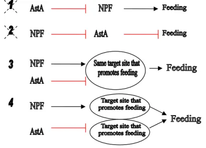
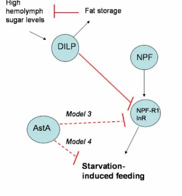
2.1 AstA neurons and NPF neurons co-regulate feeding behavior in opposing directions ... 101
2.2 Are AstA neurons involved in the regulation of feeding under normal conditions? ... 104
2.3 The regulation of starvation-induced feeding can be uncoupled from the regulation of starvation-induced hyperactivity... 108
2.4 Can we integrate molecular genetic approaches and neuroethological approaches to studying the regulation of feeding behavior?... 110
3. Are the observed feeding behavioral effects due to the action of AstA signaling?... 112
3.1 Experiments I have conducted to address this question... 113
3.2 Future experiments to address this question... 115
3.3 Orthologues of AstA signaling regulate feeding behavior ... 115
3.3.1 In arthropods... 115
3.3.2 In nematodes... 116
3.3.3 In mammals ... 116
4. What is the circuit-level mechanism that underlies the effect of AstA neuron activation to inhibit starvation induced feeding behaviors?
4.1 Neural mechanisms that regulate blowfly and
Drosophila satiety. ... 117
4.2 Does activation of AstA neurons inhibit feeding behavior by promoting proprioceptive feedback? ... 118
4.2.1 Proventriculus... 118
4.2.2 Motorneurons ... 119
4.2.3 Midgut neuroendocrine cells... 119
4.2.4 Humoral factor ... 119
4.3 How could central sites of AstA expression contribute to the behavioral effects of activation of AstA neurons? ... 120
4.3.1 Subesophageal ganglion ... 121
4.3.2 Pars intercerebralis ... 121
4.3.3 Protocerebrum ... 121
4.3.4 Optic lobes... 121
5. Other potential neuromodulators mediating the effects of activating AstA neurons 5.1 Serotonin... 122
5.2 Hugin ... 124
5.3 DILP-7 ... 125
6. Summary... 128
7. Bibliography ... 130
Appendix 1: A single population of olfactory sensory neurons mediates an innate avoidance behavior in Drosophila ...134
LIST OF ILLUSTRATIONS AND/OR TABLES
Chapter 1 Page
Figure 1. Anatomy of the digestive tract in Drosophila………..…………..5 Figure 2. A model that could explain the differential regulation of
feeding in unstarved and nutrient-deprived flies………….………..………11 Chapter 2
Figure 1. Generation and characterization of AstA-Gal4 transgenic flies……....…….39 Figure 2. Expression of AstA and AstA-Gal4 in the adult brain and
ventral nerve cord………..………..….………...……..40
Figure 3. Expression of AstA and AstA-Gal4 in the adult midgut,
hindgut, and rectum……….….………….41 .
Figure 4. Co-expession of AstA and AstA-Gal4 in the CNS of third instar larvae, ablating AstA-Gal4 cells highlights AstA+/Gal4- expression, potential AstA release sites in the brain……….……..…….….………..42 Figure S1. High-resolution images of abdominal ganglion...…….….….……..43 Chapter 3
Figure 1. Hyperexcitation of AstA neurons results in significantly
reduced feeding.….………..……….…60 Figure 2. AstA/NaChBac flies feed significantly less when tested in the CAFE
assay or fed standard fly food. Activation of NPF neurons partially
rescued the reduced feeding behavior or AstA/NaChBac flies..…….…..….61 Figure S1. Small meal sizes are easily visualized using food coloring. Activation of NPF neurons results in significantly increased food intake and
fraction of flies feeding………..…………..62 Chapter 4
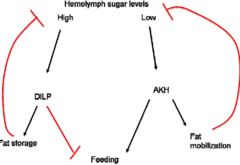
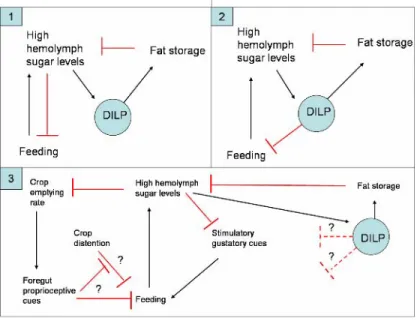
Diagram 1. Neuropeptides regulate hemolymph sugar levels……….………….65
Figure 1. Starved AstA/NaChBac flies do not have excess energy stores………92 Figure 2. AstA/NaChBac flies do not exhibit normal starvation-induced changes in sucrose responsiveness. The proboscis extension phenotype
does not account for the feeding phenotype of AstA/NaChBac flies...93 Figure 3. Starvation-induced hyperactivity of AstA/NaChBac flies was normal..…..94 Figure S1.Activity levels and locomotor agility were normal when AstA
neurons were activated...………94 Figure S2. Excessively depleting energy stores does not induce
AstA/NaChBac flies to feed.………..95 Figure S3. Starved AstA/NaChBac flies behave as though sated with
respect to the PER assay……….96 Chapter 5
Figure 1. Models of how NPF and AstA neurons regulate feeding behavior...………102 Figure 2. Possible mechanisms by which NPF and AstA neurons
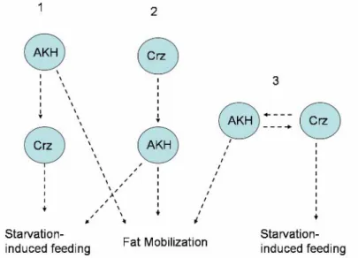
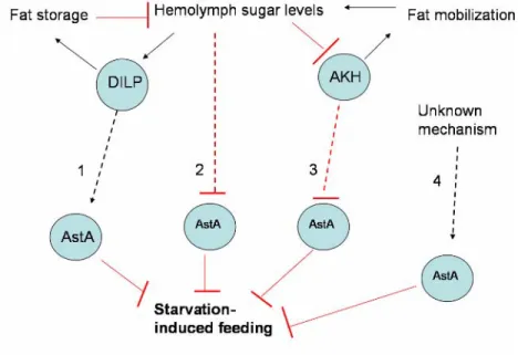
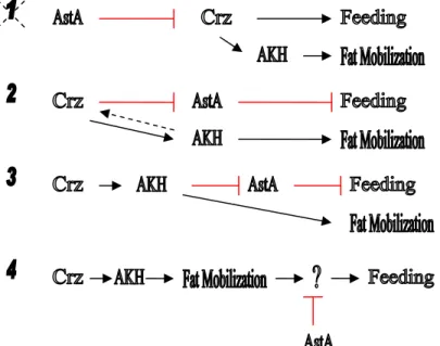
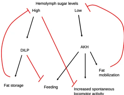
regulate starvation-induced feeding behavior……….………103 Figure 3. AKH signaling regulates fat mobilization and feeding in starved flies…...104 Figure 4. Several models that explain the interactions between Crz and AKH...…….105 Figure 5. Four potential mechanisms by which AstA neurons are regulated..….……106 Figure 6. There are several models to explain the interactions
between Crz and AstA…...……….107 Figure 7. The interactions between nutritional state, neuromodulators, energy
homeostasis, and behavior……..………109 Figure 8. Mechanisms by which feeding behavior may be regulated
in unstarved flies…...………..112 Appendix
Figure 1. Drosophila exhibits innate avoidance of odorants released
by stressed flies……….…………..………136
Figure 2. CO2 is a component of dSO. …...………..………...136 Figure 3. CO2 avoidance is mediated by ORNs that project to the V glomerulus. …..137 Figure 4. A dSO-unresponsive enhancer trap line is also defective in
its CO2 response. …...………..138
Supplementary Figure 1. Dose-response curve for avoidance
behavior to CO2. Dose-response curve for activation of GR21A+
neurons to CO2 , using the UAS-GCaMP reporter………....141
Neuoropeptides and neuropeptide receptors
AKH Adipokinetic hormone promotes fat mobilization in nutrient-deprived flies. AKH mutants do not display starvation-induced feeding or starvation-induced hyperactivity.
AKHR AKH receptor mutants do not display starvation-induced feeding but display normal starvation-induced hyperactivity.
AstA Allatostatin A has been implicated as a satiety factor and myoinhibitor in other insects.
Crz Corazonin neurons have been implicated in the regulation of stress responses and promote similar metabolic effects as AKH.
DAR-1 and -2 Drosophila AstA receptors
DILP Drosophila insulin-like peptide promotes the deposition of fat stores when hemolymph sugar levels are high. DILP is also required for normal growth and development and some studies have implicated a role for DILP in the regulation of feeding.
Hugin neurons promote feeding on novel foods in unstarved flies.
InR DILP receptor
JH Juvenile Hormone promotes the diversion of fat stores towards egg production in mated females. AstA inhibits JH synthesis in some insects, not including flies.
Leucokinin plays a role in the regulation of meal size.
NPF Neuropeptide F promotes starvation-induced feeding and foraging.
NPFR NPF receptor
Transgenic tools
Gal4/UAS This is a binary expression system used to spatially and temporally restrict transgene expression in Drosophila.
UAS-mcd8::GFP Overexpresses a membrane-tethered green fluorescent protein.
UAS-Kir2.1 Overexpresses an inwardly-rectifying potassium channel which reduces neuronal excitability.
UAS-NaChBac Overexpresses a voltage-gated bacterial sodium channel which increases neuronal excitability.
UAS-Ricin Overexpresses a biotoxin that causes cell-autonomous cell death.
UAS-TNT Overexpresses tetanus toxin which blocks synaptic transmission.
UAS-TRPA1 Overexpresses a temperature-sensitive cation channel which increases neuronal excitability at 28°C but not at 22°C.
Other
CAFE assay Capillary Feeding Assay CNS Central nervous system
DDC Dopa decarboxylase is an enzyme required for the synthesis of serotonin and dopamine.
MAN The median abdominal nerve transmits proprioceptive feedback from the crop, and is required to inhibit feeding in starved blowflies.
RN The recurrent nerve innervates the aorta, foregut, and crop. Proprioceptive feedback from the foregut, transmitted via the RN, inhibits feeding in blowflies.
SOG The subesophageal ganglion receives primary gustatory input.
SGS The stomatogastric nervous system regulates the motor patterns of feeding behavior and transmits peripheral sensory information from the gut. The SGS is analogous to the mammalian autonomic nervous system.
TH Tyrosine hydroxylase is required for dopamine synthesis.
VNC Ventral nerve cord
INTRODUCTION 1. The scientific relevance of studying satiety
Globally there are more than 1 billion overweight adults, 1/3 of which are obese, and rates of obesity are increasing worldwide [1]. Obesity is one of the leading causes of preventable deaths in the United States, responsible for 1 in 10 deaths [2]; It increases the risk factor for medical complications such as diabetes, cardiovascular disease, and some forms of cancer [3]. In the U.S., 9.1 % of total medical expenditures in 1998 were due to medical complications caused by being overweight [4]. Because of the many health and economic costs of obesity, it is important to better understand the regulation of energy homeostasis and feeding behavior.
The sheer number and diversity of life forms can be attributed to developing proficiency at: converting a variety of compounds into utilizable energy; storing excess energy for use in times of scarcity; and acquiring ways to adapt to harsh environments [5, 6]. In animals, regulatory mechanisms promote feeding on high calorie foods, promote energy storage, and minimize energy expenditure. The ever-increasing incidence of obesity in Westernized culture is fueled by an excess of high fat, high sugar foods in a technologically advanced urban world, where physical activity is a choice [6, 7]. It is medically and economically relevant to understand the neural and molecular underpinnings of hunger and satiety, how food is attributed a hedonic value, how food intake is regulated, and how the body maintains homeostasis.
In mammals, the central nervous system (CNS) integrates internal and external cues, regulates feeding behavior, and coordinates the activities of multiple peripheral organs to maintain homeostasis [5, 8]. Complex feedback loops between peripheral organs and central regulatory centers interact to maintain stable blood glucose levels and to induce feeding and foraging to replenish energy stores [5, 8]. How this barrage of information is
tracked and integrated to regulate feeding and energy homeostasis is not very well understood, and is an important and difficult challenge to scientists today [5, 8–10].
Drosophila melanogaster (fruit flies) provide an excellent model system in which to study this complex problem because many elements of feeding regulation and homeostasis are conserved between Drosophila and mammals [7, 11–13]. Over 100 years of Drosophila genetic research has resulted in the characterization of many genes, signaling pathways, physiology, and behaviors [14]. Extensive libraries of mutants and transgenics have been created, as well as tools for manipulating genes and neuronal circuits [15].
Historically, a limitation of Drosophila as a model system has been determining neuronal connectivity in the central nervous system [15]. The brain, which is the size of a poppy seed, is a dense ball of interconnected axons and dendrites (neuropile) surrounded by a shell of neuronal cell bodies [15, 16]. Although neuromodulatory neurons have been described in terms of molecular signaling and neuroanatomy, connectivity has been difficult to determine because the same neuronal processes can contain both inputs and outputs [15–17]. How “the as yet impenetrable interneuron jungle” [18] of the brain regulates behavior has been a long-standing challenge [15, 16]. Recently, transgenic tools have been developed that allow us to establish connectivity, to visualize real-time neuronal activity, and to inducibly activate and silence neuronal circuits [15, 16]. This affords us the opportunity to leverage the knowledge of Drosophila genes, signaling pathways, physiology, and behavior to tackle the questions of how the CNS coordinates homeostasis and feeding behavior.
In Drosophila, little is known about how the CNS regulates feeding behavior in response to homeostatic perturbations [9, 11, 17]. Although energy homeostasis and feeding behavior are tightly regulated [11, 19], the majority of studies have focused on energy metabolism while mostly overlooking the regulation of feeding behavior [10, 17].
In addition, most of these studies have focused on the feeding behavior of larvae, which feed continuously and are in a life stage of rapid growth [7, 12]. In contrast, adult Drosophila have exited the growth phase, are discontinuous feeders, and face more complex life decisions than larvae [7, 12]. Understanding the regulation of feeding
feeding is regulated [7, 12]. Studying insect feeding behavior could also contribute to improved methods of pest control, which could be used to target insect populations that transmit diseases and that damage agricultural crops.
2. The regulation of satiety: Lessons from the blowfly
The majority of current knowledge about the neural regulation of insect feeding comes from in-depth investigations of the feeding behavior of the blowfly, or Phormia regina, and, according to studies conducted in other insects, this knowledge can be generalized to other insects [17, 18, 20]. The mechanisms that regulate feeding behavior in starved blowflies are different from those that regulate feeding under ad libitum1 feeding conditions [19, 21]. I will focus mainly on how feeding is regulated under starvation conditions in order to better understand how a state of satiety is achieved. First, I will summarize the studies that characterized the neural regulation of feeding in starved blowflies; these studies are described in detail in [20].
In the blowfly, satiety is measured in terms of sugar responsiveness [21, 22]. A starved fly will respond to and accept a lower concentration of a sugar solution than will a fed fly [21, 22]. This can be quantified by stimulating the taste sensillae on the foreleg of a fly with stepwise increasing concentrations of a sugar solution until the fly responds by extending its proboscis2, or mouthparts. The lowest concentration to which a fly responds is designated the acceptance threshold3, which, depending on the degree of starvation, can vary over a hundred fold range of concentrations for some sugars. This method measures the relative degree of starvation either between flies or within a single fly over time. Thus a low or reduced acceptance threshold represents hungry or more nutrient-deprived flies
1 Ad libitum means “at one’s discretion” in Latin.
2 The fly proboscis is a retractrable straw-like appendage through which flies feed.
3 “Although threshold lies somewhere between the concentration that elicits extension and the one in the series immediately below it, the higher of the two is arbitrarily designated as threshold. Since the aim of practically all experiments was to obtain data for comparative analysis, this fiction was acceptable” [20].
and a high acceptance threshold represents a state of satiety.
There are several physiological mechanisms that regulate satiety in the starved blowfly. The first and most potent inhibitor of feeding is crop distention [23]. The crop is an expandable sac that arises from an invagination of the foregut ([17], see Figure 1).
When a starved fly feeds, food fills the midgut first, where the majority of digestion and nutrient absorption occurs [10], and then fills the crop, which is mostly used for food storage [17]. When food was prevented from entering the midgut, by tying off or severing the midgut, the volume of food intake of starved flies was comparable to sham operated starved flies, as was the increase in acceptance threshold [20]. Since little or no digestion occurs in the crop, this demonstrates that a state of satiety can be induced without food entering the midgut or ensuing nutrient absorption. Consistent with the hypothesis that nutrient absorption is not mediating these effects, injection of a sugar solution into the hemolymph of a starved fly was not sufficient to decrease the acceptance threshold [20].
Thus, a state of satiety can be achieved by the act of feeding, regardless of hemolymph sugar levels.
Anatomical and electrophysiological studies elucidated the mechanism by which crop distention regulates satiety. Studies demonstrated that severing the median abdominal nerve (MAN) results in hyperphagia, defined as at least a twofold increase in food intake relative to sham-operated controls [24]. The MAN contains projections from a nerve net surrounding the crop [25]. Electrophysiological recordings from this nerve revealed neurons that increased their firing rate upon crop distention, indicating that proprioceptors are monitoring crop distention. Therefore it is proposed that proprioceptive feedback from the crop is necessary and sufficient to promote satiety [26].
A sagittal view of the digestive tract illustrated within the body of Drosophila (reprinted with permission from [27]. Shown below, is an illustration of the digestive tract indicating the foregut (green), crop (yellow), midgut (red and pink), and hindgut (blue). The crop duct branches off of the foregut. The proventriculus (red) contains a valve that separates midgut from foregut contents.
A second mechanism that contributes to the promotion of satiety in starved blowflies is foregut activity. When food was prevented from entering the crop, by lesioning or tying off the crop duct, sugar acceptance threshold rose for a few hours [20]. This suggests that the crop is not the only source of inhibitory feedback. To determine whether food within the midgut is sufficient to promote satiety, food was injected into the midgut of
a starved fly [20]. Surprisingly, 80% of these flies exhibited no change in sugar acceptance threshold compared to pre-injection. These flies were dissected to ensure that no food had passed into the foregut, which is separated from the midgut by the cardiac valve. Since tying off the crop duct before feeding was sufficient to suppress the acceptance threshold, but injecting food into the midgut was not, this suggests that food passing through the foregut was responsible for the satiety effect seen in cropless flies.
Additional lesioning studies revealed the mechanism underlying this satiety effect.
When experimenters severed the recurrent nerve (RN), flies became hyperphagic [24]. The RN is part of the stomatogastric nervous system (SGS) in insects, which is analogous to the mammalian autonomic nervous system, and it innervates the aorta, foregut, crop, and hindgut [11]. To control for the possibility that severing the RN was impairing efferent motor control of the gut, several studies demonstrated that the motor patterning of food movement through the gut of these hyperphagic flies was normal [20]. First, food was able to enter the midgut and crop, and, as food was digested and the midgut emptied, the transfer of food from the crop to the midgut was normal4 in hyperphagic flies. Further evidence that the SGS is not required for the motor patterning of food digestion is the fact that a digestive tract removed from the fly (dissected out and placed in saline) will continue the pattern of midgut emptying and food transfer until the crop is empty. These results support the hypothesis that the presence or movement of food through the foregut promotes a satiety effect, and that severing the RN does not sever motor neurons that are required to move food through the gut.
Electrophysiological recordings from the RN identified neurons whose firing rate was inversely correlated with the rate of crop emptying [26]. Although chemoreceptors exist in the foregut, they do not send their projections through the RN [23]. This led to the conclusion that proprioceptors are monitoring foregut contractions and are necessary and sufficient to produce a satiety effect. It is quite surprising that neither the presence of food in the midgut nor hemolymph sugar levels is sufficient to promote a satiety effect under
4 A two-way crop valve opens and a “slug” (bolus) of food is transferred by reverse peristalsis up the crop duct and into the foregut. The cardiac valve, which separates the foregut from the midgut, opens and the slug is transferred into the midgut.
Hyperphagia resulted when either the MAN or the RN was severed, but the feeding behavior as a result of these lesions was different [23]. Severing the MAN resulted in flies taking one long continuous meal, whereas severing the RN resulted in flies taking repeated near-normal sized meals. These results suggest that crop distention (detected by the MAN) regulates meal cessation, while foregut contractions (detected by the RN) regulate meal initiation. Dramatically, when both nerves were severed, flies continued to feed until they burst.
RN and MAN lesioning in starved flies also results in polydypsia (water hyperphagia) [28]. Flies, like mammals, independently regulate water intake and food intake [19, 28]. To show that the primary effect of these lesions was not solely to promote drinking, the tastant stimulus presented to lesioned flies was varied [28]. When lesioned flies were feeding from a sucrose solution and the stimulus was switched to water, flies ceased drinking. Additional evidence was provided by switching the solution to ever increasing concentrations of solutions. When lesioned flies stopped drinking water, 100 mM sucrose was presented. This elicited feeding until the fly could not generate enough force to fill the crop any further despite continued efforts to feed. Thereupon, a 1 M sucrose solution was presented and this resulted in more vigorous pumping (sucking) until the fly burst.
There is evidence for additional mechanisms that promote satiety from ventral nerve cord (VNC) lesioning studies. Severing the cervical connective, which connects the brain to the VNC resulted in hyperphagia, and the effect of this lesion was stronger than that of severing the MAN, suggesting that the results are not merely due to a loss of the MAN (which projects to the VNC) [24]. The VNC is composed of several fused ganglia that control motor patterns, receive sensory feedback, and send and receive feedback to the brain [20]. This inhibitory feedback could be due to either loss of proprioceptive feedback from the abdomen or from locomotor centers in the VNC.
Hyperphagia as a result of lesioning the VNC may be due to loss of proprioceptive feedback from the abdomen. Experiments have demonstrated that abdomen distention is
necessary and sufficient to regulate drinking (water) [28]. “Bleeding” the fly, by nicking the cuticle and squeezing out hemolymph5, stimulated drinking, whereas when fluid was injected into the abdomen, inhibited drinking. Surprisingly, injection of either hypertonic or hypotonic solutions was sufficient to inhibit drinking, which suggests that changes in hemolymph osmolarity are insufficient to promote drinking. Instead, these results suggest that abdomen distention regulates drinking.
Lesioning the VNC may also remove inhibitory feedback from locomotor centers.
Hungry flies stop walking when they encounter a food source and during feeding, which suggests that feeding and locomotion are mutually exclusive events [29]. Indeed, it has been demonstrated that insects that are induced to fly exhibit a surprisingly high acceptance threshold despite the energy drain imposed by flight [20]. Therefore, locomotor centers in the VNC may be exerting an inhibitory influence on feeding behavior.
All of the previously described experiments were conducted on flies that were starved 24–48 hours. The lesioning studies in starved flies are provocative, and demonstrate that an empty gut and crop triggers a strong drive to feed that overrides any effects of hemolymph sugar levels or humoral factors. The crop is practically empty after 24 hours of starvation, only 5% full compared to the total capacity of the crop, and is empty after 48 hours of starvation [23]. In nature, the crop provides a safety net against starvation. Sometime between 24 and 48 hours of starvation, these flies will need to revert to internal energy reserves for energy.
These experiments demonstrate that in starved blowflies, feedback from the RN, the MAN, and the VNC is necessary to promote satiety, or reverse the drive to feed.
Recording from the RN and the MAN electrophysiologically demonstrated that these nerves carry information about gut distention. This suggests that proprioceptors monitor crop volume and foregut activity, that sensory neurons relay this information via the RN and MAN, and that this proprioceptive information is necessary to return to a state of satiety. These findings are compelling, but it must be noted that these lesions disrupt
5 Hemolymph is insect “blood”.
of this surgery is demonstrated by the high levels of morbidity: nerve-transected flies die within 1–3 days after surgery [20].
In contrast to the drastic effects of nerve transection on feeding behavior in starved flies, ad libitum fed flies starved 8 hours or less only exhibit a mild decrease in acceptance threshold upon RN transection and acceptance threshold is unaffected by MAN transection [21]. Interestingly, ad libitum fed blowflies that are supplied with carbohydrates but not protein in their diet, do become hyperphagic when the RN or MAN are severed [30]. This suggests that the regulation of satiety in blowflies is affected by beginning nutritional state.
Perhaps nutrient-deprived flies are more reliant on peripheral feedback to determine acceptance threshold, and that peripheral feedback is a requirement to return to steady state.
Nutrient deprivation may trigger a switch in behavioral responses, towards a more aggressive drive to feed, and that proprioceptive detection of food in the gut is required in order to turn off this drive to feed.
Thus far, I have focused on what is known about peripheral mechanisms that promote satiety in blowflies. Such a thorough examination of the regulation of satiety has not been reported for any other insects, though similar mechanisms of inhibitory feedback have been demonstrated in other insects [17, 18, 20]. In Drosophila, very little is known about the regulation of satiety [17]. Lesioning experiments that would demonstrate inhibitory feedback from the crop or foregut have not been reported in Drosophila melanogaster, despite the comment that “Bodenstein perfected the technique” of RN severing in Drosophila, though this reference might have been to a larger species of Drosophila [20]. Due to their small size, lesioning experiments in Drosophila melanogaster would be difficult, but analogous experiments could be performed using transgenetic tools that silence or ablate neurons.
How does the feeding behavior of blowflies compare to what is known about Drosophila feeding behavior? Similar to blowflies, Drosophila increase their acceptance of low reward food cues with increasing starvation, and the volume of food consumed also increases as a function of starvation and the stimulatory value of the food offered [19, 21].
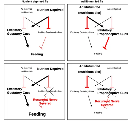
Dethier and colleagues [20] propose that feeding is regulated by gustatory cues, that satiety is regulated by proprioceptive feedback, and that the decision to feed involves a push-pull relationship between the stimulatory value of gustatory cues and the inhibitory feedback from gut distention. The feeding behavior of starved Drosophila agrees with this model, but similar to ad libitum fed blowflies, a different model is needed to describe the feeding behavior of unstarved Drosophila.
Nutritional state determines how food intake is regulated. Fed on a high concentration of sucrose (50 mM or higher), ad libitum fed Drosophila have little or no food in the crop, whereas fed on lower concentrations (10–25 mM), ad libitum fed flies store a significantly larger volume of food in the crop [19]. Here, more stimulating gustatory cues are not promoting increased food intake, and a simple model in which the decision to feed involves only inhibitory proprioceptive feedback and excitatory gustatory cues does not apply.
The neural mechanisms that regulate feeding behavior are dependent on nutritional state. A model that incorporates the influence of nutritional state on insect feeding behavior, and that is consistent with behavioral studies as well as lesioning studies, has not been reported. A model that is consistent with behavioral studies in blowflies and that incorporates the influence of nutritional state on feeding behavior is summarized in Figure 2. In nutrient-deprived flies, nutrient deprivation overrides inhibitory proprioceptive feedback, and promotes the acceptance of food. Ad libitum fed flies, fed a nutrient balanced diet, will not utilize the crop for food storage, and will only accept a highly stimulating food source. This suggests that inhibitory feedback from the gut is high in fed Drosophila. Since severing the RN and MAN in fed blowflies had little or no effect on food acceptance, suggests that these models need to be revised to agree with both blowfly and Drosophila feeding behavior. This model can account for this discrepancy, if gustatory cues and proprioceptive cues are simulateously regulated in opposite directions, depending on nutritional state. Excitatory gustatory cues and inhibitory proprioceptive cues are sensed immediately, whereas nutritional state only gradually changes. A nutrient-deprived fly (either starved or protein-deprived), has a strong drive to accept food, but when the RN is
helps to explain the hyperphagia observed in starved nerve-severed flies. On the other hand, studies found that ad libitum fed flies only had a mild phenotype from RN transection. This could be explained if there was an independent mechanism providing an ongoing inhibition of food acceptance in a “sated” fly, which would minimize the feeding effects of removing inhibitory proprioceptive feedback.
Figure 2. A model that could explain the differential regulation of feeding in unstarved and nutrient-deprived flies
Inhibitory
Proprioceptive Cues
Inhibitory
Proprioceptive Cues
Ad libitum fed (nutritious diet)
Excitatory Gustatory Cues
Nutrient Deprived
Inhibitory Proprioceptive Cues
Feeding
Ad libitum fed (nutritious diet)
Excitatory Gustatory Cues
Nutrient Deprived
Feeding
Ad libitum fed (nutritious diet)
Excitatory Gustatory Cues
Nutrient Deprived
Inhibitory Proprioceptive Cues
Feeding
Ad libitum fed (nutritious diet)
Excitatory Gustatory Cues
Nutrient Deprived
Feeding
Recurrent Nerve
Severed Recurrent Nerve
Severed + Recurrent
nerve severed
Nutrient deprived fly Ad libitum fed fly
Inhibitory
Proprioceptive Cues
Inhibitory
Proprioceptive Cues
Ad libitum fed (nutritious diet)
Excitatory Gustatory Cues
Nutrient Deprived
Inhibitory Proprioceptive Cues
Feeding
Ad libitum fed (nutritious diet)
Excitatory Gustatory Cues
Nutrient Deprived
Feeding
Ad libitum fed (nutritious diet)
Excitatory Gustatory Cues
Nutrient Deprived
Inhibitory Proprioceptive Cues
Feeding
Ad libitum fed (nutritious diet)
Excitatory Gustatory Cues
Nutrient Deprived
Feeding
Recurrent Nerve
Severed Recurrent Nerve
Severed + Recurrent
nerve severed
Nutrient deprived fly Ad libitum fed fly
3. Neuromodulators that promote satiety in other insects
Many experiments were conducted to determine whether elements of hemolymph composition can regulate feeding behavior in blowflies, and some of these experiments led to some surprising conclusions. As mentioned previously, injecting a sugar solution into
the hemolymph of a starved fly was unable to decrease the acceptance threshold at any time in the following 24 hours [20]. Regardless of whether a hypo- or hyper-osmotic solution was injected, the acceptance threshold of the fly did not increase. Further attempts were directed at identifying soluble factors that could promote satiety. When a starved fly is given a transfusion of hemolymph from a fly that had taken a meal two hours previously, the acceptance threshold of 80% of the transfused flies did not increase within the following two hours from the starting threshold [20]. Parabiosis is a more direct method to determine whether a humoral satiety cue exists in flies. Parabiosis involves surgically connecting a pair of flies so that the hemolymph is shared. A section of cuticle is removed from each fly, and paraffin is used to seal one fly onto the back of the other. (The flies
“took turns riding piggyback”). At the beginning of the experiment, both flies had been starved for 48 hours. Afterwards, only one of the two flies was fed to repletion, while the other was left unfed. The sugar acceptance threshold of the unfed fly was monitored, and no change in threshold was observed. This experiment was continued for three days, in which the fed fly was always fed to satiation, and the acceptance threshold of the unfed fly was monitored. After three days of experiments, it became clear that there was not a humoral factor present in the hemolymph that could promote satiety in an unfed fly.
Since these parabiosis studies concluded that no soluble factor (including sugars) could promote satiety, it would have been informative to characterize the volume of food intake of the fed fly. If the fed fly of the parabiosed pair of flies consumed the same volume of food compared to an unparabiosed fly, then this result would have provided further evidence that sugar levels or a humoral factor in the hemolymph is not sufficient to influence feeding behavior, since the hemolymph is shared in parabiosed flies and any nutrients absorbed by the fed fly would be diluted and any humoral factors released in response to feeding would be diluted. Alternatively, if the fed fly of the parabiosed pair of flies consumed an increased volume of food compared to a single unparabiosed fly, then this result would suggest that a humoral factor could promote feeding (but not promote satiety in an unfed fly).
Parabiosis studies suggest that a humoral factor in the hemolymph is not sufficient
products of sugar conversion do not suppress hunger. A confound of transfusion and parabiosis experiments is that these experiments involve an unnatural series of events: The nervous system never encounters this combinatorial event—nutrition without ingestion.
Nevertheless these findings, in combination with experiments discussed in the last section, suggests that at least for starved blowflies satiety is achieved solely through inhibitory proprioceptive feedback from the foregut, crop, and abdomen, and possibly also from inhibitory feedback from locomotor centers in the VNC.
In regards to the neural regulation of feeding behavior in unstarved flies, experiments characterizing the feeding behavior of blowflies starved less than 8 hours do not support the conclusion that peripheral inhibitory feedback is the only mechanism that can promote satiety. Several studies demonstrate that blowflies starved for 8 hours or less were unaffected by MAN lesioning and were only mildly affected by RN lesioning. These findings suggest that results from studies in starved blowflies involving nerve lesions or the direct manipulation of hemolymph content generate artificial scenarios (food intake without gut distention or replenished energy stores without food intake) ought to be interpreted conservatively.
How nutritional state alters the mechanisms by which feeding behavior is regulated is unknown. Gut proprioception may be involved in signaling nutritional state. This is because crop emptying rate is dependent on hemolymph osmolarity [20]. Differences in crop emptying rate would produce distinctive patterns of proprioceptive feedback, which could provide a readout of nutritional state.
In contrast to the findings that a humoral factor could not promote satiety in transfusion and parabiosis experiments, many studies have demonstrated that injected neuromodulators are able to promote satiety in blowflies and other insects. Injection of biogenic amines, including serotonin and dopamine, and neuropeptides, including sulfakinin, leucomyosuppressin, insulin-like peptides, and allatostatins have been demonstrated to decrease feeding behavior in insects.
Sulfakinin has been implicated as a “satiety factor” in several insects, including
blowflies, Tabanus nigrovittatus (salt marsh horse fly), Blattella germanica (cockroach), and Schistocerca gregaria (desert locust) [31]. In the blowfly, sulfakinin injection reduced carbohydrate but not protein intake of females (but not males) without reducing the percent of flies feeding [31]. In the locust, injection of sulfakinin reduced food intake without affecting the sensitivity of taste receptors [32]. In B. germanica, sulfakinin injection resulted in decreased food intake and sulfakinin was found to be myostimulatory [33]. It has also been shown to reduce feeding, stimulate gut contractions, and stimulate alpha- amylase secretion in the desert locust and blowflies [31, 32]. Interestingly, sulfakinin is a structural orthologue of mammalian Cholecystokinin, which is also known to decrease feeding, stimulate gut contractions, and stimulate secretion in mammals [31].
Despite being conserved as regulator of feeding behavior in other insects, in Drosophila sulfakinin is neither myostimulatory nor does it affect feeding behavior [34]. A long history of genetic studies in Drosophila have only identified few neuromodulators that promote satiety [10]. Neuromodulators that have been implicated in promoting Drosophila feeding include an insulin-like peptide, serotonin, and hugin.
Both Drosophila insulin-like peptide (DILP) and its receptor (InR) are required for normal larval feeding behavior [35–37]. Mutations in these genes caused a reduction in feeding, growth, and development, while overexpression caused an increase in feeding, growth, and development. This shows that the function of insulin has been conserved as a regulator of growth and development in animals [12]. There is also evidence for a conserved role for DILP neurons in the monitoring of internal energy stores. Transcripts of DILP are reduced during starvation and hemolymph DILP levels rise after feeding. In addition, changes in glucose or trehalose levels induce calcium release in adult DILP neurons [11].
Several lines of evidence suggest that DILP signaling is also involved in the regulation of feeding behavior independent of its metabolic/developmental effects. One study observed that the feeding deficits as a result of knocking down the DILP receptor (InR) expression preceded the developmental deficits. Furthermore, growth deficits of InR
suggests that the growth phenotype of InR mutants results from the feeding phenotype.
Since InR and serotonin have many target tissues, it remains to be determined where these two signaling pathways overlap.
In both invertebrates and mammals, serotonin signaling regulates both metabolism and feeding behavior [38]. In the blowfly, serotonin injection inhibited both sugar and protein intake [39]. Injection also resulted in an increased acceptance threshold as well as weight loss. Similar effects on feeding due to serotonin injection have been observed in other insects [40]. Serotonin can stimulate foregut and crop contractions in other invertebrates [41, 42]. A functional role for serotonin in insect feeding behavior is supported by several immunohistological studies. In Neobelliera bullata (flesh fly), immunoreactivity of serotonin in the subesophageal ganglion (SOG), which receives primary gustatory input, decreased post-feeding [40]. This suggests that serotonin was released in this region as a result of feeding. Similar conclusions were drawn from studies in Rhodnius prolixus (triatomid bug), in which serotonin release from neurosecretory cells was observed in response to feeding [43]. Further supporting a neuromodulatory role for serotonin in feeding, is the presence of serotonin in the insect stomatogastric nervous system.
Consistent with earlier suggestions that DILP signaling in serotonergic neurons promotes feeding behavior in Drosophila, are studies by Neckameyer and colleagues that demonstrated that serotonin is involved in the regulation of feeding in Drosophila as well [44]. Adult flies with a null mutation in Tph2, which is one of the enzymes required for serotonin synthesis, exhibited decreased feeding behavior [44]. Unstarved adult Tph2 mutants consumed less over a 24 hour time period, exhibited decreased activity levels, and a decreased heart rate. Since there is a paralogue of Tph in Drosophila, and since Tph is
6 Overexpressing InR in DDC neurons rescued the InR -/- growth phenotype, whereas overexpressing InR in TH neurons did not. Dopa decarboxylase (DDC) is an enzyme that is required for the synthesis of both serotonin and dopamine. Tyrosine hydroxylase (TH) is required for dopamine synthesis. The expression of DDC and TH was found to only minimally overlap.
Because neurons that coexpressed both TH and DDC were not proximal to DILP neurons, this led to the conclusion that DDC driven rescue of InR-/- occurred in serotonergic neurons and not dopaminergic neurons.
required for serotonin and dopamine synthesis, the authors demonstrated that Tph2 is exclusively expressed in serotonergic neurons in the adult, and not in dopaminergic neurons. These results support a conserved role for serotonin in regulating feeding behavior.
Conversely, Neckameyer observed that knockdown of Tph1 in the fat body resulted in increased mouthhook contractions and hyperactivity in larvae [44]. Feeding studies could not be done in Tph1 null mutants due to severe developmental deficits. Authors showed that dopamine but not serotonin is expressed in the fat body, and therefore concluded that the loss of serotonin was not responsible for the observed increased feeding behavior. Despite a previous study that demonstrated dopamine to modulate food intake and acceptance threshold [39], and numerous studies that have implicated dopamine in arousal [45, 46], the authors concluded that the feeding and hyperactivity phenotypes of fat body Tph1 knockdown larvae was due to toxic levels of tryptophan, the precursor to dopamine, and not due to serotonin or dopamine levels [44].
In Drosophila, two additional neuromodulators have been indirectly shown to modulate satiety-like behavior. Flies with null mutations in leucokinin or its receptor exhibited increased meal sizes compared to controls, though a gain of function phenotype for leucokinin was not reported. Leucokinin mutants consume larger than normal meals but compensate for the excess intake by decreasing meal frequency [47]. Given the expression of leucokinin in muscle tissue in the proventriculus it is likely that the leucokinin feeding phenotype is due to a malfunction of the cardiac valve, which separates the foregut from the midgut. A family member of leucokinin has similarly been implicated in modulating the activity of the cardiac valve. Injection of leucomyosuppressin into Blattella germanica resulted in a reduced feeding phenotype and an accumulation of food in the foregut [33].
Another study has provided indirect evidence of another potential satiety factor in Drosophila. Silencing of hugin neurons resulted in a contextual feeding phenotype: on being transferred from regular fly food to yeast paste, silencing of hugin neurons in adult flies resulted in a much shorter latency to feed than in control flies [48]. Transferred from yeast to yeast, or yeast to regular fly food, or regular food to regular food did not result in
from controls. This contextual phenotype was observed in only unstarved flies, and silencing of hugin neurons did not alter the rate of food intake in starved flies. Starvation- induced feeding was also normal. Since starvation reduces the latency to accept novel foods, and since hugin is downregulated in response to starvation, these results suggest that hugin neurons play a role in promoting the acceptance of novel foods. Another interpretation could be that hugin neurons are involved in the regulation of protein feeding, since many standard fly foods provide low levels of protein and increased acceptance of yeast paste could reflect increased protein hunger. This would explain why the other feeding conditions did not result in differences in latency to feed. Indeed, authors also reported that hugin expression was downregulated in wild-type flies fed only sugar (protein-deprived) [48]. The expression pattern of hugin further supports a role for hugin neurons in feeding behavior, as hugin is expressed in neurons that project to pharyngeal muscles and to regions of the brain where primary gustatory neurons and DILP cell bodies are located [17]. Overexpressing hugin within hugin neurons did not alter rates of food intake. This could indicate that additional neuromodulators expressed in hugin neurons are responsible for the effects observed upon silencing these neurons. Another possibility is that hugin-overexpressing flies were not tested under the proper feeding conditions (testing protein versus carbohydrate feeding). It would be interesting to determine whether hugin is involved in regulating protein metabolism. Ubiquitous overexpression of hugin resulted in growth deficits and lethality, which suggests a role for hugin development. Further studies are needed to establish the function of hugin-expressing neurons in the regulation of feeding behavior.
4. Allatostatin A
In insects, the neuropeptide Allatostatin A (AstA) is a potential satiety factor. In the cockroach, injection of AstA reduced food intake by 60% [49, 50]. Expression of AstA was shown to be anti-correlated with the feeding behavior of females. During the 7-day gonadotrophic cycle, or female reproductive cycle, food consumption was highest in the
middle of the cycle, and AstA transcript levels were highest at the beginning and the end of the cycle, consistent with a role for AstA to inhibit feeding. This observation was inconsistent with an independent study that demonstrated that peptide levels increased steadily during the gonadotrophic cycle. Additional effects of AstA injection in these studies included the inhibition of hindgut (but not foregut) contractions and an increase in the secretion of alpha-amylase, an enzyme that digests starch, in the midgut [50].
The function of AstA in adult cockroach, crickets, and termites is to inhibit Juvenile Hormone (JH) synthesis [51]. JH regulates the metabolic switch that occurs in females post-mating, by diverting diverting fat stores towards egg production. Mated females also exhibit increased feeding behavior compared to unmated females. In conditions of starvation and short photoperiod/low temperatures, AstA levels increase, inhibiting JH synthesis which, in turn, inhibits egg production and leads to the build-up of fat stores.
Thus, reproductive state and environmental conditions regulate AstA levels, and AstA inhibits the metabolic switch that occurs as a result of mating in some insects.
Another study implicated a role for AstA in the regulation of feeding, but results were difficult to interpret. In Gryllus bimaculatus, females were injected with AstA (RNAi) at emergence and tested 2 days later. Knockdown of AstA was confirmed by Q- RT-PCR [49]. Food intake of injected virgin females was 38% reduced after 30 minutes but 79% increased after 60 minutes of feeding [49, 52]. Since AstA inhibits JH synthesis in this species, and AstA levels are high in unmated females, knockdown of AstA in virgin females would be predicted to result in the disinhibiton of JH, which would lead to a diversion of fat stores to egg production and to increased feeding. This study next reported that injected mated females, which should have low levels of AstA and high levels of JH, exhibited 30% decreased alpha-amylase activity after one day, yet after two days, enzyme activity was increased by 300%. Mated females would likely have low levels of AstA and therefore knockdown of AstA would have little effect on JH synthesis. Perhaps the logic of the authors was to tease apart a dual role for AstA in the cascade of effects due to JH synthesis from a role for AstA in the gut to promote alpha-amylase release.
AstA has also been implicated in the regulation of feeding behavior in insects in
is not necessarily an indirect effect of inhibiting JH synthesis. Injection of AstA into the larvae of both L. oleracea and S. littoralis resulted in decreased feeding and growth, but only if the larval stage injected was in a feeding stage (injection during a non-feeding stage had no effect) [10].
Many neuropeptides that have been implicated in insect feeding behavior also have myoinhibitory or myostimulatory effects [10]. Perhaps their effects on feeding are by modulating proprioceptive feedback from the gut. It has been difficult demonstrate a causal role for central versus peripheral expression of neuropeptides because many are expressed both centrally and peripherally, and many are expressed at neurohemal release sites, or sites where neuromodulators can be directly released into the hemolymph [10, 53].
5. Does Drosophila have a brain-gut axis?
In mammals, the gastrointestinal tract is “the largest endocrine organ in the body”
[54]. Mechano-chemo-, and noci-ceptive information is communicated to the CNS directly via the vagus nerve and by neuromodulators via the circulatory systems. The gut can produce over 100 bioactive peptides and some of these can modulate feeding and satiety;
gut distention also affects feeding behavior in mammals.
The role of the adult stomatogastric nervous system (SGS) in the regulation of feeding behavior has been overlooked in Drosophila [10, 55]. The SGS innervates and regulates the aorta, foregut, crop, and proventriculus [11]. Little is known about the type of information afferent sensory cues transmit or how efferent cues control digestion and gut motility [55]. A number of neuropeptides that have been implicated in insect feeding behavior are either expressed in the gut or the SGS: sNPF and FMRFamide are expressed in neurons that project to the anterior midgut; FMRFamide and possibly dromyosuppressin are expressed in neurons that innervate the crop [56]; DILP3 is expressed in gut muscle in the proventriculus, foregut, and midgut [56]; leucokinin is expressed in muscle near the proventriculus [47]; and motorneurons expressing AstA, pigment dispersing factor (PDF), and proctolin innervate the posterior midgut and hindgut [51, 56]. In addition, receptors for
AstA, DILP, PDF, leucokinin, tachykinin, and diuretic hormone are expressed in the midgut and hindgut, and receptors for NPF and hugin are expressed in the crop according to microarray studies [65]. The function of neuropeptide expression in the gut and SGS has not been adequately addressed.
Neuroendocrine cells in the midgut of Drosophila express a variety of different neuropeptides, including NPF, AstA, AstB, AstC, tachykinin, diuretic hormone, FMRFamide, and possibly sulfakinin [56]. A number of these neuropeptides have been implicated in the regulation of feeding and homeostasis but there has been little attempt to address the potential contribution of gut neuroendocrine expression to feeding phenotypes.
The functions of these midgut endocrine cells could be to sense gut content, to stimulate or produce digestive enzymes, or to modulate gut motility.
Understanding the function of these various neuropeptides in digestion and feeding behavior would be informative. If proprioceptive feedback is primarily responsible for modulating feeding behavior in Drosophila, then neuropeptides with myoinhibitory and myostimulatory properties could indirectly modify feeding behavior. The satiety effects observed in other insects upon AstA injection, could be due to myinhibitory properties of AstA. If feeding behavior is regulated by gut motility that is sensed by proprioceptive feedback, then directly altering gut motility would affect feeding behavior. The myoinhibitory effects of AstA have been demonstrated in cockroach (Leucophaea maderae, Blattella germanica), moth (Manduca sexta), lobster (Homarus americanus, Homarus gammarus), and crab (Cancer borealis). Interestingly, in the crayfish, AstA has myostimulatory properties [50, 57]. Alternatively the effects of AstA on feeding behavior could be to directly modulate the firing proprerties of proprioceptive neurons. AstA is co- expressed with serotonin in the SGS of Crustacea, in both lobsters and crabs [58, 59] Co- expression has been demonstrated in stretch receptors neurons that inhibit the pyloric and gastric mill rhythms in these organisms. In the crab, bath application of both AstA and serotonin inhibited contractions and co-application had a stronger effect than either alone [60]. AstA was also shown to be co-expressed with Acetylocholine in the SGS.
Based on the co-expression of serotonin and AstA in other species, these may be
neuromodulators inhibit feeding in multiple organisms [10, 38]. The function of AstA in Drosophila is unknown [53]. Expression of AstA in feeding-related endocrine centers of Drosophila suggests that AstA might play a role in feeding behavior [61]. Drosophila eclose with food in the gut from their last meal as larvae [61]. It has been suggested that expression in the hindgut of pupae might serve to withhold the final meal of the larvae [61].
In addition, there is a conserved role for orthologues of AstA receptors in the feeding behavior of mammals and Caenorhabditis elegans [62, 63].