I hereby declare that the research results of this dissertation are the result of conducted research. Ankit Gangrade for the award of the degree of Doctor of Philosophy (Ph.D.) is an authentic report of the results obtained from the research carried out under my supervision at the Department of Biosciences and Bioengineering, Indian Institute of Technology Guwahati , India , and this work has not been submitted elsewhere for the award of any other degree.
A CKNOWLEDGEMENT
Bombyx mori Silk Fibroin Nanoparticle (BMNP) Impregnated Injectable Silk Hydrogel for Sustained Delivery of Cisplatin and its In Vitro
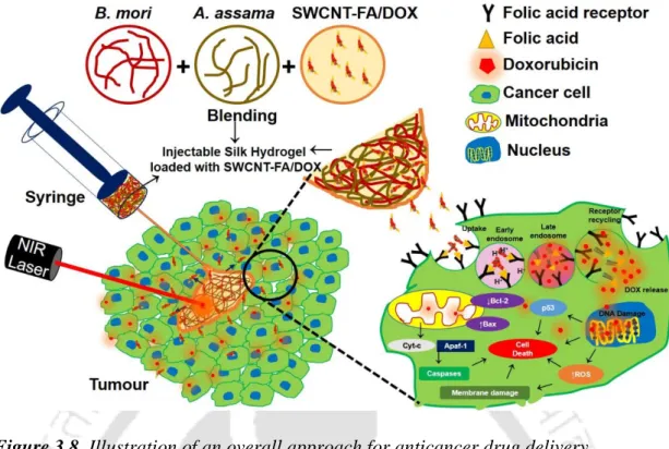
CHAPTER 3: Single-Walled Carbon Nanotube (SWCNT) Impregnated Injectable Silk Hydrogel for Folic Acid Receptor Overexpressing Oral Cancer
Silk@BMNP Bombyx mori nanoparticles embedded silk hydrogel Silk@cisplatin Anticancer drug cisplatin-loaded silk hydrogel Silk@fDOX Anticancer drug doxorubicin-loaded silk hydrogel Silk@SWCNT-. Folic acid-modified single-walled carbon nanotubes loaded with cancer drug doxorubicin embedded in silk hydrogel matrix.
L IST OF T ABLES
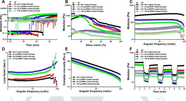
Preparation and characterization of injectable silk hydrogel through (A) degradation profile, (B) gelation time point, (C) amplitude sweep, (D) frequency sweep, (E) shear thinning, (F) thixotropy , (G) injectable hydrogel, and (H) representative images of (I) Silk@SWCNT-FA/DOX. Representative treatment group are (1) untreated, (2) treated with fDOX, (3) treated with Silk@fDOX hydrogel, and (4) Silk@SWCNT-FA/DOX without stimulation (5) Silk@SWCNT-FA /DOX with NIR laser (6) Silk@SWCNT-FA/DOX with electric field and (7) Silk@SWCNT-FA/DOX with NIR and electric field stimulation.
![Figure 1.1. (A) Biological application of CNTs (a) electrochemical sensor based on CNT, (b) CNT in thermal imaging in vivo (c) mice tumor therapy using CNT [143], (B) Intratumor injection of DOX/CNT-Gel nanocomposites into tumor mice mo](https://thumb-ap.123doks.com/thumbv2/azpdfnet/10557415.0/24.918.154.787.216.1069/figure-biological-application-electrochemical-thermal-intratumor-injection-nanocomposites.webp)
Introduction and Literature Review
Introduction and Literature Review
Introduction 1. Cancer
- Types of Cancer
- Treatment of Cancer
- Surgery
- Radiotherapy
- Chemotherapy
- Routes of cancer chemotherapy
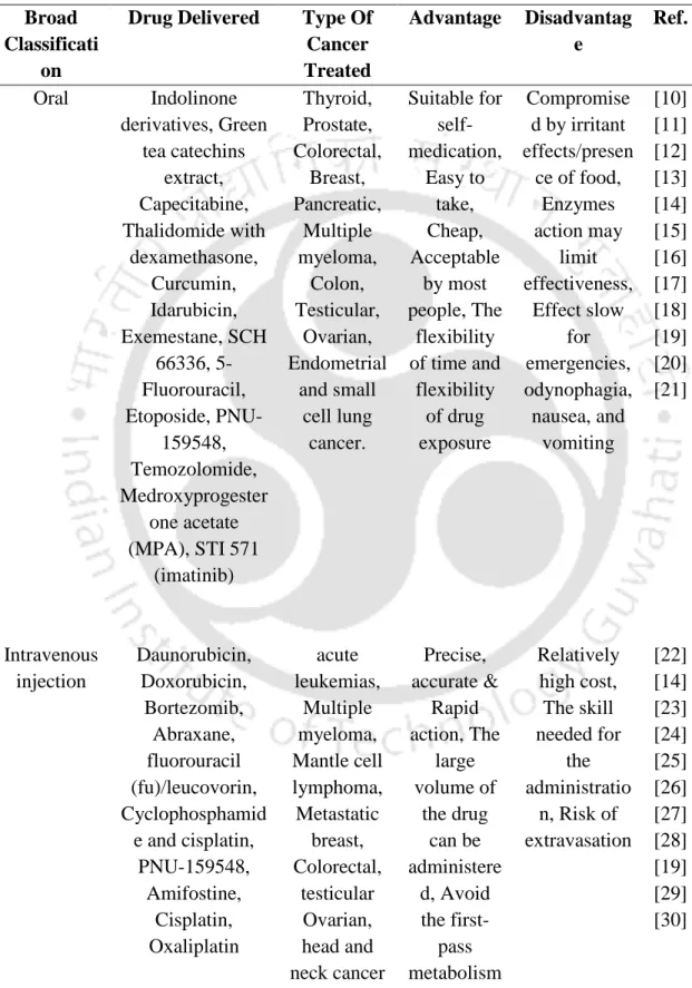
- Oral route
- Intravenous route
- Intramuscular route
- Intraperitoneal route
- Subcutaneous route
- Intraarterial route
- Inhalation
- Nanocarriers for anticancer drug delivery
This is a systemic route of drug delivery where the substances are injected into the peritoneum body cavity. It is a localized route of drug delivery in which the drug is injected into the fatty layer under the skin after crossing the cutaneous layer.
Review of Literature
- Hydrogels for localized drug delivery
- Silk fibroin
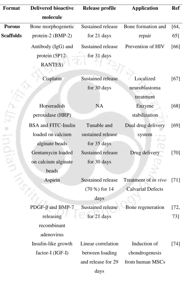
- Silk fibroin for delivery of bioactive molecules
- Nanocomposite smart hydrogels for stimuli triggered drug release
- Carbon nanotubes nanocomposites for drug delivery
Therefore, by controlling the degradation pattern of the silk structure, the kinetics of drug release can be adjusted. The authors have also suggested that drug release can be tunable by varying the concentration of MWNTs in the chitosan hydrogel [151].
MOTIVATION AND OBJECTIVES OF THE PRESENT INVESTIGATION
In connection with the aforementioned issues, we intend to produce an injectable model system for drug delivery. In one of our approaches, we aim to embed the silk fibroin nanoparticles into the silk hydrogel to assess its sustained drug delivery efficiency.
Delivery of Cisplatin and its In Vitro Functionality Assessment in 3D Cultured Stomach Cancer Cell
Model
ABSTRACT
Introduction
Hydrogels composed of various natural (collagen, gelatin, chitosan, silk fibroin, fibrin) or synthetic [polylactic-co-glycolic acid (PLGA), polyvinyl alcohol (PVA), polyethylene glycol (PEG)] polymeric materials have been extensively studied. and were reviewed for their advantages/disadvantages in drug delivery application.[156]. They are susceptible to various proteases for their biodegradation and produce biocompatible, non-toxic, absorbable amino acids as a by-product, which required the attention of researchers to use it as a biomaterial in tissue engineering and drug delivery applications.
Materials and methods
- Isolation of SF protein from Bombyx mori (BM) cocoon and Antheraea assama (AA) silkworm glands
- Preparation and characterization of BM nanoparticle (BMNP)
- Preparation and rheological characterization of BMNP loaded nanocomposite silk hydrogel
- Preparation of BMNP-cisplatin conjugate and Silk@BMNP-cisplatin nanocomposite hydrogel
- In vitro drug release studies from Silk@cisplatin and Silk@BMNP-cisplatin hydrogel
- Fabrication of tubular silk scaffold
- In vitro 3D culture of stomach cancer cell (AGS) on tubular silk scaffold The tubular silk scaffolds were washed 3-4 times with water to remove the ethanol
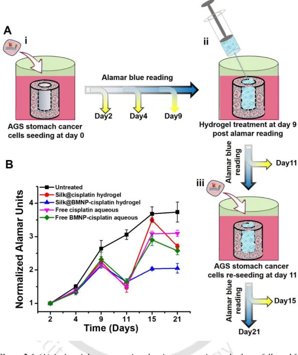
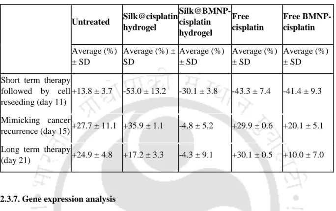
- Efficiency assessment of different anticancer therapeutic formulation (nanocomposite hydrogel) on 3D cultured AGS cells
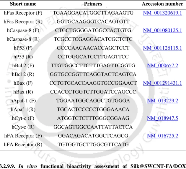
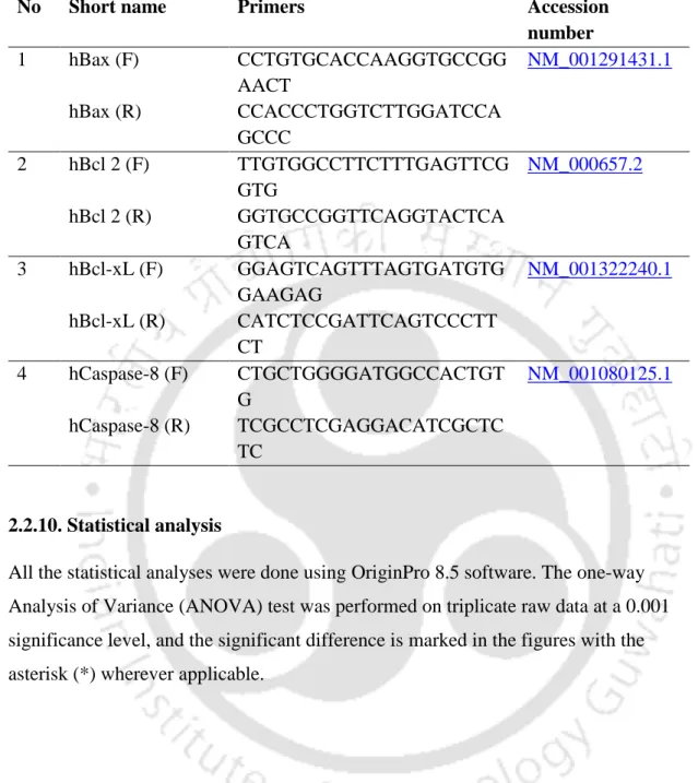
- Gene expression analysis
- Statistical analysis
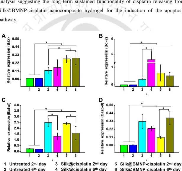
The live/dead solution containing 1 µl calcein-AM and 4 µl ethidium homodimer-1 was prepared in 2 ml PBS.[180] The cultured scaffolds were carefully washed twice with sterile PBS, followed by the addition of 300 µl live/dead solution. On the 9th day of the study, cell-seeded scaffolds were separated into five groups. The gene expression analysis of cell-seeded scaffolds was performed as previously done.[170] The cell-seeded scaffolds treated with Silk@cisplatin hydrogel and BMNP-Silk@cisplatin nanocomposite hydrogel were collected on day 2 and day 6 of treatment along with untreated control.
The scaffolds were disintegrated in Tri reagent (Sigma, USA) to isolate the ribonucleic acid (RNA).
Results
- Preparation and characterization of BMNP
- Preparation and characterization of BMNP embedded silk hydrogel
- Determination of cisplatin loading capacity of BMNP
- In vitro drug release studies from Silk@cisplatin and Silk@BMNP-cisplatin nanocomposite hydrogel
- In vitro 3D culture of stomach cancer AGS cell on the periphery of porous silk scaffold
- Assessment of nanocomposite hydrogel on 3D cultured AGS cancer cells Stomach cancer AGS cells were seeded onto tubular silk scaffold and allowed to grow
- Gene expression analysis
The FTIR spectra of pure cisplatin powder, lyophilized BMNP, and cisplatin BMNP powder are shown in Figure 2.3C. BMNP-cisplatin conjugate was mixed with BM and AA SF solution to form Silk@BMNP-cisplatin nanocomposite hydrogel with injectable properties as shown in Figure 2.3D. EDX analysis also confirmed the presence of Pt in the silk nanocomposite hydrogel (Figure 2.3E).
The red fluorescence observed in Figure 2.5Eii and Figure 2.5Ev may be due to the side autofluorescence, as shown in Figure A2.1.
Discussion
The BMNP-cisplatin-embedded silk hydrogel released cisplatin sustainably compared to the burst release of the cisplatin-embedded silk hydrogel alone at low pH, as described by the pH-responsive behavior of the SF polymer. [55, 161] Low pH causes reassembly of the silk hydrogel, allowing water to penetrate its matrix for water hydrolysis-dependent release of cisplatin. Various methods to fabricate SF hydrogel have been previously reported.[168] 3 wt% SF hydrogel is considered a standard concentration for drug delivery applications, as increasing the concentration of SF protein also increases the strength of the hydrogel, which may be difficult to inject. In this work, we mixed two different versions of the SF protein in equal mass %.
The application of injectable hydrogel system for the treatment of chronic diseases such as arthritis, cancer, skin wounds, etc., has been recognized around the world. However, the current in vitro cell culture models lack the ability to accommodate both the hydrogel and cells. together As shown in Figure 2.6, the side scaffold included both injectable nanocomposite hydrogel in the central cavity and gastric cancer spheroids around its peripheral space.
Significant findings
Single-Walled Carbon Nanotube (SWCNT)
Impregnated Injectable Silk Hydrogel for Folic Acid Receptor Overexpressing Oral Cancer Cell-Targeted
Doxorubicin Delivery
Introduction
However, the hydrophobic nature of drug molecules, inability to distinguish between normal and cancer cells, fluctuating plasma levels, development of drug resistance by cancer cells, and loss of activity in the bloodstream limit the use of these drugs in cancer treatment. Targeted drug delivery (TDD) for cancer therapy is an approach that uses the principle of identifying cancer cells based on their specific overexpression of cell markers such as receptors and their abnormal drug molecule delivery behavior. It is a 38 kDa glycosylphosphatidylinositol-anchored protein and has been considered a potent target for tumor-specific drug delivery [211].
However, no report was available that addressed the combination of SWCNTs and silk hydrogel for on-demand, localized targeted drug delivery.
Materials and methods 1. Materials
- Preparation of SWCNT-FA/DOX
- Physical characterization of prepared SWCNT-FA/DOX
- Extraction of silk fibroin (SF) protein from Bombyx mori (BM) cocoons and Antheraea assama (AA) silkworm glands
- Preparation of injectable Silk@SWCNT-FA/DOX nanocomposite and Silk@fDOX hydrogel
- Rheological studies
- Swellability and degradation of the hydrogel
- Release studies
- In vitro studies 1. Cell culture
- Cell viability assay
- Reactive oxygen species (ROS) measurement
- Lactate dehydrogenase activity assay
- Cell apoptosis assay
- Cell cycle analysis
- SWCNT-FA Specific uptake assay
- Gene expression studies
- In vitro functional bioactivity assessment of Silk@SWCNT-FA/DOX nanocomposite hydrogel
- Statistical analysis
SWCNT-FA and SWCNT-FA/DOX were diluted due to high DOX loading and folic acid modification. Preparation of injectable Silk@SWCNT-FA/DOX nanocomposite and Silk@fDOX hydrogel Silk@fDOX hydrogel. The release of SWCNT-FA/DOX due to hydrogel degradation was observed by DOX-specific fluorescence detection.
The response of SWCNT-FA/DOX and nanocomposite hydrogel to NIR laser was also measured.
Results
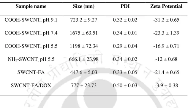
- Preparation and physical characterization of SWCNT-FA/DOX
- Preparation and characterization of injectable SWCNT loaded nanocomposite silk hydrogel
- Release studies
- In vitro cell cytotoxicity studies
- Specific uptake and uptake mechanism of SWCNT-FA/DOX
- Gene expression studies
- In vitro functional bioactivity assessment of nanocomposite silk hydrogel Both A549 Figure 3.7A and KB Figure 3.7B cells were treated with SWCNT-FA
The cytotoxicity of SWCNT-FA/DOX at all concentrations was significantly different for KB and A549 cells. In contrast, SWCNT-FA/DOX uptake by A549 cells was negligible compared to fDOX Figure 3.5CII. SWCNT-FA/DOX did not alter its expression in KB cells, while its level was increased in A549 cells. Figure 3.6G.
Both cells were also treated with 0.5 µg/ml SWCNT-FA/DOX (6) and its suspension exposed to NIR light (7).
Discussion
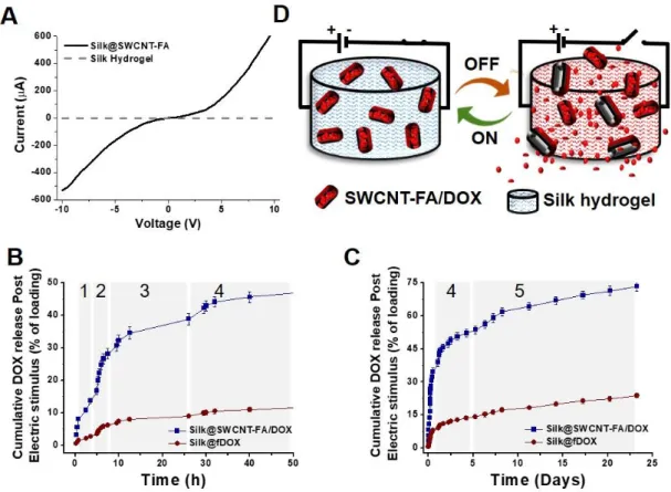
However, Figure 3.3C is a 14-day DOX release study from the Silk@SWCNT-FA/DOX nanocomposite hydrogel. The use of SWCNT-FA/DOX reduced the cytotoxic effects of DOX on A549 cells (FRs) and. In contrast, due to the smaller and interrupted supply of DOX in A549 cells treated with SWCNT-FA/DOX Bax expression level was decreased.
Due to NIR light exposure to Silk@SWCNT-FA/DOX nanocomposite hydrogel (9), some DOX was also released into the medium, causing 20% higher toxicity in both A549 and KB cells than non-NIR stimulated (8 ).
Significant findings
Assessment of Photo Electro-Active Minimally Invasive Injectable Silk Hydrogel in In Vivo Breast
Release and Tumor Therapy
Introduction
Hydrogels are preferred due to their soft injectable nature as a depot for local drug delivery, allowing their implantation directly into the solid tumor site without major surgery.[48, 264] The precise delivery of therapeutic molecules to the solid. the tumor site enhances its therapeutic effect and reduces its biodistribution to the healthy tissue, thereby minimizing its toxic side effects.[48] However, controlling the release kinetics of biomolecules from the hydrogel is still a challenging task. The application of electric field-responsive DDS in vivo was first demonstrated by Ge, Jun,. The application of 808 nm NIR laser or a small electric field or both in combination induces the release of the drug from the SWCNT-FA reservoir and diffuses to the local tumor.
To our knowledge, this is also the first in vivo report showing the combined effect of the NIR laser and electric field for cancer chemotherapy in DDS application.
Materials and methods
- Silk@SWCNT-FA/DOX nanocomposite hydrogel preparation
- Electrical conductivity test of Silk@SWCNT-FA/DOX nanocomposite hydrogel
- Recording of electric field triggered DOX release
- Recording of NIR laser-induced hyperthermia effect
- In vivo tumor studies
- Histology
- TUNEL assay
- Western blot assay
- Echocardiography of heart
- Statistical Analysis
The software created the digital slices of 0.1 mm step size of the 3D video of the tumor. Animals of group (1) were untreated, (2) were treated with free DOX (fDOX), (3) were treated with Silk@DOX hydrogel, and (4) to (7) were treated with Silk@SWCNT-FA/ DOX. On the treatment day, animals from group (2) were administered intraperitoneally with fDOX, while animals from groups (3) to (7) were administered locally (near tumor) with the above-mentioned hydrogels.
The scheme of the study and the arrangement of the external NIR laser and the electric field is shown in Figure A4.1.
Results
- Electric field conductivity of Silk@SWCNT-FA/DOX hydrogel triggers the drug release
- NIR laser induces hyperthermia in Silk@SWCNT-FA hydrogel and triggers the drug release
- In vivo tumor regression study
The NIR laser induces hyperthermia in the Silk@SWCNT-FA hydrogel and triggers the release of the drug-releasing drug. Time-temperature plot of Silk@SWCNT-FA hydrogels exposed to NIR laser for (A) single 20 min ON/OFF cycle and (B) series of five 5 min ON/OFF cycles (n=3, Mean ± SD ). Its cleavage by caspase 3 inactivates its function, which leads the cells to apoptosis.[279] The expression level of caspase 8 was more or less constant in all groups (Figure 4.4A & 4.4C).
The representative treatment groups are (1) untreated, (2) fDOX treated, (3) Silk@fDOX hydrogel treated, and (4) Silk@SWCNT-FA/DOX without stimulation (5) Silk@SWCNT-FA/DOX with NIR laser (6) Silk@SWCNT-FA/DOX with electric field and (7) Silk@SWCNT-FA/DOX with both NIR and electric field stimulation.
Discussion
In such a scenario, the localized therapy may be beneficial.[262] The solid tumor prolonged exposure to adjacent available hydrogel-encapsulating anti-cancer drugs weakens it and offers a great chance to cure this chronic disease. In this regard, Silk@SWCNT-FA/DOX hydrogel was applied close to the tumor (group 4 to 7) and exposed to the external stimulus (group 5 to 7). Furthermore, the simultaneous application of NIR laser and the electric field (group 7), was more effective as it allows the tumor to infiltrate SWCNT-FA/DOX deep into it.
Furthermore, the SWCNT-FA/DOX embedding into the silk fibroin hydrogel matrix formed its nanocomposites.
Significant findings
SUMMARY AND FUTURE PERSPECTIVES
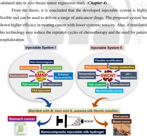
Injection System I was evaluated for its long-term functionality in vitro by culturing gastric cancer cells in specially designed scaffolds. However, the II injection system was developed to target and precisely deliver an anticancer drug to folic acid receptor overexpressing cancer cells. The thesis shows that the developed injection system is highly flexible and can be used to deliver various anti-cancer drugs.
The injection system developed here is a flexible system that can be changed according to requirements.
Bibliography
Kaplan, Sustained-release silk fibroin discs: antibody and protein delivery for HIV prevention, Journal of Controlled Release. Zhang, Nanocomposite hydrogels and their applications in drug delivery and tissue engineering, Journal of biomedical nanotechnology. Kundu, Silk fibroin/gelatin multilayer films as a model system for controlled drug delivery, European Journal of Pharmaceutical Sciences.
Alisaraie, Functionalized Single-Walled Carbon Nanotubes: Cellular Uptake, Biodistribution and Applications in Drug Delivery, International Journal of Pharmaceutics (2017).
APPENDIX
A PPENDIX
![Figure 1.1. (A) Biological application of CNTs (a) electrochemical sensor based on CNT, (b) CNT in thermal imaging in vivo (c) mice tumor therapy using CNT [143], (B) Intratumor injection of DOX/CNT-Gel nanocomposites into tumor mice](https://thumb-ap.123doks.com/thumbv2/azpdfnet/10557415.0/53.892.118.731.174.1047/figure-biological-application-electrochemical-thermal-intratumor-injection-nanocomposites.webp)