This is to certify that the work has been incorporated into the thesis entitled "Antimicrobial peptides from membrane-interactive regions of bacterial proteins". Antimicrobial peptides (AMPs) happen to be one of the key components of such defense arsenal and play an important role in warding off pathogens [ 1 ].
Introduction and Literature Review
Introduction
Gradually, multidrug-resistant TB strains have evolved into extensively drug-resistant (XDR) that cause XDR-TB, a form of TB that is resistant to at least four major anti-TB drugs. Antimicrobial peptides, commonly abbreviated as AMPs, are indigenous components of the innate immune system in a wide variety of organisms such as insects, plants, animals, and microorganisms to defend against pathogens [ 2 , 12 - 15 ].
Distinctive features of antimicrobial peptides
Malanovic and Lohner recently performed a statistical analysis of the known AMPs to understand the selectivity of AMP against bacteria [16]. Conformation: The sequences of AMPs are so diverse that they are rarely classified based on their sequences.
A historical perspective
It shows activity against both Gram-positive and Gram-negative bacteria, as well as mammalian cells [44]. In 1999, the first mammalian theta-defensin was discovered from the leukocytes of rhesus monkey (Macaca mulatta) [62].
Sequences and structures of antimicrobial peptides
- Alpha-helical peptides
- β-sheet peptides
- Circular peptides
- α-β structured peptides
- Non- α-β structured peptides
- Peptides rich in specific amino acids
- Anionic peptides
For example, polyphemusin I isolated from the horseshoe crab Limulus polyphemus and tachyplesin I isolated from horseshoe crab Tachypleus tridentatus are composed of antiparallel β-strands connected by a turn. Drosomycin is the first inducible antifungal protein isolated from insects, and its structure consists of a single helix and twisted three-stranded β-sheet stabilized by four disulfide bridges [94].
![Figure 1.2 α-helical antimicrobial peptides. [A] magainin 2 (PDB ID: 2MAG), [B] LL-37 (PDB ID: 2K6O), [C] cecropin P1 (PDB ID: 2N92), and [D] melittin (PDB ID: 2MLT)](https://thumb-ap.123doks.com/thumbv2/azpdfnet/10550743.0/24.892.101.781.114.980/figure-helical-antimicrobial-peptides-magainin-pdb-cecropin-melittin.webp)
Antimicrobial peptide databases
Data are collected from published experiments, which include information regarding protein fusion, expression, cleavage and yield of the peptide [112]. This database is a large source of information on peptide sequences, their conformations, antimicrobial activity, physicochemical properties, patents, clinical trials and references [4].
Mechanism of action
- Membrane-disruptive peptides
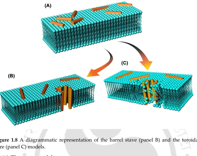
- The barrel stave model
- The toroidal pore model
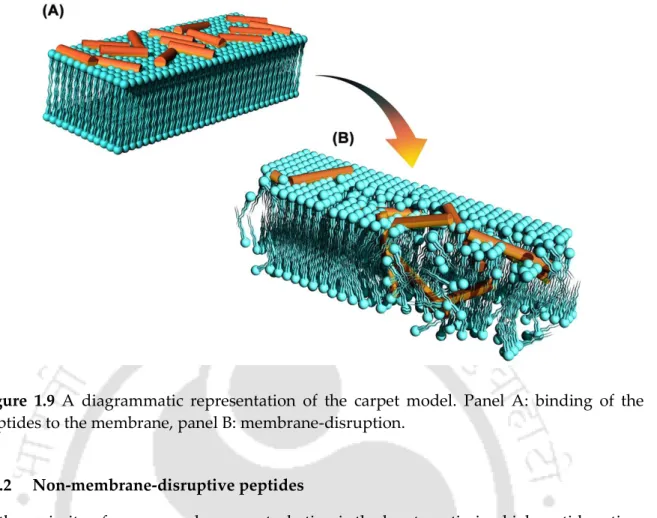
- The carpet model
- Non-membrane-disruptive peptides
Once the peptide molecules gain access to the inner membrane (cytoplasmic membrane), their modes of action branch out; membrane-disrupting peptides orient and disrupt the cytoplasmic membrane; non-membrane-disrupting peptides, on the other hand, translocate the membrane and target intracellular receptors [120]. Polar head groups are forced aside and the hydrophobic region of the peptide interacts with the membrane.
Selectivity of antimicrobial peptides towards Gram-positive or Gram- negative bacteria
Immunomodulatory functions of antimicrobial peptides
HBD-2 brings dendritic cells and peripheral T cells (CD4+/CD45RO+) to the site of inflammation. Upon invasion by pathogens, the circulating hemocytes move to the site of injury by chemotaxis and release AMPs.
Antimicrobial resistance against antimicrobial peptides
In addition, the multiple peptide resistance factor MprF inserts L-lysine into membrane phospholipids that reduces the net negative charge on the membrane, conferring resistance to S. In addition to structural changes in the cell membrane, AMP activity is also influenced by the energy of membrane.
Challenges in developing AMPs as antibiotics
Furthermore, the activity of histatin-5 was reduced when the wild-type strain was treated with inhibitors of the electron transport chain. These results suggested that the antimicrobial activity of histatin-5 is dependent on the energy of the target cell.
Antimicrobial peptides as therapeutics
It is in phase II clinical trials and exhibits an inhibitory effect on scar formation by reducing infections, inhibiting inflammation and promoting fibrinolysis [200]. Iseganan hydrochloride is the salt of synthetic protegrin and is currently in phase III clinical trials [203].
![Table 1.6 List of antimicrobial peptides in clinical trials [26, 199]](https://thumb-ap.123doks.com/thumbv2/azpdfnet/10550743.0/48.892.80.739.136.1106/table-list-antimicrobial-peptides-clinical-trials-26-199.webp)
Hypothesis: membrane-binding stretches in microbial proteins can turn out to be potential AMPs
Materials and Methods
- Materials
- Peptides
- Peptide synthesis
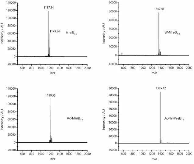
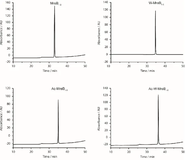
- Peptide purification and characterization
- Peptide solutions and concentration estimation
- Surface activity of peptides
- Preparation of small unilamellar vesicles
- Steady-state tryptophan fluorescence
- Tryptophan fluorescence quenching
- Outer membrane permeabilization assay
- Inner membrane permeabilization assay
- Circular dichroism spectroscopy
- Antibacterial assay .1 Colony count method
- Broth microdilution method
- Antimycobacterial assay
- Antifungal assay .1 Colony count method
- Broth microdilution method
- Salt sensitivity assay .1 Colony count method
- Broth microdilution method
- Hemolytic assay
- FESEM analysis
- Killing kinetics assay
Each amino acid was coupled twice, with the first coupling performed with a threefold molar excess (3X) and the second coupling performed with a twofold molar excess (2X) of amino acid over the resin substitution (X). After stabilization of the surface pressure, peptides were gently injected into the subphase (final concentration of peptide in the subphase = 10 μM) through the hole in the trough without disturbing the monolayer. Twofold concentrated peptides were serially diluted from a range of 128 µM to 1 µM in microtiter wells; the final concentration of the cultures in the wells was 5 x 10 CFU/ml.
Twofold concentrated peptides were serially diluted from a range of 128 µM to 1 µM in microtiter wells (50 µl volume). Mid-log phase microbial cells were diluted in 10 mM phosphate buffer, pH 7.4, to obtain a cell concentration of 10 CFU/ml. The cells were washed twice with 10 mM phosphate buffer, pH 7.4, loaded onto glass slides and dried at room temperature.
Antimicrobial Peptides from
Summary
MreB is a bacterial cytoskeleton protein present in non-spherical cells localized under the cell membrane in the form of filaments [211]. The peptide was predicted to seek out the membrane and was then investigated for its antimicrobial properties. Extension with a tryptophan residue at the N-terminus drastically improved the activity of the peptides at flight concentrations.
Several studies reported that introduction of a tryptophan moiety increases the antimicrobial efficacy of peptides, as tryptophan effectively interacts with the interfacial region of the membrane and facilitates partitioning of the peptide through the lipid bilayer [212]. End-tagging of peptide with hydrophobic amino acids increases antimicrobial potency even in the presence of salts and serum [213, 214]. Lipid binding studies and electron microscopic analyzes of the peptide-treated microbes suggest membrane disruption as the killing mechanism.
Results .1 Peptides
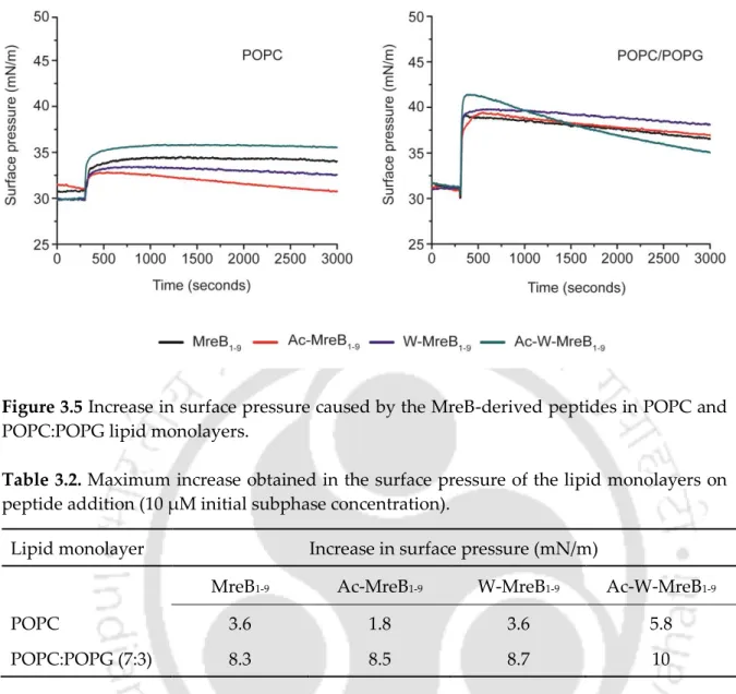
- Surface activity and membrane binding of the peptides
- Tryptophan fluorescence
- Tryptophan fluorescence quenching
- Antimicrobial assay
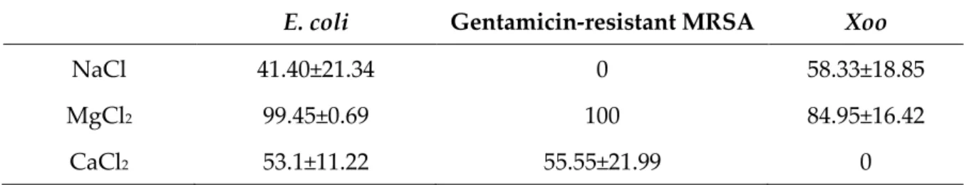
- Salt sensitivity
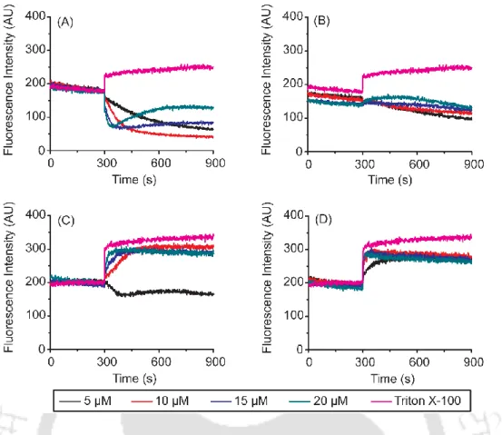
- Outer membrane permeabilization assay
- Inner membrane permeabilization assay
- Circular dichroism spectroscopy
- Hemolytic assay
- FESEM analysis
All peptides caused a significant increase in surface pressure (≥5 mN/m), even at a peptide concentration of 2 μM (Figure 3.4). In the presence of POPC:CHL SUVs, both peptides exhibit emission maxima of ~346–348 nm at a peptide/lipid ratio of 1:100. Stern-Volmer plots for the peptides in the presence of SUVs fit linearly and the determined KSV values are shown in Table 3.3.
The antimicrobial activity of the peptides was investigated against Gram-positive and Gram-negative bacteria and against fungi. All four peptides exhibit a positive ellipticity of ~195–197 nm in the presence of POPE:POPG SUVs, indicating folding of the peptides. Ac-MreB1-9 and W-MreB1-9 fold into a distinct α-helical conformation in the presence of negatively charged SUVs (panels B and C).
Conclusion
Antimicrobial Peptides from
- Summary
- Results .1 Peptides
- Tryptophan quenching
- Antimicrobial assay
- Salt sensitivity assay
- Outer membrane permeabilization assay
- Inner membrane permeabilization assay
- Circular dichroism
- Hemolytic assay
- FESEM analysis
- Conclusion
The peptides exhibit preferential binding to the negatively charged lipid vesicles and kill all organisms tested. In the presence of POPC:CHL SUVs, FtsA10 and Ac-FtsA10 show no noticeable change in the emission spectra. However, in the presence of POPE:POPG vesicles, a clear blue shift with enhancement of the fluorescence intensity is observed, indicating binding to the lipid vesicles.
The activity of FtsA10 and Ac-FtsA10 decreased in the presence of salt and divalent cations. MIC of Ac- FtsA11 and FtsA13 against gentamicin-resistant MRSA is the same in the presence of salt and divalent cations (32 μM). Ac-FtsA10 shows a gradual increase in the fluorescence intensity indicating permeabilization of the inner membrane (panel B).
![Figure 4.1 Helical wheel representation of FtsA13 peptide derived from E. coli Mean hydrophobicity (<H>) and mean hydrophobic helical moments (<μH>) were calculated using Heliquest web server [215]](https://thumb-ap.123doks.com/thumbv2/azpdfnet/10550743.0/86.892.328.600.107.347/figure-helical-representation-ftsa13-hydrophobicity-hydrophobic-calculated-heliquest.webp)
Aromatic Residue-Rich
Summary
Bacterial AMPs, referred to as bacteriocins, are produced by a large number of bacteria to protect themselves from invading pathogens. The high thermodynamic stability of LCI is thought to be conferred by aromatic stacking interactions, cation–π interactions, and aromatic–backbone amide interactions. The distribution of cationic and aromatic residues along the sequence is noteworthy; the N-terminal 21-residue stretch harbors only 2 Phe and 1 Lys residues.
This implies that ~86% of the cationic residues and 80% of the aromatic residues lie in the C-terminal stretch of 26 residues. We synthesized the C-terminal amidated peptide (KWIFKSKYYDSSKGYWVGIYEVWDRK-NH2), hereafter referred to as LCI22-47, and examined its antibacterial activity against E.
Results .1 Peptide
- Antibacterial activity
- Salt sensitivity assay
- Steady state tryptophan fluorescence
- Tryptophan fluorescence quenching
- Membrane permeabilization assay
- Circular dichroism
- Hemolytic assay
- FESEM analysis
Complete loss of activity in the presence of salt or divalent cations does not mean that the peptide cannot kill the microbe. However, in the presence of POPE:POPG vesicles, a clear blue shift of about 8 nm with slight increase in fluorescence intensity is observed, suggesting binding of the peptide to lipid vesicles. The spectra were recorded in the absence of lipid (black trace) and in the presence of SUVs such that the peptide/lipid ratio is 1:50 and 1:100.
The peptide is unusually rich in aromatic residues and the 230 nm band is assigned to the aromatic clusters in the folded peptide. The peptide also shows a negative band around 205 nm, which also suggests the presence of disordered conformation. The peptide structure is largely the same in the presence of POPC:CHL vesicles, suggesting little, if any, interaction with lipids.
Conclusion
This loss in activity, however, does not imply complete inactivity of the peptide; this implies that the peptide exhibits antibacterial activity at higher concentrations. The structure of LCI suggests that the peptide is not amphipathic, an aspect similar to the rabbit kidney defensin, RK-1 [229]. Such behavior may arise due to the following possibilities: (i) formation of passage pores that allow only small molecules to pass through denying peptide molecules access to the inner membrane or (ii) translocation of peptides across the membrane internal without interrupting it.
The data suggest that membrane permeabilization may not be the only mechanism of bacterial death induced by LCI22-47. Further investigations with LCI and LCI22-47 are needed to gain mechanistic insights into their antimicrobial activity. Nevertheless, the LCI22-47 fragment is a promising AMP against both Gram-negative and Gram-positive bacteria.
Conclusion, Discussion, and
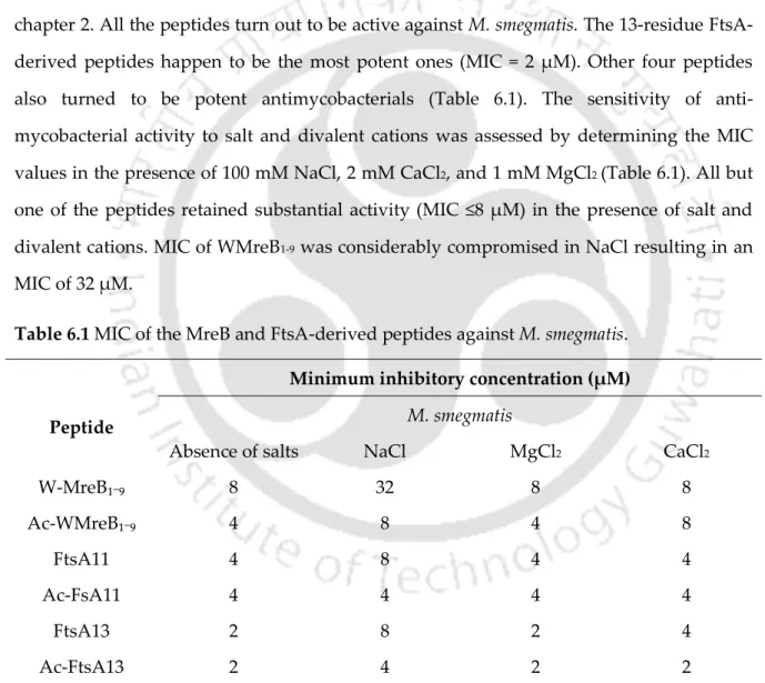
This thesis deals with the antimicrobial properties of peptides derived from proteins of bacterial origin. The low hydrophobicity of the FtsA-derived peptides could be responsible for conferring them with selectivity for Gram-negative bacteria. It was therefore imperative to test the activity of the peptides in the presence of salts and divalent cations at physiologically relevant concentrations.
Some of the highlights of the FtsA-derived peptides include: (i) MICs of 11- and 13-residue peptides are within 8 µM against E. None of the peptides resulted in any noticeable hemolysis at their lethal concentrations or MICs. All but one peptide retained significant activity (MIC ≤8 μM) in the presence of salt and divalent cations.
Nakamura, T., et al., Tachyplesin, a class of antimicrobial peptides from the hemocytes of the horseshoe crab (Tachypleus tridentatus). Pasupuleti, M., et al., Tryptophan end-labeling of antimicrobial peptides for increased potency against Pseudomonas aeruginosa.
Highly potent antimicrobial peptides from N-terminal
OPEN
The kinetics of killing in the presence of lethal concentration of peptides was examined (Fig. 2). Stern-Volmer plots for peptides in the presence of SUVsfit linearly and determined KSV values are shown in Table 2. Steady-state tryptophan fluorescence spectra for peptides (1 μM) were recorded in HEPES-buffered saline without and with SUV at peptide to lipid molar ratios of 1:50 and 1:100 as previously described [ 15 ].
Steady-state tryptophan fluorescence spectra for peptides (1 μM) were recorded in HEPES-buffered saline in the presence of SUVs (peptide:lipid ratio 1:100). The KSV values of the peptides decrease in the presence of lipid vesicles suggesting membrane association. It is therefore imperative to test the activity of peptides in the presence of salts and divalent cations at physiologically relevant concentrations.
![Table 1. Sequences and physicochemical properties of the peptides. [a] ‘Ac-’ at N-terminus represents acetylated amino-terminus, while ‘-am’ at C-terminus represents C-terminal amide](https://thumb-ap.123doks.com/thumbv2/azpdfnet/10550743.0/138.892.236.683.69.159/sequences-physicochemical-properties-peptides-terminus-represents-acetylated-represents.webp)
![Figure 1.3 Antimicrobial peptides with β-sheet structure. [A] human α-defensin 1 (PDB ID:](https://thumb-ap.123doks.com/thumbv2/azpdfnet/10550743.0/26.892.90.746.345.831/figure-antimicrobial-peptides-sheet-structure-human-defensin-pdb.webp)
![Figure 1.4 Cyclic antimicrobial peptides. [A] enterocin AS-48 (PDB ID: 1E68), [B] circulin A (PDB ID: 1BH4), [C] kalata B1 (PDB ID: 1KAL), [D] rhesus θ-defensin 1 (PDB ID:](https://thumb-ap.123doks.com/thumbv2/azpdfnet/10550743.0/28.892.152.735.315.791/figure-cyclic-antimicrobial-peptides-enterocin-circulin-kalata-defensin.webp)
![Figure 1.5 Cyclization by disulphide bridges. [A] polyphemusin I (PDB ID: 1RKK)](https://thumb-ap.123doks.com/thumbv2/azpdfnet/10550743.0/29.892.179.739.106.713/figure-cyclization-disulphide-bridges-polyphemusin-pdb-id-1rkk.webp)
![Figure 1.6 α-β structured antimicrobial peptides. [A] human β-defensin-1 (PDB ID: 1E4S), [B] heliomicin (PDB ID: 1I2U), [C] plectasin (PDB ID: 1ZFU), and [D] drosomycin (PDB ID:](https://thumb-ap.123doks.com/thumbv2/azpdfnet/10550743.0/30.892.169.743.101.629/figure-structured-antimicrobial-peptides-defensin-heliomicin-plectasin-drosomycin.webp)