Anatomy of Flowering Plants
Understanding plant anatomy is not only fundamental to the study of plant systematics and palaeobotany, but is also an essential part of evolutionary biology, physiology, ecology, and the rapidly expanding science of developmental genetics. In the third edition of her successful textbook, Paula Rudall provides a
comprehensive yet succinct introduction to the anatomy of flowering plants. Thoroughly revised and updated throughout, the book covers all aspects of comparative plant structure and development, arranged in a series of chapters on the stem, root, leaf, flower, seed and fruit.
Internal structures are described using magnification aids from the simple hand-lens to the electron microscope.
Numerous references to recent topical literature are included, and new illustrations reflect a wide range of flowering plant species. The phylogenetic context of plant names has also been updated as a result of improved understanding of the relationships among flowering plants. This clearly written text is ideal for students studying a wide range of courses in botany and plant science, and is also an excellent resource for professional and amateur horticulturists.
Paula Rudallis Head of Micromorphology(Plant Anatomy and Palynology) at the Royal Botanic Gardens, Kew. She has published more than 150 peer-reviewed papers, using comparative floral and pollen morphology, anatomy and embryology to explore evolution across seed plants.
Anatomy of
Flowering Plants
An Introduction to
Structure and Development
PAULA J. RUDALL
Cambridge University Press
The Edinburgh Building, Cambridge CB2 8RU, UK
First published in print format
ISBN-13 978-0-521-69245-8 ISBN-13 978-0-511-29453-2
© Paula J. Rudall 2007
2006
Information on this title: www.cambridge.org/9780521692458
This publication is in copyright. Subject to statutory exception and to the provision of relevant collective licensing agreements, no reproduction of any part may take place without the written permission of Cambridge University Press.
ISBN-10 0-511-29453-0
ISBN-10 0-521-69245-8
Cambridge University Press has no responsibility for the persistence or accuracy of urls for external or third-party internet websites referred to in this publication, and does not guarantee that any content on such websites is, or will remain, accurate or appropriate.
Published in the United States of America by Cambridge University Press, New York www.cambridge.org
paperback eBook (EBL) eBook (EBL)
paperback
Contents
Preface ix
Taxonomic overview xi
1 Organs, Cells and Tissues 1
1.1 Organs 1
1.2 Cells 2
1.3 Cell Inclusions 5
1.4 Secretory Ducts and Laticifers 7
1.5 Transfer Cells 9
1.6 Tissues 9
1.6.1 Parenchyma 10
1.6.2 Aerenchyma 10
1.6.3 Collenchyma 10
1.6.4 Sclerenchyma 11
1.7 Epidermis 13
1.7.1 Stomata 13
1.7.2 Trichomes 15
1.8 Ground Tissue 17
1.9 Vascular Tissue 18
1.9.1 Xylem 18
1.9.2 Phloem 19
1.10 Meristems 21
1.10.1 Apical Meristems 21
1.10.2 Lateral Meristems 22
1.10.3 Meristemoids and Asymmetric
Cell Division 22
2 Stem 23
2.1 Shoot Apex 23
2.2 Primary Stem Structure 24
2.3 Primary Vascular System 26
2.4 Nodal Vasculature 27
2.5 Vascular Cambium 29
2.6 Secondary Xylem 31
2.7 Secondary Phloem 35
2.8 Primary and Secondary Thickening
Meristems 36
2.9 Periderm 40
3 Root 43
3.1 Primary Root Structure 43
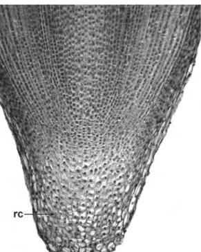
3.2 Root Apex 43
3.3 Root Cap 45
3.4 Root Epidermis and Hypodermis 46 3.5 Root Cortex and Endodermis 48 3.6 Pericycle and Vascular Cylinder 49 3.7 Initiation of Lateral and Adventitious
Roots 50
3.8 Secondary Growth in Roots 51 3.9 Roots Associated with
Micro-Organisms 53
3.10 Haustoria of Parasitic Angiosperms 54
4 Leaf 57
4.1 Leaf Morphology and Anatomy 57
4.2 Leaf Development 60
4.3 Leaf Epidermis 61
4.3.1 Pavement Epidermal Cells 61
4.3.2 Stomata 62
4.3.3 Trichomes and Papillae 63
4.3.4 Cuticle and Wax 66
4.4 Extrafloral Nectaries 66
4.5 Mesophyll 68
4.6 Sclerenchyma and Idioblasts 69
4.7 Leaf Vasculature 70
4.8 Bundle Sheath and Kranz Anatomy 72
5 Flower 75
5.1 Floral Organs 75
5.2 Floral Vasculature 77
5.3 Perianth 79
5.4 Androecium 81
5.5 Pollen 84
5.6 Gynoecium 87
5.6.1 Stigma and Style 87
5.6.2 Ovary 89
5.7 Ovule 90
5.8 Embryo Sac 93
5.9 Pollen-Tube Growth 94
5.10 Floral Secretory Structures 96
6 Seed and fruit 99
6.1 Seed Coat 99
6.2 Pericarp 101
6.3 Grass Caryopsis 102
6.4 Endosperm 104
6.5 Perisperm 106
6.6 Embryo 107
6.7 Seedling 109
Glossary 111
References 128
Index 139
Contents vii
Preface
In the twenty-first century, plant anatomy remains highly relevant to systematics, paleobotany, and the relatively new science of developmental genetics, which interfaces disciplines and utilizes a combination of techniques to examine gene expression in growing tissues. Modern students need to consider information from an increasingly wide range of sources, most notably inte- grating morphological and molecular data. The third, thoroughly revised, edition of this book presents an introduction to plant anatomy for students of botany and related disciplines.
Although the simple optical lens has been used for centuries to examine plant structure, detailed studies of plant anatomy originated with the invention of the compound microscope in the seventeenth century. Nehemiah Grew (1641 1712) and Marcello Malpighi (1628 1694), physicians working independently in England and Italy respectively, were early pioneers of the microscopical examination of plant cells and tissues. Their prescient work formed the foundation that eventually led to the development of our understanding of cell structure and cell division27. Other early outstanding figures included Robert Brown (1773 1858), who discovered the nucleus, and the plant embryologist Wilhelm Hofmeister (1824 1877), who first described the alternation of generations in the life cycle of land plants. In the nineteenth and twentieth centuries plant anatomy became an important element of studies of both physiology and systematic biology, and an integral aspect of research in the
developing field of anatomical paleobotany, led by such luminaries as Dukinfield Henry Scott (1854 1934). The physiologist Gottlieb Haberlandt (1854 1945) utilized anatomical observa- tions in his ground-breaking work on photosynthetic carbon metabolism. One of the most notable plant anatomists of the twentieth century was Katherine Esau (1898 1997), recognized particularly for her work on the structure and development of phloem and her influential textbooks on plant anatomy30. Other important textbooks include works on paleobotany, morphology, anatomy and embryology13,34,68,106
.
The invention of the transmission electron microscope (TEM) in the mid twentieth century allowed greater magnification than any optical microscope, and hence revitalized studies in cell ultra- structure and pollen morphology98. The subsequent invention of the scanning electron microscope (SEM) provided greater image clarity and much greater depth of focus than light microscopes, and thus further increased accessibility of minute structures, including seeds, pollen grains and organ primordia28,98. More recent innovations, including fluorescence microscopy, differen- tial interference contrast (DIC) microscopy and confocal imaging, have allowed enhanced visualization of tissue structure. Others, including nuclear magnetic resonance (NMR) imaging and high-resolution X-ray computed tomography (HRCT) facilitate enhanced visualization of three-dimensional objects.
Taxonomic Overview
In textbooks published before 1990, extant angiosperms were consistently subdivided into two major groups dicotyledons (dicots) and monocotyledons (monocots), based partly on the number of cotyledons in the seedling. This dichotomy was long considered to represent a fundamental divergence at the base of the angiosperm evolutionary tree. Other features marked this distinction, including the absence of a vascular cambium and presence of parallel leaf venation in monocots. However, the expansion of molecular phylogenetics through the early 1990s indicated that some species that were formerly classified as primitive dicots do not belong to either category, though the monophyly of monocots was confirmed2,3,103. Thus, although the dicot/monocot distinction remains useful for generalized descrip- tions of angiosperm groups, current evidence suggests that it does not represent a wholly natural classification. It is now widely accepted that several relatively species-poor angiosperm lineages (here termed early-divergent angiosperms or magnoliids) evolved before the divergence of the two major lineages that led to the monocots and the remaining dicots (now termed eudicots, or sometimes tricolpates).
Early-divergent angiosperms (including magnoliids) are a small but highly diverse assemblage of taxonomically isolated lineages that probably represent the surviving extant members of their respective clades, accounting for only about 1% of extant species.
They possess some morphological features in common with both
monocots and eudicots, and include the New Caledonian shrub Amborella, the water lilies (Nymphaeaceae), woody families such as Magnoliaceae and Lauraceae, and herbaceous or climbing families such as Piperaceae and Aristolochiaceae. Monocots account for approximately a quarter of all flowering plants species. They dominate significant parts of world ecosystems, and are of immense economic importance, including the staple grass food crops (wheat, barley, rice and maize) and other important food plants such as onions, palms, yams, bananas and gingers. Eudicots represent about 75% of extant angiosperm species, and encompass a wide range of morphological diversity, especially in the two largest subclades, Rosidae (rosid eudicots) and Asteridae (asterid eudicots).
1
Organs, cells and tissues
1.1 Organs
Plants consist of several organs, which in their turn are composed of tissues. Broadly, vegetative organs support plant growth, and reproductive organs enable sexual reproduction. The three main types of vegetative organ are the root, stem and leaf. Roots typically occur underground, and extract moisture and nutrients from the soil, though there are many examples of plants with aerial roots. The stem and leaves together comprise the shoot (Fig. 1.1). Stems occur both above and below ground. Some stems are modified into underground perennating or storage organs such as corms or rhizomes. Leaves typically occur above ground level, though some underground stems possess reduced scale leaves, and underground bulbs possess swollen leaves or leaf bases.
Primary organs and tissues develop initially from the shoot and root apical meristems and from cell divisions in meristems closely adjacent to them, such as the primary thickening meristem.
Secondary tissues such as secondary xylem (wood) develop from lateral meristems such as the vascular cambium. Organs such as adventitious roots develop from differentiated cells that have retained meristematic capacity. At the onset of flowering, the shoot apical meristem undergoes structural modification from a veg- etative to a reproductive apex and subsequently produces flowers (chapter 5). Flowers are borne on an inflorescence, either in groups or as solitary structures. A group of inflorescences borne on a single plant is termed a synflorescence121(Fig. 1.2).
1.2 Cells
Plant cells typically have a cell wall containing a living protoplast (Fig. 1.3). The layer that contacts the walls of adjacent cells is termed the middle lamella. Following cessation of growth, many cells develop a secondary cell wall which is deposited on the inside surface of the primary wall. Both primary and secondary walls consist of cellulose microfibrils embedded in a matrix and ori- ented in different directions. Secondary cell walls consist mostly of cellulose, but primary walls commonly contain a high proportion of hemicelluloses in the gel-like matrix, affording a greater degree of plasticity to the wall of the growing cell. The secondary wall can also contain deposits of lignin (in sclerenchymatous cells) or suberin (in many periderm cells), and often appears lamellated.
Thin areas of the primary wall, which usually correspond with thin areas of the walls of neighbouring cells, are primary pit fields, and usually have protoplasmic strands (plasmodesmata) passing through them, connecting the protoplasts of neighbouring cells36. The connected living protoplasts are collectively termed the sym- plast. Primary pit fields often remain as thin areas of the wall even after a secondary wall has been deposited, and are then termed Figure 1.1 Hyptis ditassoides(Lamiaceae), transverse section of vegetative bud near apex, showing three successive pairs of leaf primordia surrounding central stem. Scale¼100mm.
pits, or pit-pairs if there are two pits connecting adjacent cells. Pits may be simple, as in most parenchyma cells, or bordered, as in tracheary elements. In simple pits the pit cavity is of more or less uniform width, whereas in bordered pits the secondary wall Figure 1.2 Salvia involucrata(Lamiaceae), dissected developing synflorescence showing flower clusters, each consisting of three flowers enclosed within a bract; younger stages towards apex. b¼bract. Scale¼500mm.
Cells 3
arches over the pit cavity so that the opening to the cavity is rela- tively narrow. Through a light microscope the outer rim of the primary pit field appears as a border around the pit opening.
The cell protoplast is contained within a plasma membrane.
It consists of cytoplasm that encloses bodies such as the nucleus, plastids and mitochondria, and also non-protoplasmic contents such as oil, starch or crystals. The nucleus, which is bounded by a nuclear membrane, often contains one or more recognizable bodies (nucleoli) together with the chromatin in the nuclear sap. During cell division the chromatin becomes organized into chromosomes. Most cells possess a single nucleus, but examples of multinucleate cells (coenocytes) include the non-articulated laticifers found in many plant families (chapter 1.4). Such cells elongate and penetrate established tissues by intrusive tip growth, Figure 1.3 Diagram of a generalised plant cell illustrating details of protoplasmic contents.
in which the cell apices secrete enzymes that dissolve the middle lamellae of neighbouring cells; bifurcation occurs when they encounter an obstacle36.
Mitochondria and plastids are surrounded by double mem- branes. Plastids are larger than mitochondria, and are classified into different types depending on their specialized role.
For example, chloroplasts are plastids that contain chlorophyll within a system of lamellae that are stacked to form grana; this is the site of photosynthesis. Chloroplasts occur in all green cells, but are most abundant in the leaf mesophyll, which is the primary photosynthetic tissue (chapter 4.5). Membranes occur widely throughout the cytoplasm, sometimes bounding a series of cav- ities. For example, the endoplasmic reticulum is a continuous membrane-bound system of flattened sacs and tubules, sometimes coated with ribosomal particles. Dictyosomes are systems of sacs associated with secretory activity. Vacuoles are cavities in the cytoplasm; they are usually colourless and contain a watery fluid.
Their size and shape varies in different cell types, and also changes during the life of a cell.
1.3 Cell Inclusions
Many cells possess non-protoplasmic contents such as oils, mucilage (slime), tannins, starch granules, calcium oxalate crystals and silica bodies. Both oil and mucilage are produced in secretory idioblasts which are often larger than adjacent parenchymatous cells. Tannins are phenol derivatives which are common in plant cells; they are amorphous, and appear yellow, red or brown in colour in cells of sectioned material. Cystoliths are cellulose bodies encrusted with calcium carbonate that occur in epidermal cells in some species (Fig. 4.4); the body is attached to the cell wall by a silicified stalk36.
Starch is especially common in storage tissues such as endosperm or in parenchyma adjacent to a nectary. Starch granules
Cell inclusions 5
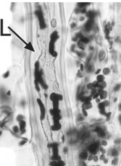
are formed in plastids (amyloplasts). They often appear layered due to the successive deposition of concentric rings, and may possess characteristic shapes. For example, in species ofEuphorbia, starch grains in laticifers are elongated and sometimes rod-shaped or bone-shaped compared with the more rounded starch grains of neighbouring parenchyma cells (Fig. 1.4)70.
Calcium oxalate crystals (Figs 1.5, 1.13) are borne in crystal idioblasts that can occur in almost every part of the plant, includ- ing both vegetative and reproductive organs82. They are often present near veins, possibly due to transport of calcium through the xylem, and are sometimes associated with air space formation;
some aquatic plants possess calcium oxalate crystals projecting into air spaces. Crystals form within vacuoles of actively growing cells and are usually associated with membrane chambers, lamellae, mucilage and fibrillar material. Crystal sand is relatively amor- phous and represents fragmented non-nucleated crystalline particles. Druses (cluster crystals) are aggregated crystalline struc- tures that have precipitated around a nucleation site. Raphides are bundles of needle-like crystals that are borne in the same cell; they occur commonly in monocots. In the monocot family Araceae,
Figure 1.4 Monadenium ellenbeckii (Euphorbiaceae). Elongated I-shaped starch grains in laticifer (L); ovoid starch grains present in adjacent parenchyma cells. Scale¼20mm.
raphides are characteristically grooved and sometimes barbed.
Styloid crystals are typically solitary, larger and needle-like or rhomboidal; they are highly characteristic of some families, such as Iridaceae91.
Opaline silica bodies are also a characteristic feature of some plant groups83. They occur in all plant parts, often associated with sclerenchyma, though they are rare in roots. In many dicot species they occur in the ray or axial parenchyma cells in secondary xylem.
Some families, such as grasses (Poaceae), sedges (Cyperaceae), orchids (Orchidaceae) and palms (Arecaceae), possess character- istic silica bodies contained in well-defined cells, either in the epidermis (e.g. in grasses: Fig. 4.3B) or in vascular bundle sheath cells (e.g. in palms and orchids).
1.4 Secretory Ducts and Laticifers
In many plants, substances such as oils, resins and mucilage are secreted internally, often into specialized ducts formed either by Figure 1.5 Crocus cancellatus(Iridaceae), longitudinal section of leaf showing crystal idioblast containing styloid crystal (sc). Scale¼50mm.
Secretory ducts and laticifers 7
cell wall separation (schizogenous ducts) or cell wall degradation (lysigenous ducts), or a combination of the two processes (schizo- lysigenous ducts)13. Some angiosperms, especially eudicots such asEuphorbia andFicus, produce latex from specialized cells (latici- fers) that permeate their tissues (Figs 1.4, 1.6). In Euphorbia, the laticifers are derived from a small group of initial cells in the cotyledonary node of the embryo; these cells are coenocytes, since they undergo repeated nuclear divisions without corresponding wall formation. They grow intrusively between cells of surround- ing tissues, and often branch and eventually ramify throughout the entire plant31,71,87,90
. Coenocytic laticifers are termed non-articulated laticifers. By contrast, laticifers of a few species (e.g. Hevea brasiliensis, the source of commercial rubber) undergo cell-wall formation, and thus consist of linked chains of cells; these are termed articulated laticifers. Laticifers of the opium poppy Figure 1.6 Euphorbia eyassiana(Euphorbiaceae), longitudinal section of stem showing branched non-articulated laticifers in parenchyma. Scale¼50mm.
(Papaver somniferum) are always associated with vascular bundles122; the alkaloids produced in the latex of these cells are the source of narcotic analgesics such as morphine.
1.5 Transfer Cells
Transfer cells occur at the interface between tissues; they are specialized cells that facilitate transport (absorption or secretion) of soluble substances across tissue boundaries. For example, they can occur at the junction of the megagametophyte and mega- sporophyte, in companion cells in phloem tissue (especially at the node of a stem), in root nodules, in the haustoria of parasitic plants, and in the epidermis of water plants80. Several cells of the embryo sac and seed, including synergids, antipodals and specialized endosperm cells, have been identified as transfer cells in different species. Transfer cells are typically characterized by numerous cell-wall ingrowths protruding into their protoplasts or those of adjacent cells; these ingrowths are sometimes visible using light microscopy. Secretory cells, such as those of glandular hairs and nectaries, also frequently possess wall ingrowths.
The plasma membrane of the transfer cell follows the contour of the wall ingrowths, thus increasing the surface area.
1.6 Tissues
Simple tissues, such as parenchyma, collenchyma and scleren- chyma, consist of a single cell type, though they may be interspersed with other, isolated, cell types (idioblasts). Complex tissues consist of multiple cell types, and can be divided into three main groups: dermal tissue (epidermis), ground tissue and vascular (conducting) tissue, each distributed throughout the plant body, and often continuous between the various organs.
Complex tissues often include elements of several different simple tissue types; for example, secondary xylem includes not only vascular tissue, but also parenchyma and sclerenchyma.
Tissues 9
1.6.1 Parenchyma
Parenchyma cells are typically thin-walled and often polyhedral or otherwise variously shaped, sometimes lobed. Cells with living contents that do not fit readily into other categories are often termed parenchyma cells. They are the least specialized cells of the mature plant body and often resemble enlarged meristematic cells.
Parenchyma cells may occur in primary or secondary tissues.
Relatively specialized types of parenchyma include certain secretory tissues and chlorenchyma, which contains chloroplasts for photosynthesis. Parenchymatous cells may be tightly packed or may be interspersed with intercellular air spaces.
Callus tissue is a cellular proliferation that is often produced at the site of a wound by divisions in parenchyma cells that have retained the ability to divide at maturity. A single isolated callus cell can be used to artificially grow a new plant using tissue culture methods.
1.6.2 Aerenchyma
Aerenchyma is a specialized parenchymatous tissue that often occurs in aquatic plants (hydrophytes). It possesses a regular, well- developed system of large intercellular air spaces (Fig. 1.7) that facilitates internal diffusion of gases. In leaves, stems and roots of some water plants (e.g.Hydrocharis), aerenchyma is associated with a system of transverse septa or diaphragms that provide mechanical resistance to compression. These septa are uniseriate layers of parenchyma cells that are thicker-walled than neigh- bouring aerenchyma cells.
1.6.3 Collenchyma
Collenchyma consists of groups of axially elongated, tightly- packed cells with unevenly thickened walls. This tissue has a strengthening function and often occurs in the angles of young stems, or in the midribs of leaves, normally in primary ground tissue. Collenchyma cells differ from fibres in that they often retain
their contents at maturity and do not generally have lignified walls, though they may ultimately become lignified.
1.6.4 Sclerenchyma
Sclerenchyma, also a supporting or protective tissue, consists of cells with thickened, often lignified, walls, which usually lack contents at maturity. Sclerenchyma cells occur in primary or secondary tissue, either in groups or individually as idioblasts interspersed in other tissue types. They are categorized as either fibres or sclereids, though transitional forms occur.
Fibres are long narrow cells that are elongated along the long axis of the organ concerned; they normally occur in groups. Bast Figure 1.7 Cyperus papyrus(Cyperaceae), longitudinal section of leaf showing aerenchyma. a¼air space. Scale¼100mm.
Tissues 11
fibres are extraxylary cortical fibres which can be of economic use, as in flax and hemp.
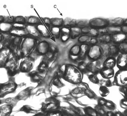
Sclereids are variously shaped and may occur throughout the plant33. Brachysclereids (stone cells) are isolated, approximately isodiametric cells dispersed among parenchyma cells; they develop thick secondary walls as the plant matures. Astrosclereids are highly branched cells with projections that grow intrusively into surrounding intercellular air spaces or along middle lamellae during the growth phase of the organ. Their shapes are to some extent dictated by the nature of the surrounding tissues; for example, they are often star-shaped (astrosclereids: Fig. 1.8) or bone-shaped (osteosclereids).
Figure 1.8 Camellia japonica(Theaceae), transverse section of leaf midrib showing branched sclereid(s) in ground parenchyma. c¼cuticle, e¼epidermis, s¼sclereid. Scale¼100mm.
1.7 Epidermis
The epidermis, the outermost (dermal) cell layer, is a complex tissue that covers the entire plant surface. The epidermis is a pri- mary tissue derived from the outermost layer of the apical meri- stem. It includes many specialized cell types, such as root hairs (chapter 3.4), stomata, trichomes and secretory tissues such as nectaries, both floral and extrafloral (chapters 4.4, 5.10). The aerial plant surface is covered with a non-cellular cuticle and some- times with epicuticular waxes (chapter 4.3.4). Undifferentiated epidermal cells are termed pavement cells. In a developing plant the protodermal cells may give rise to trichomes, stomata or pavement cells, depending on their relative position. InArabidopsis most stomata develop over the junction between underlying cortical or mesophyll cells, and most root hairs develop over the junction of hypodermal cells52.
In growing organs, anticlinal divisions (at right angles to the surface) may occur in mature epidermal cells to accommodate stem or root thickening. In older stems and roots the epidermis often splits and peels away following an increase in thickness, and is replaced by a periderm (chapter 2.9; Fig. 2.15). In some roots the epidermis is worn away by friction with soil particles, and is replaced by an exodermis, which is formed by cell-wall thickening in the outer cortical layers.
1.7.1 Stomata
Stomata are specialized pores in the epidermis through which gaseous exchange (water release and carbon dioxide uptake) takes place. They occur on most plant surfaces above ground, especially on green photosynthetic stems and leaves, but also on floral parts. Each stoma consists of two guard cells surrounding a central pore (Fig. 1.9). Cuticular ridges extend over or under the pore from the outer or inner edges of the adjacent guard cell walls. Guard cells (Fig. 4.3) are either kidney-shaped (in most plants) or dumbbell-shaped (in Poaceae and Cyperaceae). Stomata
Epidermis 13
may be sunken or raised, and are often associated with a sub- stomatal cavity in the mesophyll, which is caused by differential expansion between the guard cell mother cell and the developing underlying mesophyll cells42.
The epidermal cells immediately adjacent to the guard cells are termed subsidiary cells if they differ morphologically from surrounding epidermal cells. Classifications of stomatal types are based either on the arrangement of mature subsidiary cells, or on their patterns of development. Types of mature stomata include anomocytic, anisocytic, diacytic and paracytic124. Anomocytic stomata lack subsidiary cells entirely; anisocytic stomata possess three unequal subsidiary cells; diacytic stomata possess one or more pairs of subsidiary cells with their common walls at right angles to the guard cells; and paracytic stomata possess one or Figure 1.9 Arabidopsis thaliana(Brassicaceae), SEM abaxial leaf surface, showing a single stomatal pore. Scale¼10mm.
more subsidiary cells at either side of the guard cells. However, different developmental pathways may lead to similar stomatal types, so this classification could group types that are non- homologous.
Ontogenetic stomatal types include agenous, mesogenous and perigenous85,112. During development, a protodermal cell under- goes an unequal mitotic division to produce a larger daughter cell and a meristemoid (guard cell mother cell). In the agenous devel- opmental type the meristemoids give rise directly to the guard cells, and there are no subsidiary cells. In the perigenous type, the meristemoid gives rise directly to the guard cells, and subsidiary cells are formed from neighbouring cells, often by oblique divi- sions. In the mesogenous developmental type the guard cells and subsidiary cells have a common origin; the meristemoid under- goes a further mitotic division into two cells, of which one further subdivides to form the guard cells, and the other usually forms one or more subsidiary cells. In mesoperigenous stomatal complexes the guard cells and subsidiary cells are of mixed origin. Subsidiary cells derived from the meristemoid are termed mesogene cells, whereas those derived from neighbouring cells are termed perigene cells, though in some cases mesogene cells are not distinct from surrounding epidermal cells at maturity.
1.7.2 Trichomes
Trichomes are epidermal outgrowths that occur on all parts of the plant surface (Fig. 1.10). They vary widely in both form and function, and include unicellular or multicellular, branched or unbranched forms, and also scales, glandular (secretory) hairs, hooked hairs and stinging hairs. Papillae are generally smaller than trichomes and unicellular, though the distinction is not always clear. In cases where there are several small outgrowths on each epidermal cell, these outgrowths are termed papillae, but where there is only one unicellular outgrowth per cell, the distinction is dependent on size.
Epidermis 15
Glandular trichomes usually possess a unicellular or multi- cellular stalk and a secretory head with one to several cells.
Secreted substances such as volatile oils collect between the secretory cells and a raised cuticle, which later breaks to release them. There are many different types of glandular hair, and they secrete a variety of substances, including essential oils and salt;
some carnivorous plants’ digestive juices contain proteolytic enzymes31. Leaf glandular hairs ofCannabis sativasecrete a resinous substance containing the mild hallucinogen tetrahydrocannabinol.
Glandular hairs of Drosophyllum and Drosera secrete both sticky mucilage and proteolytic enzymes. The stinging hairs of Urtica dioica (stinging nettle) are rigid, hollow structures that contain a poisonous substance (Fig. 1.11). The spherical tip of the hair is readily broken off in contact with an outside body, and the remaining sharp point may then penetrate the skin and release Figure 1.10 Salvia involucrata(Lamiaceae), trichomes on petal surface.
n¼nonglandular trichome, g1¼glandular trichome with unicellular head, g4¼glandular trichome with four-celled head. Scale¼50mm.
the fluid. Other examples of specialized hair types include water-absorptive leaf scales in many Bromeliaceae, and salt- secreting glands of species ofAvicennia60.
1.8 Ground Tissue
Ground tissue, sometimes termed packing tissue, forms the bulk of primary plant tissue and occupies the areas that are not taken up by vascular tissue or cavities. It has a mechanical function, and may be concerned with storage or photosynthesis. Ground tissue typically consists of parenchyma, sclerenchyma or collen- chyma, and is often interspersed with idioblasts and secretory cells or canals. Ground tissue is initially formed at the apical meristems but may be supplemented by intercalary growth, and in monocots by tissues differentiated from primary and secondary thickening meristems. In dicots the ground tissue of secondary xylem (wood), formed by the vascular cambium, consists of fibres and axial parenchyma (chapter 2.6). In older stems the central area of ground tissue (pith) often breaks down, leaving a cavity (Fig. 2.2).
Figure 1.11 (left)Urtica dioica(Urticaceae), intact tip of stinging hair. Scale¼10mm.
Ground tissue 17
1.9 Vascular Tissue
Vascular tissue consists of xylem and phloem, and may be primary or secondary in origin. Primary vascular tissue is derived from procambium, itself produced by the apical meristems, and also by the primary thickening meristem in stems of monocots (chapter 2.8). Secondary vascular tissue is derived from the vascular cambium in dicots, and from the secondary thickening meristem in a few monocots (Fig. 2.13). Both xylem and phloem are complex tissues, composed of many different cell types. Xylem is primarily concerned with water transport and phloem with food transport. Distribution of vascular tissue varies considerably between different organs and taxa.
1.9.1 Xylem
The primary function of xylem is as a water-conducting tissue.
Xylem is a complex tissue composed of several cell types. The water-conducting cells are termed tracheary elements, and are typically linked to form axial chains (vessels). They have thickened lignified cell walls and lack contents at maturity. Two basic types of tracheary element can be recognized: tracheids and vessel elements; an evolutionary series from tracheids to vessel elements is widely recognized7. Vessel elements possess large perforations in their end walls adjoining other vessel elements, whereas tracheids lack these perforations. The perforations may have one opening (simple perforation plate) or several openings which are divided either by a series of parallel bars (scalariform perforation plate: Fig. 2.7) or by a reticulate mesh (reticulate perforation plate). In some species tracheary elements possess wall thickenings (Fig. 2.8) that are arranged either in a series of rings (annular rings), helically or in a scalariform or reticulate mesh. Annular and helical thickenings are the types most commonly found in the first-formed (protoxylem) elements. Later-formed primary tracheary elements (metaxylem) and also secondary tracheary elements typically possess bordered pits in their lateral walls.
These pits vary considerably in size, shape and arrangement;
they may be oval, polygonal or elongated (scalariform pitting), organized in transverse rows (opposite pitting) or in a tightly packed arrangement (alternate pitting).
1.9.2 Phloem
Phloem has complex roles in translocation and messaging within the plant. Primary phloem is formed by the apical meristem and secondary phloem by the vascular cambium. Phloem may develop precociously in regions that require a copious supply of nutrients, such as developing sporogenous tissue.
Phloem is a complex tissue that consists of conducting cells (sieve elements) and associated specialized parenchyma cells (companion cells) (Figs. 1.12; 1.13); these two closely inter- dependent cell types are produced from a common parent cell but develop differently. Angiosperm sieve elements lack nuclei and most organelles at maturity, but retain plastids and phloem-specific
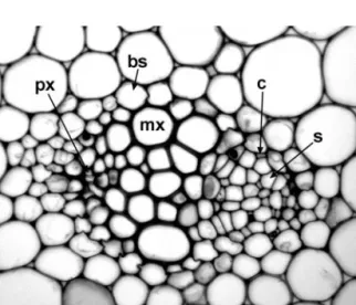
Figure 1.12 Lilium tigrinum(Liliaceae), transverse section of stem vascular bundle. bs¼bundle sheath, c¼companion cell, mx¼metaxylem vessel, px¼protoxylem vessel, s¼sieve tube element. Scale¼100mm.
Vascular tissue 19
proteins (P-proteins) which occur in several morphological forms (amorphous, filamentous, tubular and crystalline) that are often highly characteristic for particular plant families, and thus of systematic and evolutionary value14,116. Sieve-element plastids are classified according to their inclusions: starch (S-type plastids), protein (P-type plastids), or both. By contrast, companion cells are densely cytoplasmic, retaining nuclei and many active mitochondria.
Sieve elements are linked axially to form sieve tubes. The two basic types of sieve element, sieve cells and sieve-tube elements, are differentiated by their pore structure; most angiosperms exclusively possess sieve-tube elements. The walls of sieve ele- ments are thin and possess characteristic regions (sieve areas) that connect adjacent sieve elements; sieve areas consist of groups
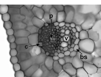
Figure 1.13 Crocus cancellatus(Iridaceae), transverse section of leaf vascular bundle. bs¼bundle sheath, c¼crystal, v¼metaxylem vessel, p¼phloem.
Scale¼50mm.
of pores and associated callose. In sieve cells the sieve areas are distributed throughout the cell wall, but in sieve-tube elements they are mainly localized on the adjoining end walls, forming sieve plates that link two axially linked elements of a sieve vessel.
Sieve plates can be simple or compound.
1.10 Meristems
Meristematic tissue consists of thin-walled, tightly packed living cells which undergo frequent divisions. Meristematic cells undergo cell division and wall formation followed by differential cell expansion. After nuclear division there is progressive deposi- tion of membranes in the cytoplasm into a cell plate that is located in the equatorial zone between the two daughter nuclei108. The cell plate extends to join the cell walls, thus depositing a new wall. Most of the plant body is differentiated at the meristems in well-defined zones, though cells in other regions may also occasionally divide. There are some remarkable examples of fully- differentiated cells giving rise to entire plantlets, notably on leaves of Crassulaceae, such asKalanchoe102.
1.10.1 Apical Meristems
Apical meristems are located at the shoot apex (Fig. 2.1), where primary stem, leaves and flowers differentiate, and at the root apex (Figs 3.1, 3.2), where primary root tissue is produced. Subsequent elongation of the shoot axis may occur by random cell divisions and growth throughout the youngest internodes. This region of diffuse cell division is termed an uninterrupted meristem, and is continuous with the apical meristem. However, in some plant stems, particularly in grasses, most cell divisions contributing to stem elongation occur in a limited region, usually at the base of the internode, which is then termed an intercalary meristem.
Both intercalary and uninterrupted meristems represent growth in regions of already differentiated tissues.
Meristems 21
1.10.2 Lateral Meristems
Lateral meristems are located parallel to the long axis of a shoot or root, most commonly in the pericyclic region, at the junction between vascular tissue and cortex. Examples of lateral meristems include primary and secondary thickening meristems (PTM and STM) and vascular cambium. Primary and secondary thicken- ing meristems produce both ground tissue and vascular bundles (chapter 2.8; Figs 2.13, 2.14). Vascular cambium produces secondary xylem and phloem (chapter 2.5; Fig. 2.5).
Adventitious roots are typically formed in the root pericycle; in these cases the pericycle could be termed a lateral meristem.
The phellogen (cork cambium) is a lateral meristem that occurs in the stem or root cortex, where it forms a protective corky layer (chapter 2.9; Fig. 2.15). A phellogen may also develop in the region of a wound, or at the point of leaf abscission.
1.10.3 Meristemoids and Asymmetric Cell Division Meristemoids are individual cells that are responsible for the differentiation of distinct structures. In many cases meristemoids represent the smaller, densely cytoplasmic, daughter cell that results from an unequal (asymmetric) cell division; the larger daughter cell is less active. Asymmetric divisions are caused by cell polarization resulting from organized arrays of actin filaments in the dense cytoplasm during determination of cell plate alignment42. Examples of unequal cell divisions include cleavage of the microspore into a larger vegetative cell and smaller generative cell, formation of a root hair initial (trichoblast), a protophloem division to form a larger sieve tube element and smaller companion cell, and division of an epidermal cell into two cells of unequal sizes, the smaller of which is the meristemoid that will divide to form the guard cells of a stoma.
2
Stem
2.1 Shoot Apex
The vegetative shoot apex contributes to extension growth of the shoot and initiates leaf primordia. Most shoot apices are inde- terminate, though some (e.g. shoot thorns) become determinate.
The vegetative shoot apical meristem is typically dome-shaped and partitioned by distinct zones of activity (Fig. 2.1). In many species, the outermost two (sometimes more) cell layers (L1 and L2, collectively termed the tunica) are maintained predominantly by anticlinal cell divisions. The corpus (L3), in which cell divisions are randomly oriented, is the region proximal to the tunica. Thus, the outer layers contribute to surface growth and the inner layers to an increase in volume, though there is often slight intergradation between the two layers20.
The central regions of both tunica and corpus are sometimes larger and more highly vacuolated than those on either side. The central region underlying the corpus layer is a rib meristem; this gives rise to files of cells that later become the pith. This central region is surrounded by a peripheral flank meristem that produces the procambium, cortical region and leaf primordia.
Reproductive shoot apices are complex examples of deter- minate growth. During the transition to the flowering phase (termed floral transition), the shoot apex commonly undergoes profound morphological change, though the tunica/
corpus structure is maintained77. In general, at floral transition there is an overall increase in mitotic activity at the shoot apex,
but a proportionally greater increase among the axial apical cells than among the peripheral cells43.
2.2 Primary Stem Structure
The plant stem is generally cylindrical, or sometimes ridged or quadrangular (Fig. 2.2). Primary vascular tissue typically consists of either a complete cylinder or a system of discrete vascular bundles.
The cortex is the region of ground tissue between the vascular tissue and the epidermis; the junction between the cortex and vascular region is termed the pericyclic region, from which endogenous adventitious roots can arise (Fig. 3.4). The pith is the central region of ground tissue, though in many stems it breaks down to form a central hollow cavity. The stem epidermis often bears stomata and trichomes, as in the leaf epidermis (chapter 4.3).
The stem primary ground tissue is basically parenchymatous but can be modified into various tissue types or interspersed with Figure 2.1 Diagram of angiosperm shoot apical organisation.
fibres and sclereids, and parenchyma cells frequently become lignified as the plant ages. Ridged or angled stems often possess strengthening collenchyma at the angles, immediately within the epidermis. Many stems are photosynthetic organs with a chlorenchymatous cortex, particularly in leafless (apophyllous) plants, which normally occur in nutrient-poor habitats.
Some plant stems possess secretory cells or ducts in the ground tissue. For example, many species of Euphorbia possess branched networks of laticifers in the cortex (Fig. 1.6), which extend throughout the ground tissue of the stem and leaves. Plants with succulent stems, such as many Cactaceae, typically possess regions of large thin-walled cells that contain a high proportion of water.
Some stems (e.g. corms of Crocus) are specialized as storage or perennating organs; they store food reserves in the form of starch granules, most commonly in the inner cortex. Sometimes the layer of cortical cells immediately adjacent to the vascular tissue is distinct from the rest of the cortex, and may be packed with starch granules; this is termed a starch sheath, or sometimes an endodermoid layer or endodermis, though the component cells Figure 2.2 Vicia faba(Fabaceae), transverse section of stem. Scale¼100mm.
Primary stem structure 25
usually lack the Casparian thickenings that are typically found in the root endodermis (chapter 3.5).
2.3 Primary Vascular System
The primary vascular system is mostly derived from the pro- cambium near the shoot apex. Primary vascular bundles possess both xylem and phloem, arranged either adjacent to each other (in collateral vascular bundles: Fig. 1.12), or with strands of phloem on both sides of the xylem (bicollateral vascular bundles), or with xylem surrounding the phloem (amphivasal vascular bundles). In woody angiosperms, internodal stem vasculature is typically arranged either in a continuous cylinder, or in a cylinder of separate or fused collateral bundles, with the phloem external to the xylem (Fig. 2.2). In some stems the bundles may be bicollateral; for example in species of Cucurbita internal phloem is present in addition to the external phloem. The vascular cambium, which produces secondary vascular tissue in woody species, is initially situated between the xylem and phloem within vascular bundles, but eventually extends between the vascular bundles to form a complete vascular cylinder. Some stems also possess cortical or medullary (pith) bundles, which can be associated with leaf vasculature.
In monocots, which lack a vascular cambium, the stem vascu- lar bundles are typically scattered throughout the central ground tissue (Fig. 2.3), or sometimes arranged in two or more distinct rings. Vascular bundles may be collateral, bicollateral or amphi- vasal. Cortex and pith are frequently indistinct from each other, though the cortex may be defined by an endodermoid layer, or a distinct ring of vascular bundles, or in some stems, particularly inflorescence axes, by a cylinder of sclerenchyma that encloses the majority of vascular bundles. The monocot vascular system is often extremely complex128. Each major bundle, when traced on an upward course from any point in the stem, branches or forms
bridges with other bundles at several points before passing into a leaf. One of its major branches then continues a similar upward course towards the apex. Some palms possess literally thousands of vascular bundles in a single transverse section of the stem, though in most other monocots the number is much smaller.
2.4 Nodal Vasculature
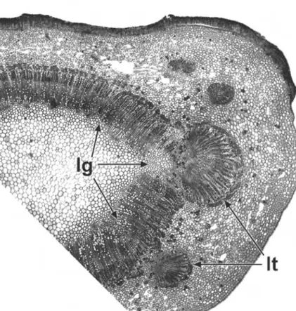
At regions of leaf insertion on the stem (nodes), the vasculature of the leaf and stem are connected. Openings (termed lacunae or leaf gaps) occur in the stem vascular cylinder beneath their point of contact (Fig. 2.4). In eudicots and magnoliids, nodal anatomy is often characteristic of taxonomic groups, particularly the number and arrangement of leaf traces and leaf gaps.
Figure 2.3 Monocot stem anatomy:Lilium tigrinum(Liliaceae), transverse section of inflorescence axis, showing cortex (c), surrounding central region with numerous distinct vascular bundles. sc¼sclerenchymatous layer. Scale¼100mm.
Nodal vasculature 27
Nodes may be unilacunar, trilacunar or multilacunar, depending on the number of leaf gaps in the stem vascular cylinder. This feature is most obvious in stems in which there is otherwise a continuous vascular cylinder, especially where a limited amount of secondary thickening has taken place; as a result, nodal anatomy has been studied far more extensively in woody than herbaceous plants54. Sometimes the number of leaf gaps per node varies within a species or individual, usually increasing with increased plant size and age67.
Figure 2.4 Prunus lusitanica(Rosaceae), transverse section of stem at node, showing connection of petiole vasculature to main vascular cylinder of stem.
lg¼leaf gap, lt¼leaf trace. Scale¼100mm.
The number of leaf traces departing from each gap is also generally characteristic of a species, but may vary within a plant, especially in species with unilacunar and trilacunar nodes.
For example, in Clerodendrum two traces typically depart from a single gap, and inPrunus a single trace departs from each of three gaps in the central vascular cylinder (Fig. 2.4). In Quercus up to five traces depart through a trilacunar node. Normally, leaf trace bundles are initiated acropetally from the stem procambial system near the shoot apex, to serve developing primordia67. However, in some species (e.g.Populus deltoides) subsidiary vascular bundles are initiated at the base of each developing primordium, and grow basipetally to meet the stem procambial trace.
Nodal vasculature is further complicated by the axillary bud vascular traces, which are connected to the main stem vascu- lature immediately above the leaf gaps. In most species two traces diverge to supply each bud or branch.
In large woody trees, the junction of the trunk and its branches is characterized by a complex arrangement of secondary vascular tissue, which typically forms a collar around the base of the branch99. This branch collar is enveloped by a trunk collar, which links the vascular tissue of the trunk above and below the branch. There is no direct connection of xylem from the trunk above a branch into the branch xylem, as the tissues are oriented perpendicular to each other. If a branch dies, a protection zone forms around its base to prevent spread of infection into the trunk, and the branch is often shed.
2.5 Vascular Cambium
Increase in height, achieved by growth at the apical meristem, is inevitably followed by at least some degree of increase in stem thickness. This is achieved by different types of meristems in different species. In woody eudicots and most magnoliids (but not monocots), secondary vascular tissue (both xylem and phloem)
Vascular cambium 29
is produced by the vascular cambium (Fig. 2.5), which usually becomes active at a short distance behind the stem apex. The vascular cambium is initiated between xylem and phloem within vascular bundles, but soon consists of an unbroken cylinder of meristematic cells. It typically generates secondary xylem (wood) at its inner edge and secondary phloem at its outer edge, though plants with anomalous secondary growth do not always follow this pattern. The amount of secondary vascular tissue produced is extremely variable, depending on the habit of the plant. Vascular cambium is absent in monocots and some herbaceous eudicots (e.g.Ranunculus) and magnoliids (e.g.Saururus).
The vascular cambium is a single cell layer (uniseriate) or several cell layers (multiseriate) if xylem and phloem mother cells are included55. It is a complex tissue consisting of both Figure 2.5 Prunus communis(Rosaceae). Transverse section of stem in region of vascular cambium, with secondary phloem (above) and secondary xylem (below). cc¼companion cell, r¼ray, s¼sieve element, vc¼vascular cambium, ve¼vessel element.
fusiform initials and ray initials, which form the axial and radial systems respectively. Both fusiform and ray initials are vacuolate (unlike most meristematic tissue) and plastid-rich. Fusiform initials are axially elongated cells with tapering ends. They divide periclinally to form the axial elements of secondary tissues:
tracheary elements, fibres and axial parenchyma in secondary xylem, and sieve elements, companion cells and fibres in second- ary phloem. Ray initials are isodiametric cells that divide peri- clinally to form ray parenchyma cells in both xylem and phloem.
Fusiform initials sometimes give rise to new ray initials as the stem increases in circumference and new rays are formed.
2.6 Secondary Xylem
Secondary xylem (wood) varies considerably between species.
The texture and density of a particular type of wood depend on the size, shape and arrangement of its constituent cells73. Wood is composed of a matrix of cells (Fig. 2.6), some arranged parallel to the long axis (fibres, vessels and chains of axial parenchyma cells), and others (ray parenchyma cells) forming the wood rays that extend radially from the vascular cambium towards the pith. The precise cellular arrangement in wood is often characteristic of species or genera. To observe their structure, woods are sectioned transversely (transverse section: TS) and in two longitudinal planes: along the rays (radial longitudinal section: RLS) and perpendicular to the rays (tangential longi- tudinal section: TLS). In some woods the vessels are solitary when viewed in transverse section (Figs 2.6, 2.10), but in other woods they are arranged in clusters or radial chains (Fig. 2.9). Axial parenchyma cells may be independent of the vessels (apotracheal) or associated with them (paratracheal), and sometimes occur in regular tangential bands. The relative abundance of axial paren- chyma varies in different species, from sparse (or even completely absent) to rare cases such as Ochroma pyramidale (balsa), in which
Secondary xylem 31
axial parenchyma cells are often more abundant than fibres, making this type of wood soft and easy to carve.
Rays are termed uniseriate if they are one cell wide tangentially, and multiseriate if they are more than one cell wide, viewed in TS and TLS. Sometimes both uniseriate and multiseriate rays occur in the same wood, as inQuercus. Ray cells vary in shape (best viewed in RLS); homocellular rays are composed of cells of similar shapes, whereas in heterocellular rays the cells are of different shapes.
Other aspects of variation in the structure of hardwoods include the presence of either axial or radial secretory canals in some woods (Fig. 2.10), the storied (stratified) appearance of various elements, particularly rays, or the fine structure of the vessel walls (intervascular pitting, perforation plates and wall thickenings:
chapter 1.7.1). For example, in Tilia cordata (Fig. 2.8), the vessel Figure 2.6 Secondary xylem:Quercus robur(Fagaceae), block of wood at edge of transverse and tangential longitudinal surfaces, showing large early (spring) wood vessels.
element walls are helically thickened, and in many Fabaceae the pit apertures are surrounded by numerous warty protuberances, termed vesturing19. Perforated ray cells, an unusual feature of some woods, are ray cells that link two vessel elements and themselves resemble and function as vessel elements, with perforation plates corresponding to those of the adjacent vessel elements. However, like other ray cells, perforated ray cells are formed from ray initials rather than from fusiform initials, like vessel elements.
In many woody temperate plants cambial activity is seasonal (usually annual), which results in the formation of growth rings.
The secondary xylem formed in the early part of the season (early wood or spring wood) is generally less dense and consists of thinner-walled cells than the xylem formed later in the growing season (late wood or summer wood). In ring-porous woods the vessels are considerably larger in early wood than in late wood (Fig. 2.11). In diffuse porous woods the main distinction between early and late wood is in size and wall thickness of the fibres (Fig. 2.9). As woody plants age and their trunks increase in Figure 2.7 Betula utilis(Betulaceae). Wood in (A) tangential longitudinal section (TLS) and (B) radial longitudinal section (RLS). b¼bar of scalariform perforation plate, r¼ray. Scale¼100mm.
Secondary xylem 33
girth, the central area becomes non-functional with respect to water transport or food storage, and the vessels frequently become blocked by tyloses. Tyloses are formed when adjacent parenchyma cells grow into the vessels through common pit fields. The central non-functional area of the trunk, the heartwood, is generally darker than the outer living sapwood.
In some woody angiosperms, particularly climbing plants (lianas) such as many Bignoniaceae (Fig. 2.12), secondary growth does not fit the ‘‘normal’’ pattern of xylem and phloem production, and is termed anomalous secondary growth. For example, some plants develop regions of phloem (included or interxylary phloem) embedded in the xylem, either in islands (e.g. in Avicennia) or in alternating concentric bands. Other examples have irregularly divided or deeply fissured areas of xylem and phloem, or stems that are flattened or otherwise Figure 2.8 Tilia olivieri(Tiliaceae), SEM inside surface of vessel element showing wall thickenings and intervascular pitting.
irregularly shaped73. Such anomalous forms are achieved either by the formation of new vascular cambia in unusual positions or by the unusual behaviour of the existing cambium in producing phloem instead of xylem at certain points.
2.7 Secondary Phloem
Secondary phloem is also a product of the vascular cambium in woody species. As in secondary xylem, secondary phloem consists of both axial and radial systems, formed from the fusiform and ray initials respectively. Phloem rays are radially continuous with xylem rays, and may be similarly uniseriate or multiseriate, though in transverse section they often appear dilated towards the cortex as a result of cell divisions to accommodate increase in stem thickness (Fig. 2.11). At their outer periphery, the Figure 2.9 Alnus glutinosa(Betulaceae), wood, transverse section.
Scale¼100mm.
Secondary phloem 35
parenchymatous ray cells are often difficult to distinguish from cortical cells. Older ray cells sometimes become lignified to form sclereids. The axial system of the phloem consists of sieve elements and companion cells, as in primary phloem (chapter 1.9.2). It also typically includes fibres, sclereids and axial parenchyma cells.
In some species fibres are formed in groups at regular intervals, resulting in characteristic tangential bands of fibres alternating with groups of sieve elements and parenchyma cells.
2.8 Primary and Secondary Thickening Meristems In monocots, which lack a vascular cambium, increase in stem diameter is typically relatively limited. However, most monocots Figure 2.10 Shorea resina-nigra(Dipterocarpaceae), wood, transverse
section showing vessels (v) and axial secretory canals (sc), r¼ray.
Scale¼100mm.
and a few other thick-stemmed angiosperms, especially species with short internodes and crowded leaves, possess a primary thickening meristem (PTM) near the vegetative shoot apex88. The PTM (Fig. 2.13) is situated in the pericyclic region. It consists of a narrow multiseriate zone of meristematic cells that produces radial derivatives, usually a limited amount of parenchyma towards the outside (centrifugally), and both parenchyma and discrete vascular bundles towards the inside (centripetally). In addition to primary stem thickening, the PTM is responsible for formation of linkages between root, stem and leaf vasculature.
Also, it frequently retains meristematic potential further down Figure 2.11 Tilia olivieri(Tiliaceae), transverse section of twig with slightly more than three years growth, c¼cortex, p¼pith, ph¼phloem, vc¼vascular cambium. Scale¼100mm.
Primary and secondary thickening meristems 37
the stem and is the site of adventitious root production in some species.
The PTM normally ceases activity at a short distance behind the apex, and subsequent stem thickening is limited. Tree-forming palms possess an extensive PTM that forms a large sunken apex;
considerable further stem thickening occurs by subsequent division and enlargement of ground parenchyma cells. This is termed diffuse secondary growth.
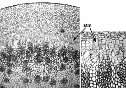
In some woody monocots in the order Asparagales (e.g. Agave, Aloe, Cordyline, Yucca) further increase in stem thickness is achieved by means of a secondary thickening meristem (STM) (Fig. 2.14).
The STM is essentially similar to the PTM in that it is located in the pericyclic region and produces radial derivatives. However, it is active further from the primary apex and produces second- ary vascular bundles that are often amphivasal and radially elongated. In some species (e.g.Nolina recurvata, Cordyline terminalis) Figure 2.12 Tynanthus elegans(Bignoniaceae). Transverse section of woody stem showing anomalous secondary growth: xylem region with four deep fissures of phloem. Scale¼1 mm.
the PTM and STM are axially discontinuous104,105, whereas in others (e.g.Yucca whipplei) they are axially continuous26. Apart from the distance from the apex, there are no precise criteria for distinguishing between derivatives of the two meristems, and transitional forms exist. Thus, they are perhaps best regarded as developmental phases of the same meristem.
The PTM and STM are not homologous with the vascular cambium, because the vascular derivatives are arranged in different ways. The vascular cambium produces phloem centri- fugally and xylem centripetally, whereas most derivatives of the PTM and STM are centripetal, and consist of a parenchyma- tous ground tissue and discrete vascular bundles containing both xylem and phloem. Furthermore, the PTM originates in ground tissue, is a tiered meristem, and is often fairly diffuse, especially near the shoot apex (Fig. 2.13). By contrast the vascular cambium is typically uniseriate and initially originates within vascular tissue, though it later extends between bundles.
Figure 2.13 Primary thickening meristem (PTM): diagram of longitudinal section of the crown of a typical thick-stemmed monocot, showing orientation and extent of radial PTM derivatives. Vascular strands not shown.
(Adapted from DeMason 1983).
Primary and secondary thickening meristems 39
2.9 Periderm
Periderm is a protective tissue of corky (suberinized) cells that is produced either as a response to wounding or in the outer layers of the cortex of a stem or root that has increased in thickness. The periderm consists of up to three layers: phellogen, phellem and phelloderm. The phellogen is a uniseriate meri- stematic layer of thin-walled cells that produces phellem to the outside, and (in some cases) phelloderm to the inside. The phellem cells constitute the corky tissue. They are tightly-packed cells that lack contents at maturity. They possess deposits of suberin and sometimes lignin in their walls, and form an impervious layer to prevent water loss and protect against injury.
Phelloderm cells are non-suberinized and parenchymatous, and contribute to the secondary cortex.
Figure 2.14 Secondary thickening in monocots:Dracaena indivisa (Ruscaceae), transverse section of stem showing secondary thickening meristem (STM) and radial internal vascular derivatives. Scale¼100mm (left hand image).
A periderm commonly occurs in the cortex of secondarily thickened stems, to replace the epidermis, which splits and peels away (Fig. 2.15). The phellogen may originate either adjacent to the epidermis (or even within the epidermis) or deeper in the cortex. Sometimes several phellogens form almost simultaneously.
The pattern of periderm formation largely dictates the appearance of the bark of a woody plant. For example, the smooth papery bark of a young silver birch tree (Betula pendula) is formed because the periderm initially expands tangentially with the increase in stem diameter, but later flakes off in thin papery sheets as Figure 2.15 Sambucus nigra(Caprifoliaceae). Transverse section of stem surface, showing periderm forming in outer cortical layers. c¼cortex, e¼epidermis, le¼lenticel, p¼periderm, ph¼secondary phloem, pi¼pith, vc¼vascular cambium, xy¼secondary xylem. Scale¼100mm.
Periderm 41
abscission bands of thin-walled cells are formed. In the trunk of cork oak (Quercus suber), the initial phellogen may continue activity indefinitely, and produces seasonal growth rings. In the commer- cial process it is removed after about 20 years to make way for a second, more vigorous phellogen, which produces the commercial cork.
Many species possess lenticels in the bark (Fig. 2.15); these are areas of loose cells in the periderm, which are often initially formed beneath stomata in the epidermis, and are thought to be similarly concerned with gaseous exchange.