No part of this publication may be reproduced, stored in a retrieval system, or transmitted in any form or by any means, electronic, mechanical, photocopying, recording, scanning, or otherwise, except as permitted by Section 107 or 108 of the 1976 United State copyright law, without either prior written permission from the publisher or permission through payment of the appropriate fee per copy to Copyright Clearance Center, Inc., 222 Rosewood Drive, Danvers, MA by fax or on the Internet at www.copyright. com. Limitation of Liability/Disclaimer of Warranty: Although the publisher and author have used their best efforts to prepare this book, they make no representations or warranties as to the accuracy or completeness of the contents of this book and specifically disclaim any implied warranty of merchantability or fitness for a specific purpose.
APPLIED TOXICOLOGICAL NEUROPATHOLOGY
CONTRIBUTORS
Reilly, DVM, DACVP, Department of Pathology, Microbiology and Immunology, School of Veterinary Medicine, University of California–Davis, Davis, California. Vernau, DVM, MSc, DACVIM (Neurology), Department of Surgical and Radiological Sciences, School of Veterinary Medicine, University of California–.
PREFACE
INTRODUCTION
The techniques described in Part 3 provide an excellent basis for an evaluation of the nervous system. Direct delivery is an increasingly common way to get various test items, especially proteins, antibodies, lipophobic drugs, and stem cells, to the central and peripheral components of the nervous system.
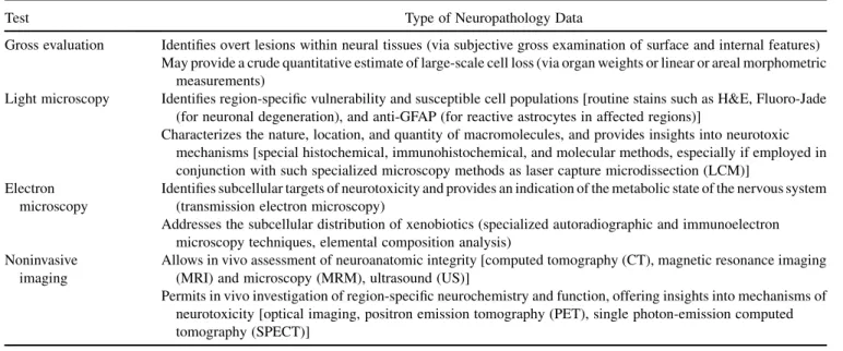
FUNDAMENTALS OF NEUROBIOLOGY
FUNDAMENTAL NEUROPATHOLOGY
AN INTRODUCTION
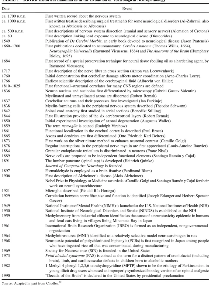
1838 Myelin-forming cells in the peripheral nervous system described (Theodor Schwann) 1842 The anatomy of the spinal cord is studied for the first time in serial sections (Benedikt Stilling). 1861 Functional localization in the cerebral cortex is described (Paul Broca) 1865 Axons and dendrites are first differentiated (Otto Friedrich Karl Deiters).
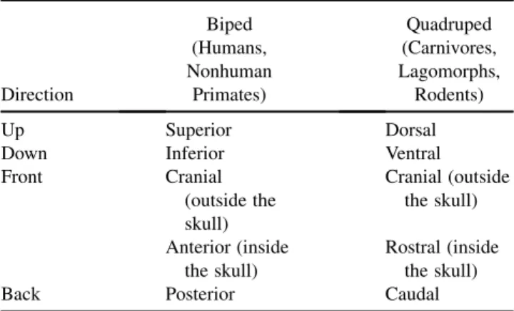
FUNCTIONAL NEUROANATOMY
The unmyelinated white matter adjacent to the gray matter connects the different segments of the spinal cord. Fibers running in the medial part of the optic tract project directly into the pretectal area.

ATLAS OF COMPARATIVE NEUROANATOMY
The cross-sections of the chicken brain are shown separately (Figures 22 to 28) because of their greater deviation from the mammalian pattern of regional brain organization. DRC1, dorsal root of the spinal nerve of C1; FL, longitudinal cerebral fissure; LF, lobus frontalis; LO, lobus occipitalis; LP, lobus parietalis; LT, temporal lobe; M, myelencephalon; T, telencephalon; TeO, tectum mesencephali (tectum opticum). NF, nucleus fastigii; NI, nuclear interpositus; P, pyramid; PC, pedunculus cerebelli caudalis; PF, paraflocculus; S, spine of the trigeminal nerve; SN, nucleus of the spinal canal of the trigeminal nerve;
CH, cerebellar hemispheres; CP, fourth ventricle choroid plexus; CV, cerebellar vermis; F, flakes; H, nucleus motorius nervi hypoglossi;. NC, nucleus cuneatus; NG, nucleus gracilis; S, spinal tract of trigeminal nerve; SN, nucleus of the spinal tract of the trigeminal nerve. Manocha SL, Shantha TR, Bourne GH. Stereotaxic brain atlas of the Cebus monkey (Cebus apella).

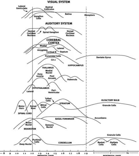
PRINCIPLES OF COMPARATIVE AND CORRELATIVE NEURODEVELOPMENT AND THEIR IMPLICATIONS
The remainder of this section outlines the basic mechanisms that help delineate the basic structure of the nervous system. Ari€ens Kappers CU, et al. The Comparative Anatomy of the Vertebrate Nervous System. Conel JL. The Cortex of the Newborn: The Postnatal Development of the Human Cerebral Cortex, Vol I.
Selected aspects of the anatomy and response to injury of the chicken (Gallus domesticus) nervous system. Cell proliferation in the sub-ependymal layer of the postnatal marmoset, Callithrix jac- chus. Dev Brain Res. The sequence of myelination in the brainstem of the rat monitored by myelin basic protein immunohistochemistry.Brain Res.
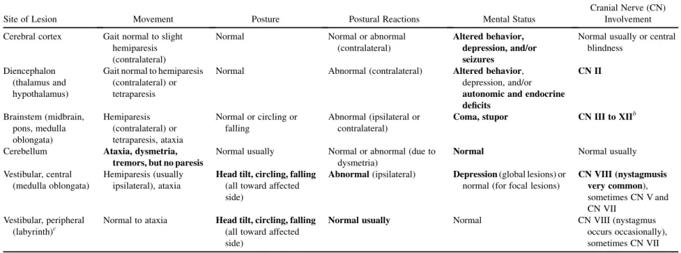
LOCALIZING NEUROPATHOLOGICAL LESIONS USING NEUROLOGICAL FINDINGS
Paresis/paralysis (UMN): one side (usually contralateral) or all limbs Abnormal posture or proprioception (position). Abnormality of touch (generalized) Medial geniculate nucleus Hearing loss (no vestibular signs) Lateral geniculate nucleus Visual loss (pupillary reflexes normal). Abnormality of touch (localized: the usual pattern is normal sensation above the lesion, decreased sensation below it, $local increase in sensation above the lesion).
Paresis/paralysis (resulting in drooping ears, eyes and/or lips) CN VIII (cochlear division) and/or. Altered perception: touch abnormality (localized: reduced or absent sensation$altered sensation [paresthesia]) in the dermatome innervated by the affected nerve. Peripheral somatic nerves (localized) Ptosis [hanging of the upper eyelid (Horner's syndrome)] Sympathetic system: CN III Altered perception.
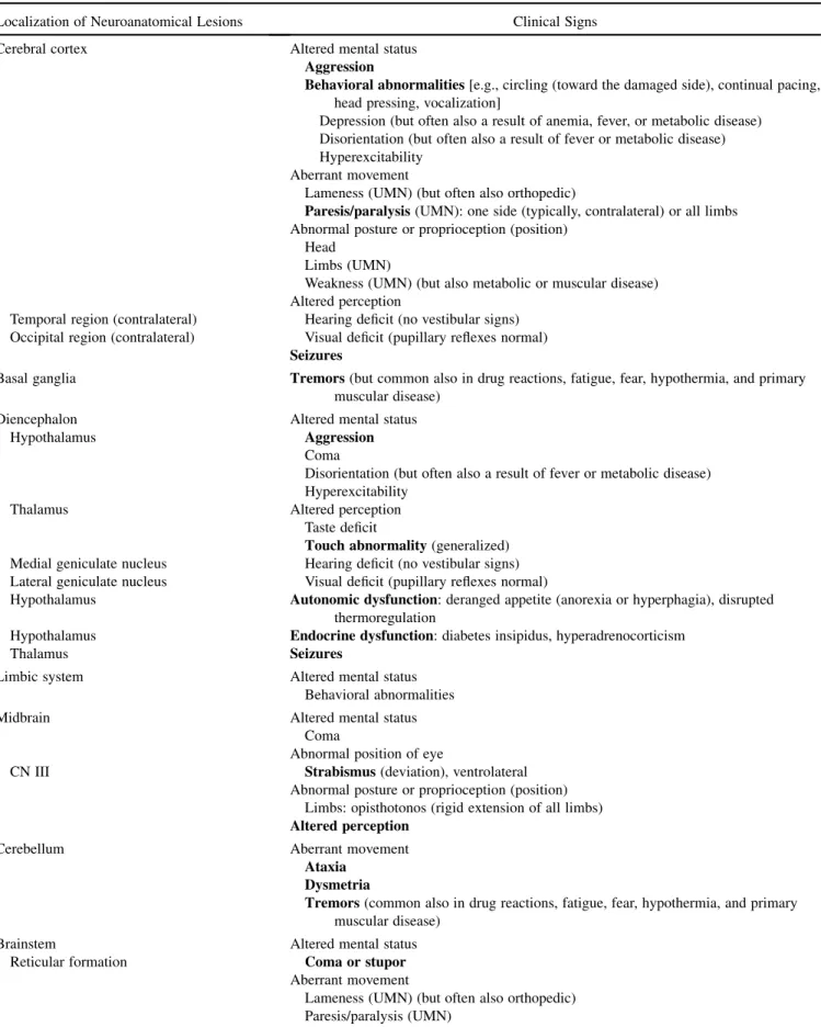
Syncope (usually cardiovascular or metabolic disease) Brain stem
Toxic substances that produce vestibular syndrome usually do so by causing hair cell degeneration in the sensory epithelium of the vestibular labyrinth.32–34. If the lesion is cranial to the origin of the phrenic nerve, death will result from respiratory paralysis. Upper cervical syndrome is typically caused by cervical trauma, developmental malformations of the associated vertebrae, inflammation (bacterial, protozoal, or viral etiologies), or the presence of a space-occupying mass.
The neurologic deficits that develop in PNS typically induce clinical signs related to only one PNS domain (autonomic parasympathetic, autonomic sympathetic, somatic motor, and/or somatic sensory), and often affect only part of the affected domain. These conditions are called paroxysmal syndromes because of the sporadic and temporary nature of the clinical deficits. Triphenyl phosphite and diisopropyl phosphorous fluoride produce separate and distinct patterns of axonal degeneration in the rat central nervous system. Fundam Appl Toxicol.
BEHAVIORAL MODEL SYSTEMS FOR EVALUATING NEUROPATHOLOGY
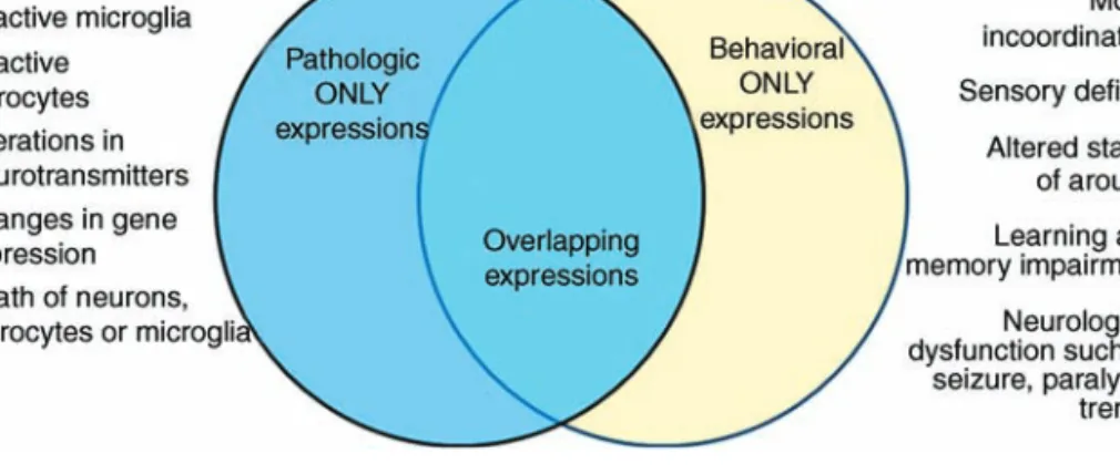
Any change in the structure or function of the central and/or peripheral nervous system should be considered an adverse effect. A comparison of the acute behavioral effects of alkylbenzenes using a functional observational battery in rats. Fundam Appl Toxicol. Effects of 3-acetylpyridine on the central nervous system of the rat as shown by silver methods.
Characterization of the effects of N-hydroxy-IDPN on the auditory, vestibular and olfactory systems in rats. Morphology of axonal swelling in the muscular branch of the posterior tibial nerve and spinal cord. Effects of the neurotoxin 3,30-iminodipropionitrile on acoustic startle and locomotor activity in rats: a comparison of functional observational and automated startle assessment methods. Neurotoxicol Teratol.

COGNITIVE ASSESSMENTS IN NONHUMAN PRIMATES
The frontal cortex, parietal cortex, anterior cingulate, and parts of the basal ganglia appear crucial in working memory function. It has been shown that tasks in the OTB are differentially sensitive to the acute effects of drugs from different pharmacological classes and that performance of each task is not highly correlated with performance of other tasks. The learning task used in the NTB NCTR is referred to as FIGURE 1 Schematic of the operant test panel.
As part of the NCTR OTB, this task is presented for a maximum of 5 min per test session, but typically generates more than 100 choice trials. An increase in the negative slope of the decay line with no change in the origin (position on the y-axis) is said to represent an increase in rate. Changes in response duration distribution characteristics are expected to provide insight into timing mechanisms.
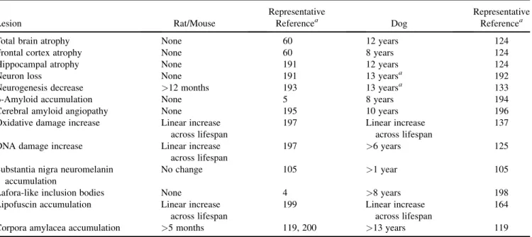
IMPACT OF AGING ON BRAIN STRUCTURE AND FUNCTION IN RODENTS AND CANINES
For example, transgenic mice that develop age-related amyloid deposits in the brain have been used to. Phases of Abeta deposition in the human brain and its relevance to the development of AD. Histochemical accumulation of oxidative damage products is associated with Alzheimer-like pathology in the dog. Amyloid.
Topographical relationship between senile plaques and cerebrovascular amyloidosis in the brains of older dogs.J Vet Med Sci. Role of metal ions in Abeta oligomerization in Alzheimer's disease and other neurological disorders. Neurogenesis in the dentate gyru of the adult rat: age-related decrease in neuronal progenitor cell proliferation.
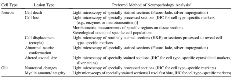
FUNDAMENTALS OF NEUROTOXICITY DETECTION
At one end of the spectrum, there are no detectable changes in the CNS (safe), and at the other end of the spectrum are permanent changes in the CNS (unsafe). The main distinct regions highlighted in the sagittal section of the brain (Figure 3) include the cerebral cortex. Of course, not all major regions can be represented at a single level, but major divisions are often mentioned in the brain.
The location of damage in the brain is unpredictable One of the most common shortcomings in safety assessments is to only look at areas where damage is expected. Some time later, the pathology resolves as the brain clears the debris and removes evidence of the insult. However, at mid-cycle, evidence of cell disintegration is more striking and detection is more reliable.
TOXICOLOGIC NEUROPATHOLOGY: METHODOLOGY
PRACTICAL NEUROPATHOLOGY OF THE RAT AND OTHER SPECIES
At the very least, pressure from extraction instruments can darken neurons or destroy areas critical to brain evaluation. In the rat, Bregma 0.0 mm corresponds to a coronal section at the level of the optic chiasm. Note that some of the section planes intentionally deviate (tilt) from the true coronal plane.
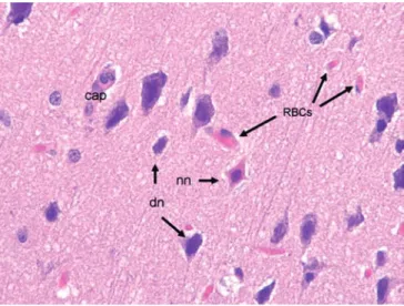
To minimize displacement of the brain within the apparatus, all discs must remain in the matrix until all blades are inserted. The most rostral and most caudal 2- to 4-mm portions of the brain will not be included. In (A), note degenerating neuron (nn), few Purkinje neurons overlying the granule cell (gc) layer, and vacuolation (v) of the overlying molecular layer.
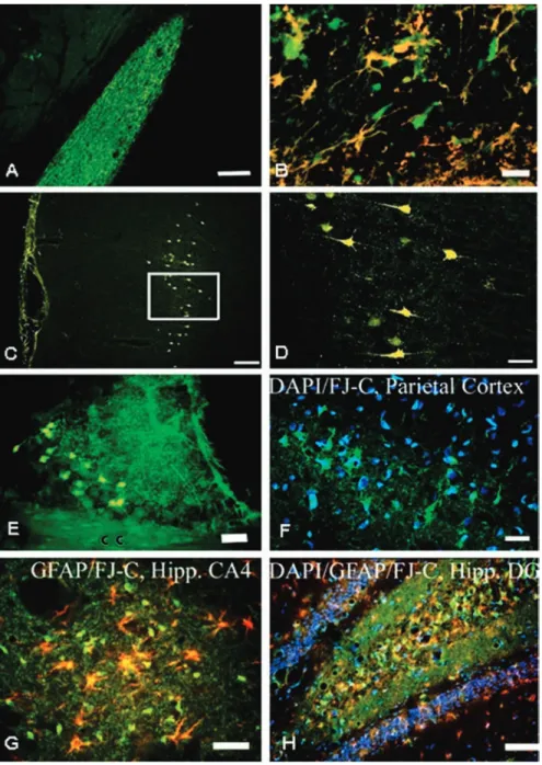
FLUORO-JADE DYES: FLUOROCHROMES FOR THE HISTOCHEMICAL LOCALIZATION
For staining with fluorine jade dyes, the following procedures should be followed to achieve optimal results. This is accomplished by making a stock solution of 0.01% of the counterstain, which is then diluted 1:100 with the Fluoro-Jade staining solution. Avoiding pretreatment with potassium permanganate allowed extensive GFAP labeling of astrocytes, but increased background staining with Fluoro-Jade.
The main advantages of Fluoro-Jade stains are their simplicity, sensitivity, reliability and specificity for localizing degenerating neurons in the brain. The Fluoro-Jade dyes are specifically designed to stain only degenerating neurons (and their processes) in the brain after all types of neurotoxic insults or physical trauma. These decarboxylated polyamines are expected to exhibit high affinity for the polyanionic fluoro-jade dyes.