저작자표시-비영리-변경금지 2.0 대한민국 이용자는 아래의 조건을 따르는 경우에 한하여 자유롭게
l 이 저작물을 복제, 배포, 전송, 전시, 공연 및 방송할 수 있습니다. 다음과 같은 조건을 따라야 합니다:
l 귀하는, 이 저작물의 재이용이나 배포의 경우, 이 저작물에 적용된 이용허락조건 을 명확하게 나타내어야 합니다.
l 저작권자로부터 별도의 허가를 받으면 이러한 조건들은 적용되지 않습니다.
저작권법에 따른 이용자의 권리는 위의 내용에 의하여 영향을 받지 않습니다. 이것은 이용허락규약(Legal Code)을 이해하기 쉽게 요약한 것입니다.
Disclaimer
저작자표시. 귀하는 원저작자를 표시하여야 합니다.
비영리. 귀하는 이 저작물을 영리 목적으로 이용할 수 없습니다.
변경금지. 귀하는 이 저작물을 개작, 변형 또는 가공할 수 없습니다.
이학 석사 학위 논문
근육유래인자 Lumican의 근감소증 치료효과 및 기전연구
Anti-sarcopenic effects of muscle-derived lumican and their mechanisms
울 산 대 학 교 대 학 원 의 학 과
김 다 애
[UCI]I804:48009-200000171656
[UCI]I804:48009-200000171656
[UCI]I804:48009-200000171656
근육유래인자 Lumican의 근감소증 치료 효과 및 기전연구
지 도 교 수 고 정 민
이 논문을 이학 석사 학위 논문으로 제출함
2018년 12월
울 산 대 학 교 대 학 원 의 학 과
김 다 애
김다애의 이학석사 학위 논문을 인준함
심사위원 고 정 민 (인) 심사위원 김 경 곤 (인) 심사위원 유 현 주 (인)
울 산 대 학 교 대 학 원
2018년 12월
국문 요약
연구배경
골격근의 양과 근력이 감소하는 질환인 근감소증은 삶의 질 저하뿐만 아니라 사망률 증가로 이어진다. 근감소증의 여러 원인 중 가장 중요하게 여겨지는 것이 노화이기 때문에, 근감소증은 대표적인 노인성 질환으로 분류되고 있다. 따라서 근감소증은 현대 고령화 사회에서 그 중요성은 점점 더 커질 것으로 여겨지고 있으나, 아직까지 공인된 치료제가 없는 것이 현실이다. 2008년 근육에서 IL-6가 분비되는 것이 처음 밝혀진 이후, myokine이라고 불리는 다양한 분비인자들이 근감소증 치료 표적으로 발굴되고 있다.
연구목적
본 연구에서는 myokine인 Lumican이 근육에 미치는 영향과 작용기전을 규명함으로써 근감소증의 새로운 치료 표적으로서 Lumican의 가능성을 확인하고자 하였다.
연구방법 및 내용
• Myokine으로서의 Lumican 발현 및 근육량과의 상관관계를 조사하였다.
• C2C12 세포에서 근분화, 증식, 이동능, 근분화 관련 전사인자들의 발현 및 autophagy에 대한 Lumican의 영향을 관찰하였다.
• Lumican의 수용체를 규명하고자 하였다.
• Lumican의 주요 신호전달경로를 조사하였다.
• 근감소증 동물모델을 통하여 Lumican의 생체 치료 효과를 관찰하였다.
연구결과
• 운동으로 활성화된 근육에서는 Lumican의 발현이 증가하였다. 반면 근육에서의 Lumican 발현은 노화와 성호르몬 결핍에 영향을 받지 않았다.
• Lumican은 근분화를 촉진하지만, 세포 증식, 이동, 미토콘드리아의
biogenesis, autophagy와는 관련이 없었고, Lumican은 단백질 합성과 관련이 깊은 Akt-mTOR signaling pathway를 활성화시켰다.
• 각종 inhibitor 실험을 통해 Lumican의 수용체로서 Integrin a2/b1과 Integrin a5/b3를 특정하였다.
• 근감소증 동물 모델인 난소절제 마우스에서 Lumican의 혈관주사는 근육량을 증가시키고 근력을 향상시켰다.
결론
근육유래인자인 Lumican은 Akt-mTOR pathway를 통해 근분화 (myogenesis)를 촉진하고, 동물 모델에서 근감소증 치료 효과를 관찰하여 Lumican이 근감소증 치료에 대한 신규 표적이 될 수 있을 것으로 판단된다.
목 차
국문요약 ··· i
표 차례 ··· iv
그림 차례 ··· v
서론 ··· 1
연구재료 및 방법 ··· 4
연구 결과 ··· 11
고찰 ··· 17
결론 ··· 20
참고문헌 ··· 46
영문요약 ··· 56
List of Tables
Table 1. Primer sequences ··· 21
List of Figures
Figure 1. Muscle-derived factors increase muscle mass in mice ··· 22
Figure 2. Changes of Lumican expression and secretion during myogenesis
··· 23
Figure 3. Exercise, but not aging nor estrogen-deficiency, stimulates Lumican expression in muscle
··· 24
Figure 4. Effect of Lumican on in vitromyotube formation ··· 26
Figure 5. Effect of Lumican on myoblast proliferation
··· 29
Figure 6. Effect of Lumican on myoblast migration
··· 30
Figure 7. Effects of Lumican on myogenesis-related genes
··· 31
Figure 8. Effect of Lumican on mitochondrial DNA content in myoblasts
··· 33
Figure 9. Effects of Lumican on autophagy of myoblasts
··· 34
Figure 10. Possible receptors for Lumican in myoblasts
··· 35
Figure 11. Possible signalings of Lumican-stimulated myogenesis
··· 40
Figure 12. Systemic administration of Lumican increases muscle mass and strength in mice
··· 44
서론
근감소증은 1989년 Rosenberg에 의해 연령 관련 근육량 감소를 설명하기 위한 'sarcopenia' (그리스어 sarx '또는 flesh +'penia '또는 loss)라는 용어로 정의되었다(1, 2). 근감소증은 주로 노인의 질병이지만, 근감소증의 발달은 위축, 영양실조 및 악액질과 같이 고령자에게만 독점적으로 나타나는 것이 아니며, 근육 질량 감소는 힘의 상실, 낙상의 가능성 증가 및 자율성 상실로 인한 기능 장애를 직접적으로 초래한다(3, 4). 근육 질량과 근육 기능은 35세부터 감소하지만 근력과 힘은 근육 질량보다 더 빨리 감소한다(5). 근육량의 점진적인 감소는 40대 이후부터 70대까지 10년에 8%의 감소가 일어난다고 추정하고 있으며, 그 이후로는 더욱 급격한 감소가 발생하여 10년마다 15%까지 발생할 수 있다고 알려져 있다(6). 많은 추적 연구를 통해서 노인에서 발생하는 근육감소에 의한 생리적 변화는 다양하다는 것이 밝혀졌다. 하지만 연령 증가에
따른 골다공증에 대한 연구와 비교하여 근육량 감소로 인한 근감소증
(sarcopenia)에 대한 인용이 50년 전 골다공증의 인용 정도와 비슷한 정도로 매우 적다(7).
매년 sarcopenia가 증가함에 따라 골다공증, 당뇨병 및 일부 암과 같은 다른 질병의 발생 위험이 증가하고 있다(8). 또한 근감소증은 다른 노인성 질환처럼 완치되지 않는 만성 질환이기 때문에 고령화 사회에서의 경제적 손실 또한 큰 문제가 되고 있다. 현재 우리나라뿐만 아니라 세계적으로 고령화가 중요한 문제로 대두되고 있기 때문에 근감소증에 대한 사회적 관심은 더욱 커져가고 있으며, 이를 반영하듯 2016년 10월 미국은 세계보건기구 (WHO)의 질병분류체계 (ICD-10-CM)에서 노인성 근감소증에 질병코드 (M62.84)를 부여하였다. 하지만 현재까지 승인된 치료제는 전무하고, 개발 과정 중에 있는 약물도 비교적 적은 실정이다. 더구나 myostatin-activin-follistatin system과 androgen receptor를 표적으로 개발 중인 대부분의 약물들은 치료 효과가 크지 않거나 부작용의 한계를 나타내고 있기 때문에, 근감소증에 대한 치료 표적
발굴이 매우 중요하다. 근육의 양을 증가시키는 방법으로는 현재까지 운동이 가장 효과적이다. 근육은 secretory organ으로서 다양한 근육유래 분비인자 (myokine)들을 분비하는데, 최근 RNA sequencing을 통해 사람의 근육에서 운동에 의해 조절되는 100여종의 myokines이 보고되었다(9). 또한 운동에 의해 유도되는 myokine인 apelin의 투여가 age-associated sarcopenia를 개선함이 보고되었다(10). 이를 바탕으로 근육에서 분비되는 물질 (myokine)이 근육량 증진 효과를 가져올 수 있다는 가설을 세울 수 있다.
Lumican은 피부, 근육, 연골, 및 각막기질 등 여러 세포의 세포 외 기질 (extracellular matrix, ECM)의 주요 구성성분 중 하나인 leucine-rich keratan sulfate proteoglycan이다(11, 12). Lumican은 닭 각막에서 중요한 각질 황산 (keratan sulfate) 함유 proteoglycans 중 하나로써 처음 보고되었다. Lumican 결핍 마우스는 조직이 얇고 콜라겐 섬유로 가득 차 있기 때문에 각막 혼탁과 피부가 부서지기 쉽다고 보고되어 있다(13-18). 각막 이외에도 Lumican은 피부, 동맥, 폐, 추간판, 신장, 뼈, 대동맥, 관절 연골, 직장, 췌장 등 다양한 인간의 조직이나 기관에서 발현된다고 보고된 바 있다(19-30). 사람 Lumican 유전자는 338 개의 아미노산으로 이루어진 단백질을 코딩한다(12).
Lumican은 여러 matricellular 기능을 가지고 있다. Lumican은 각막 염증 시 CXCL1을 격리함으로써 투명 각막의 형성과 유지에 관여하고, 또한 다른 많은 결합 조직, 예를 들어 공막, 피부(31-34) 및 chemokine gradient maker의 완전성을 위한 콜라겐 섬유 형성의 조절자 역할을 한다(35-38). Lumican은 각막 상피의 상처 치유를 촉진(39)하고 손상된 렌즈의 치유 과정에서 상피 간엽의 전이를 조절하며, 각막 간질 세포의 유전자 발현 프로파일을 조절한다고 보고되었다(40-42). Lumican은 상처 치유의 두 가지 실질적인 측면인 염증 세포 침투(43)와 혈관 신생(44)에 관여한다는 것이 입증되었고, 각막 상처 모델의 상피화에 관여하는 것으로 제안되었다. 뿐만 아니라 Lumican은 종양 기질 조직 및 암 세포-기질 상호 작용의 주요 조절자로도 보고되었다(45).
본 연구에서는 운동에 의해 조절되는 myokines으로 보고된 Lumican(49)이 근육에 미치는 영향과 작용 기전을 규명하고, 이를 통해 근감소증의 새로운 치료 표적으로서 Lumican의 가능성을 확인하고자 하였다.
연구재료 및 방법
연구재료
세포배양에 사용한 Dulbecco's modified Eagle's medium (DMEM)은 Welgene (경산시, 대한민국)에서 구입하였고, fetal bovine serum (FBS), trypsin-EDTA, penicillin/streptomycin (P/S)은 Gibco (Waltham, MA, USA)에서, horse serum (HS)은 HyClone (Logan, UT)에서 구입하였다. Anti- Lumican antibody는 Abcam (Cambridge, UK)에서 구입하였다. 그리고 β- actin과 -tubulin, myosin heavy chain (MyHC)에 대한 antibody와 bovine serum albumin (BSA)는 Sigma Chemical Co. (St Louis, MO, USA)에서, p62, LC3B, phosphor(p)-Akt, Akt, p-mTOR, mTOR, p-Smad 1/5/9, Smad 1, p- Smad 2/3, Smad 2/3, Smad 4에 대한 antibody와 horseradish peroxidase (HRP)-conjugated anti-mouse 혹은 anti-rabbit IgG antibody는 Cell signaling (Beverly, MA, USA)에서 구입하였다. Alexa Fluor 488 혹은 Alexa Fluor 555 goat anti-mouse-IgG antibody는 Invitrogen (Carlsbad, CA, USA)에서 구입하였다. Westernblot analysis에 사용한 nitrocellular membrane은 GE Healthcare (Piscataway, NJ, USA)에서, X-Omat X-ray film은 Kodak (Rochester, NY, U.S.A)에서 구입하였으며, Western Lightning Plus-ECL (Enhanced Chemiluminescence Substrate)는 PerkinElmer (Waltham, MA, USA)에서 구입하였다.
C2C12 세포 배양 및 myotube로의 분화
본 연구에 사용한 C2C12 세포는 American Type Culture Collection (ATCC, Rockville, MD, USA, Catalog No: CRL-1772)로부터 구입하였고, 10%
FBS와 1%의 P/S을 포함하는 DMEM 배지로 37°C 습윤한 CO2 incubator (5%
CO2/p5% air)에서 배양하였다. 세포가 60% confluent해지면, phosphate- buffered saline (PBS, pH 7.4)으로 세포의 단층을 씻어낸 후, 0.25% trypsin-
C2C12 myoblasts가 90% confluent 해지면 2% HS이 포함된 DMEM 배지 (분화배지)로 4~5일동안 배양하였으며, 배지는 1일마다 교환하였다.
Integrin complex 및 Akt-mTOR pathway를 inhibition하기 위하여 다양한 inhibitor [Echistatin (R&D Systems, Minneapolis, MN, USA), TC-I15 (R&D Systems), LY294002 (Sigma), Rapamycin (Sigma), 혹은 Integrin a7 blocking antibody (MBL, Nagoya, Japan)]가 포함된 분화배지로 C2C12 myoblast를 1시간동안 전처리하였다. 이후 이들 inhibitor와 Lumican이 포함된 분화배지로 교체하여 2~3일동안 배양하였다.
siRNA transfection
C2C12 세포를 Lipofectamin 2000(Invitrogen)을 이용하여 Integrin a7 siRNA를 transfection하였다. C2C12 myoblasts가 90% confluent 해지면 Lumican이 포함된 분화배지로 2일동안 배양하였다. 실험에 사용한 Integrin a7 siRNA의 sequence는 다음과 같고, 대조군에는 Qiagen (Valencia, CA, USA)에서 commercial하게 판매하는 Negative Control siRNA를 구입하여 transfection하였다.
Integrin a7 siRNA: 5’-CTGGAGGTCAATGAACTTGAA-3’
면역형광염색법 (Immunofluorescence staining)
Lumican의 근분화 촉진효과를 관찰하기 위하여 C2C12 근육세포를 5×10⁴ cells/well의 density로 60 μ-Dish (Ibidi, Martinsried, Germany)에 부착시켰다. 세포가 90% confluent해지면, 다양한 농도의 Lumican이 포함된 분화배지로 3일 동안 배양하였다. 형성된 myotubes는 MyHC antibody로 다음의 방법을 통해 염색하여 가시화하였다. 세포를 4% paraformaldehyde로 15분 동안 고정시킨 후, 0.1% Triton X-100 (Sigma) 용액으로 10분간 처리하였다. PBS로 세척한 후, 2% BSA/PBS으로 1시간 동안 blocking하였다.
이 후 MyHC antibody (1:500)가 첨가된 1% BSA/PBS로 4°C에서 12시간 동안 반응시켰다. PBS로 세척한 후, Alexa Flour 488 또는 Alexa Flour 555가
결합된 secondary antibody로 상온에서 1시간 동안 반응시켰다. DAPI (4′,6- diamidino-2-phenylindole) 시약이 들어있는 Vectashied mounting (Vector Laboratories, Burlingame, CA, USA) 시약으로 mounting한 후, 형광현미경 (Carl Zeiss Axio Imager microscdope, Carl Zeiss, Oberkochen, Germany)을 이용하여 형광 이미지를 얻었다. 형성된 myotube의 면적은 ZEN 2 (blue edition) software (Carl Zeiss)를 이용하여 측정하였다.
살아있는 세포의 수 및 세포증식의 측정
C2C12 세포에 대한 Lumican의 영향을 조사하기 위하여, C2C12 세포를 500 cells/well의 density로 96-well plate에 분주하였다. 다음날 다양한 농도 (0, 1, 10 및 100 nM)의 Lumican이 첨가된 배양액으로 배지를 교환하여 세포를 24시간 혹은 48시간 동안 배양하였다 살아있는 세포의 수는 Cell Counting Kit-8 (CCK-8, Kumamoto, Japan)을 이용하여 측정하였다. 이를 간략히 기술하면 10 μL의 CCK-8 시약을 각 well에 첨가하여 37℃에서 1시간 incubation한 후, Infinite M200 Pro (Tecan, San Jose, CA, USA)를 이용하여 450 nm에서 흡광도를 측정하였다.
세포증식에 대한 Lumica의 영향은 Cell Proliferation ELISA (Roche, Mannheim, Germany)를 이용하여 측정하였다. 이를 기술하면, C2C12 세포 2×10³를 96 well plate에 분주하여 하루 동안 부착시킨 후, 5% FSB가 포함된 DMEM에 다양한 농도 (0, 1, 10 및 100 nM)의 Lumican을 첨가하여 세포를 24시간 동안 배양하였다. 이 후 배양액을 제거하고 1×BrdU labeling reagent를 100 μL씩 첨가하여 37°C에서 3시간 반응시켰다. 그리고 용액을 제거하여 FixDent를 200 μL씩 첨가하고 상온에서 30분 방치하였다. 이후 용액을 제거하고 Anti-BrdU-POD를 100 μL씩 첨가하여 상온에서 90분 동안 반응시켰다. Washing solution으로 3회 세척 후, 100 μL의 substrate solution을 첨가하여 발색시키고, 25 μL의 stop solution (1 M H2SO4)을 첨가하여 반응을 멈춘 후, 450 nm에서 흡광도를 측정하였다.
Total RNA 추출과 실시간 중합효소 연쇄 반응 (real-time reverse transcriptase-polymerase chain reaction, real-time RT-PCR)
각각의 시료 (세포 및 동물조직)에서 TRIzol (Invitrogen, Carlsbad, CA)을 이용하여 제조사에서의 매뉴얼에 따라 total RNA를 추출하였다.
SuperScript Ⅲ First-Strand synthesis system (Invitrogen)을 이용하여 4 μg total RNA로부터 cDNA를 합성하였고, 이후 cDNA는 SYBR Green (Roche, Mannheim, Germany) 혹은 Taqman probes (Invitrogen)을 이용하여 근분화 관련 단백질들의 mRNA 발현을 조사하였다. Taqman probes (18S, Hs03928990_m1; Integrin a5, Mm00434486_m1; Integrin b3, Mm00443980_m1)는 Invitrogen에서 구입하였고, SYBR Green을 이용한 Real-time RT-PCR시 사용한 primer의 염기서열 정보는 Table 1에 나타내었다.
Western blot analysis
C2C12 세포는 proteinase inhibitor 및 phosphatase inhibitor가 포함된 RIPA buffer (Enzo Life Sciences Farmingdale, NY)를 이용하여 용해시킨 후, 원심분리 (13,000 rpm, 10 min, 4℃)를 통해 상층액 (total cell lysates)를 수집하였다. Protein 양은 BCA protein assay kit (Pierce, Rockford, IL, USA)를 사용하여 정량하였다 수집한 total cell lysates 및 conditioned media (CM)은 sodium dodecyl sulfate-polyacrylamide gel electrophoresis (SDS-PAGE)를 통해 단백질의 크기에 따라 분리한 후, nitrocellular membrane에 transfer하였다. Membrane은 5% milk-TBST (20 mM Tris-HCl, 150 mM NaCl, 0.1% Tween-20, pH 7.5)로 1시간동안 blocking한 다음, 상온에서 TBST로 10분간 3회 씻어내었다. 각각의 antibody를 5% BSA/TBST로 희석한 후, membrane를 overnight incubation (4℃)하였다. Antibody의 host에 따라 anti-rabbit 또는 anti-mouse HRP-conjugated antibody로 membrane를 1시간동안 incubation하고, TBST로 10분간 3회 씻어내었다. Antibody에 결합된 단백질들의 signal은 Western Lightning Plus-ECL (Enhanced
Chemiluminescence Substrate)를 사용한 chemiluminescence 방법을 통하여 가시화하였다.
Transwell migration assay
세포의 이동능은 Transwell (Corning Costar, Cambridge, MA, USA)을 이용하여 조사하였다. C2C12 세포 (5×10⁴ cells/100 μL)를 Transwell에 분주한 후, 세포가 이동 할 수 있도록 4시간 동안 incubation하였다.
Transwell의 filter로 이동하지 않은 세포들은 면봉을 이용하여 제거한 후, Crystal Violet Fixing solution을 이용하여 filter에 이동한 세포들을 염색하였다.
증류수를 이용한 세척 후, 현미경 (Olympus)을 이용하여 이동된 세포의 이미지를 얻었으며, 또한 Crystal Violet에 염색된 세포는 0.1% SDS solution으로 용해하여 정량화하였다.
Lumican의 생체 치료 효능 검증 (1) 동물 사육
연구에 사용된 C57BL/6 (male, female) 마우스는 오리엔트바이오 (성남, 대한민국)을 통해 구입하여 아산생명과학연구원 실험동물연구실의 specific pathogen-free 조건에서 사육하였다. 사용된 마우스의 사육 및 실험방법에
관련된 사항들은 아산생명과학연구원의 동물실험윤리위원회의 승인하에
진행하였다.
(2) 동물 모델
본 연구에서는 다음과 같은 다양한 동물 모델을 사용하였다.
Young mouse vs Old mouse
3개월령의 젊은 마우스와 20개월령의 노화 마우스는
한국생명공학연구원 (오창, 대한민국)에서 구입하였다.
Unloading vs Exercise
9주령의 male C57BL/6 마우스는 체중에 따라 2군 (Unloading group, Exercise group)으로 분류하였다. Unloading group (무부하 운동군)의 마우스는 테이프를 이용하여 사육케이지에 마우스의 꼬리를 매달았다. 앞다리는 깔집 (사육케이지의 바닥)에 닿을 수 있지만 뒷다리는 공중에 띄워진 상태로, 사료와 음수 섭취에는 문제가 없는 높이로 꼬리를 매달았다. Exercise group (운동군)은 하루 30분씩, 주 5회로 Treadmill (기울기: 5°, 12 m/min)을 이용하여 운동시켰다. Unloading과 Exercise는 3주동안 진행하였다.
난소절제 유도 근감소증 모델
8주령의 female C57BL/6 마우스는 가짜수술 (Sham surgery, Sham) 혹은 난소절제 (Ovariectomy, OVX)를 시행한 후, 4주 동안 사육함으로써 호르몬 결핍성 근감소증을 유도하였다. 이 후 다양한 용량 (0, 0.4, 2 μg)의 Lumican 혹은 세포로부터 수집한 conditioned media (CM)을 마우스의 꼬리정맥으로 주 5회, 4주간 처치하였다.
(3) Kondziella’s inverted screen test
마우스의 근력 측정은 Kondziella's inverted screen test(46)를 이용하여 측정하였다. 각각의 마우스를 wire mesh screen (1 mm 직경의 wire를 이용하여 제작하였으며, 12 mm2 면적의 정사각형 48개가 이어진 형태)의 가운데에 위치하게 한 후, screen을 서서히 뒤집었다. 이 시점부터 마우스가 screen에서 떨어지는 시간을 타이머 (stopwatch)를 이용하여 측정하고, 이를 마우스 근력의 지표로 활용하였다.
(4) 마우스의 희생 및 sarcopenic index의 측정
각 동물모델의 마우스들은 해부 시, 체중을 측정한 후 CO2를 이용하여 안락사하였다. 이 후, 마우스의 상지와 하지근육을 채취하여 무게를 측정하였다.
Sarcopenic index는 체중에 대한 근육의 비율로 다음의 식을 이용하여 계산하였다.
Sarcopenic index (%) = muscle weight / body weight × 100
통계분석
본 연구의 모든 실험 분석 결과는 각 실험군의 평균과 표준오차로 계산하였다. 두 군의 차이 비교 분석에는 Student’s t-test를, 각 실험 군들의 평균치간의 유의성은 analysis of variance 분석 후 Duncan's multiple range test로 p < 0.05 수준에서 검증하였다. 통계분석에는 SAS statistical software (SAS Institute, Cary, NC, USA)를 이용하였다.
연구 결과
근육유래인자의 근육량 증가 효과
비분화 근모세포 (myoblast, MB)와 근육세포 (myotube, MT)의 conditioned media (CM)를 수집하여, 난소절제로 유도한 근감소증 마우스의 꼬리정맥으로 주 5회, 4주간 처치하였다. 이 후 마우스를 희생하여 하지근육의 무게를 측정하고, 이는 체중에 대한 근육의 비율, 즉 sarcopenic index (muscle weight / body weight × 100%)로 나타내었다. Figure 1에서 보는 바와 같이, 가짜 수술 (sham surgery, Sham)를 시행한 마우스와 비교하여 난소절제 (ovariectomy, OVX)를 시행한 마우스에서 하지 근육인 extensor digitorum longus (EDL) muscle에서의 sarcopenic index가 감소하는 것을 관찰하였다.
이는 난소절제 동물모델이 근감소증 모델로 이용될 수 있음을 의미한다. 특히 비분화 근모세포의 배양액 (MB-CM)과 비교하여 근육세포의 배양액 (MT- CM)을 마우스에 주사하였을 때에 OVX에 의해 감소된 EDL muscle에서의 근육량이 유의적으로 증가되는 것을 관찰하였다. 이 결과는 비분화 근모세포가 근육세포로 분화되면, 근육량 증가 효과를 가지는 물질을 분비함을 제시한다.
다양한 생리활성이 보고된 Lumican은 근육을 포함한 체내 다양한
조직에서 발현함이 보고된 바 있다(11-12, 19-30). 본 연구에서는 비분화
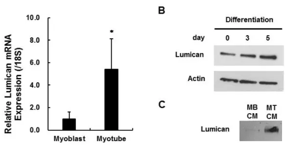
근모세포에서 Lumican의 mRNA 발현을 확인하였으며, 특히 근육세포로 분화되었을 때에 Lumican의 mRNA 발현이 크게 증가하는 것을 관찰하였다 (Figure 2A). 또한 비분화 근모세포가 근육세포로 분화됨에 따라 Lumican의 단백질 발현이 증가 (Figure 2B)할 뿐만 아니라 세포배양액으로 분비되는 Lumican의 양 (Figure 2C) 또한 크게 증가함을 확인하였다. 이 실험 결과를 통해 근육에서 분비되는 물질 (근육유래인자)은 근육량 증가 효과를 나타내며, 근육유래인자로서 Lumican이 근육량과 상관관계가 있는 것으로 사료된다.
Lumican과 근감소증의 관련성을 조사하기 위하여, 먼저 대표적인 근감소증 모델인 노화마우스의 근육에서 Lumican의 mRNA 발현을 측정하였다.
Figure 3A에서 보는 바와 같이 젊은 마우스 (young mouse)와 비교하여 노화
마우스 (old mouse)의 EDL muscle에서 sarcopenic index가 감소함으로서 muscle mass가 감소함을 알 수 있으나, EDL muscle에서의 Lumican mRNA 발현에는 변화가 없었다 (Figure 3B). 또한 난소 절제로 인한 성호르몬 결핍성 근감소증 모델에서도 Lumican mRNA 발현은 가짜 수술 (Sham)군과 난소절제 (OVX)군 사이에서의 변화를 관찰하지 못하였다 (Figure 3D). 반면 무부하 마우스 (unloading), 다시 말해 운동을 제한한 마우스와 비교하여 운동시킨 마우스 (exercise)의 EDL muscle에서는 sarcopenic index가 증가 (Figure 3E)함과 동시에 Lumican mRNA 발현이 증가 (Figure 3F)함을 관찰하였다. 이 결과는 Lumican의 발현이 노화와 성호르몬 결핍이 아닌 운동 (muscle contraction)에 의해 영향을 받는 것을 의미한다.
Lumican의 근분화 (myogenesis) 촉진 효과
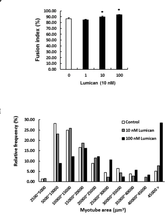
근육유래인자인 Lumican이 근육세포 분화에 미치는 영향을 조사하기 위하여 murine myoblast cell line인 C2C12 세포를 이용하였다. 2% Horse serum을 포함하는 분화배지로 C2C12 세포의 근분화를 유도함과 동시에 다양한 농도 (1, 10, 혹은 100 nM)의 Lumican을 처치하였다. 분화를 유도한지 2일 후, 형성된 myotube를 myosin heavy chain (MyHC) antibody를 이용한 immunocytochemical staining (ICC)을 통해 염색하였다. Figure 4에서 보는 바와 같이 Lumican은 10 nM 이상의 농도에서 myotube 형성을 증가시킴을 알 수 있다. MyHC+ myotubes의 분석 결과, 10 nM 이상의 Lumican의 처치는 myotube area (Figure 4C)와 fusion index (Figure 4D)를 증가시켰다. 특히 도수분포를 확인한 결과, 10 nM Lumican의 처치는 작은 면적의 myotube의 비율은 감소시키고 큰 면적의 myotube의 비율을 증가시켰으며, 이는 100 nM Lumican의 처치시에 더욱 두드러졌다 (Figure 4E). 즉 Lumican의 처리는 myoblasts의 fusion을 증가시키고 muscle hypertrophy를 유도함을 알 수 있다.
Lumican이 세포의 증식과 이동에 미치는 영향
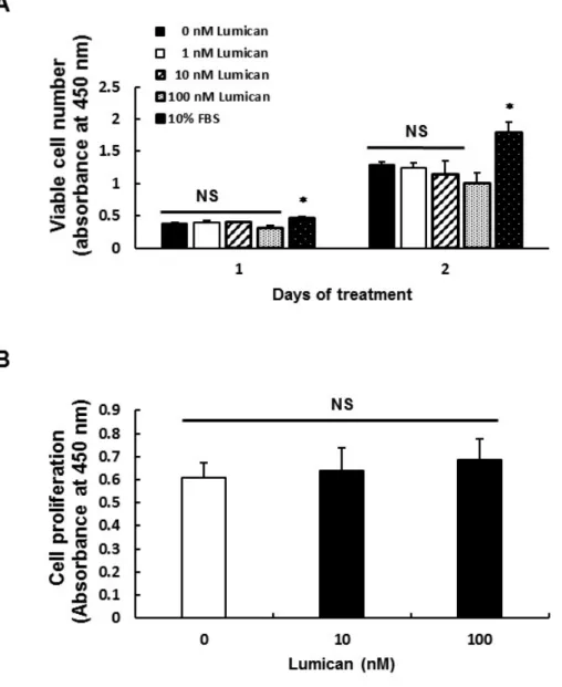
Cell viability assay를 수행한 결과, 분화 유도 2일에 100 nM Lumican을 처리하였을 때에 살아있는 세포의 수가 감소하였다 (Figure 5A).
하지만 분화 유도 1일에는 Lumican 처리에 의해 살아있는 세포 수에 큰 변화가 없었으며, BrdU assay를 통한 세포 증식도 1일 동안의 Lumican 처리에 의해 유의적으로 변화하지 않았다 (Figure 5B). 근분화 시에 세포의 증식이 제한되기 때문에 분화 유도 2일에서 100 nM Lumican에 의한 살아있는 세포 수 감소는 Lumican에 의한 근분화 촉진의 영향으로 판단된다.
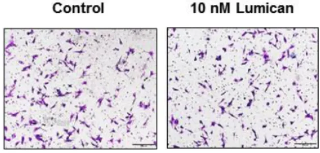
근분화 단계에서는 근세포의 융합을 위해 세포의 이동이 필요하기 때문에, Lumican이 세포의 이동에 관여하는지를 조사하였다. Transwell migration assay를 수행한 결과, Lumican은 근모세포의 이동에는 영향을 미치지 않았다 (Figure 6).
Lumican이 근분화 관련 전사인자들의 발현에 미치는 영향
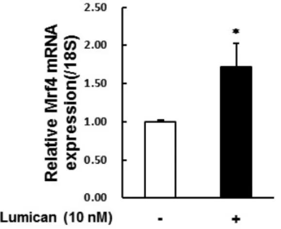
Lumican이 근분화를 매개하는 전사인자들의 발현에 미치는 영향을 조사하기 위하여 Lumican이 포함된 분화 배지로 24시간 동안 C2C12 세포의 분화를 유도하였다. Total RNA를 수집하여 real-time RT-PCR을 통해 다양한 전사인자들의 발현을 조사하였다 (Figure 7). MyHC mRNA 발현을 통해 myotube로의 분화가 Lumican에 의해 촉진됨을 확인하였다. 근분화 초기에 관여하는 MyoG, MyoD, Myf5의 mRNA 발현이 Lumican 처리하였을 때 증가하였다. 또한 근분화 중기에서 후기에 관여하는 Myocyte Enhancer Factor (Mef) 중, Mef2A와 Mef2C의 mRNA 발현 뿐만 아니라 근분화 후기에 관여하는 Mrf4도 Lumican 처리에 의해 증가하였다. 이렇게 Lumican 처치에 의해 모든 근분화 관련 전사인자들의 mRNA 발현이 증가함은 Lumican에 의한 작용이 이들 전사인자들의 발현 조절을 통해 나타나기 보다는 Lumican에 의해 촉진된 근분화의 영향인 것으로 판단된다.
Lumican이 미토콘드리아와 autophagy에 미치는 영향
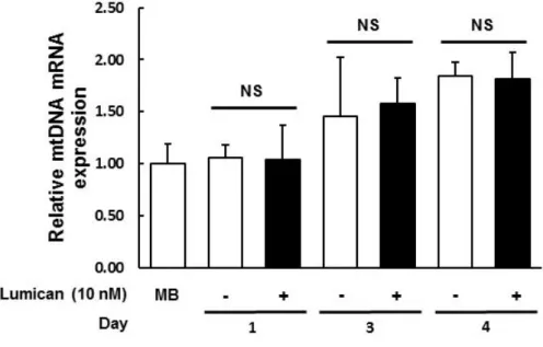
에너지 대사에 중요한 세포소기관인 미토콘드리아 함유량은 운동시에 골격근에서 증가함이 잘 알려져 있다. 하지만 Lumican 의 처리는 mitochondrial DNA (mtDNA)의 발현에는 영향을 미치지 않음을 real-time RT-PCR 을 통해 관찰하였다 (Figure 8).
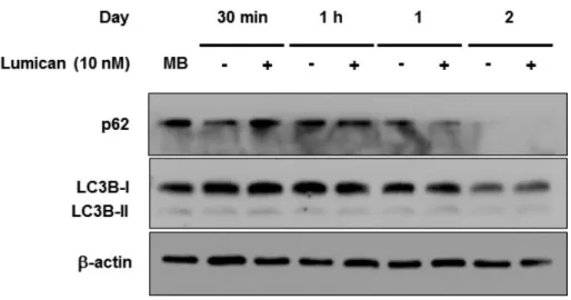
세포 내 분해 과정인 자가 포식(autophagy)은 세포의 발달과 분화에서 중요한 역할(47)을 하며, 최근 muscle differentiation 시에 유도되는 autophagy 가 myotube fusion에 필요함이 제시되었다(48). Autophagy의 대표적인 indicator인 p62와 autophagy marker Light Chain 3 (LC3)의 단백질 수준은 Lumican 처치에 의해 변화하지 않았다 (Figure 9).
Integrin을 통한 Lumican의 작용 기전 (Lumican 수용체로서의 Integrin)
많은 연구를 통해 Lumican의 수용체로 Integrin이 대두되고 있으나, 아 직까지 Lumican의 수용체로서 Integrin의 isotype이 명확하게 알려지고 있지 않다. 본 연구에서는 우선 근육세포에서 Integrin isotype의 mRNA 발현 조사를 통해 Lumican에 의해 촉진되는 근분화와 관련된 Integrin을 찾고자 하였다 (Figure 10A). 분석 결과, Integrin a2와 a5의 발현은 근분화시에 감소하고 Integrin a7은 증가하였다. 하지만 Lumican 처리에 의한 유의적인 차이를 관찰 하지 못하였다. Integrin b1은 근분화시에 감소하고 Integrin b3는 변화가 없었으 며, 분화 유도 2일에 Lumican 처리에 의해 Integrin b1의 발현만이 증가하였다.
Real-time RT-PCR 결과에서 각 isotpye의 delta CT value는 a2가 23.2, a5 가 6.5, a7이 8.2, b1이 7.2, 그리고 b3이 10.2였다. 즉 근육세포에서 Integrin a5, a7, b1, b3 에 비해 a2의 발현양이 적음을 의미한다.
다음으로 각각의 Integrin isotype의 inhibition을 통해 Lumican의 작용 을 매개하는 Integrin의 isotype을 특정하고자 하였다. Integrin a2/b1 inhibitor 인 TC-I15와 Integrin a5/b3 inhibitor인 echistatin의 처리는 Lumican에 의해 촉진된 근분화를 감소시켰다 (Figure 10B, 10C). 반면 Integrin a7 blocking Ab 의 처리는 Lumican에 의해 촉진된 근분화를 오히려 증가시켰으며, Integrin a7
기법으로 Integrin a7의 발현을 억제하였을 때에도 이와 유사한 결과를 관찰할 수 있었다 (Figure 10F, 10G). 이 결과들을 종합하면, Lumican의 수용체는 Integrin a2/b1과 Integrin a5/b3로 예상된다.
Lumican의 signaling pathway
Lumican에 의한 근분화 촉진 효과의 기전을 조사하고자 하였다. 다양한 시간 (5, 10, 30분) 동안 Lumican을 처리하여 수집한 total cell lysates를 이용 하여 Western blot analysis를 통해 근분화와 관련된 signaling pathway의 활성 화를 조사하였다. 근분화 및 muscle hypertrophy의 대표적인 signaling pathway인 Akt-mTOR (mammalian target of rapamycin) pathway를 조사한 결과, Lumican의 처리는 10분에서 Akt와 downstream target인 mTOR의 인산 화를 증가시켰다 (Figure 11). 다른 주요 signaling pathway인 Smad signaling 을 조사한 결과, Lumican의 처리는 Smad 1/5/9와 Smad2/3, 그리고 partner인 Smad4의 인산화 혹은 단백질 발현에는 영향을 미치지 않았다. 즉 Lumican의 작용은 Smad signaling이 아닌 Akt-mTOR signaling의 활성화를 통해 나타남 을 알 수 있다. 특히 Akt의 upstream인 PI3K의 inhibitor (LY294002)와 mTOR inhibitor (Rapamycin)의 처리는 Lumican에 의해 촉진된 근분화를 억제 (Figure 11B-11E)함은 Akt-mTOR signaling pathway가 Lumican의 근분화 촉진 작용의 주요 기전임을 뒷받침해 준다.
Lumican의 생체 치료 효능
이상의 세포실험을 통하여 Lumican 의 근분화 촉진 효과와 그 작용 기전을 규명하였다. 그렇다면 실제로 Lumican 이 생체에서도 근분화 촉진
효과를 나타내어 근감소증 치료 효과를 나타낼 수 있는지를 시험하고자,
근감소증 동물 모델인 난소절제 마우스에 Lumican 을 처치하였다. 8 주령의 암컷 C57BL/6 마우스는 가짜수술 (Sham) 혹은 난소절제 (OVX)를 시행한 후, 4 주동안 사육함으로써 성호르몬 결핍성 근감소증을 유도하였다. 이 후 다양한 용량 (0, 0.4, 2 μg)의 Lumican 을 마우스의 꼬리정맥으로 주 5 회, 4 주간 처치하였다. 이 후 마우스를 희생하고 상지와 하지 근육의 무게를 측정하여
sarcopenic index 를 측정한 결과, OVX 에 의해 모든 근육에서 sarcopenic index 가 감소하였으며, 2 μg 의 Lumican 처치는 상지 근육인 triceps 와 하지 근육인 quadriceps, soleus 에서 sarcopenic index 를 유의적으로 증가시켰다 (Figure 12A). 뿐만 아니라 Kondziella’s inverted screen test 를 통해 마우스의 근력을 조사한 결과, 가짜 수술군과 비교하여 난소절제 마우스에서 inverted screen 에 매달리는 시간이 감소, 즉 근력이 감소하였으며, 2 μg 의 Lumican 의 처치는 마우스의 근력을 유의적으로 향상시킴을 관찰하였다 (Figure 12B). 이 결과를 통하여 Lumican 이 근육의 양을 증가시키고 근력을 향상시킴을 생체 수준에서 확인하였다.
고찰
Sarcopenia의 근본적인 치료법은 아직 없지만 일반적으로 식이조절과 운동을 들 수 있다. 여러 연구에 의하면 단백질 섭취와 근육량이 관계 있는 것으로 알려져 있다(49-52). 단백질 섭취량이 권장량 미만인 노인에서 근육량과 근력의 유의한 감소가 있었고, 노인에서는 근육량 감소를 막기 위해서 권장량 이상의 단백질을 섭취해야 한다는 보고가 있었다(53). 골격근 질량은 단백질 합성과 분해 사이의 동적 균형에 의해 결정(54, 55)되는데, 골격근의 단백질 합성은
아미노산(56, 57), 글루코스(58), 운동(59), 인슐린 유사 성장 인자-1 (IGF-I)과
인슐린(60) 등과 같은 다양한 영양 및 생리학적 요인에 의해 영향을 받는 것으로
알려져 있다. 영약학적 접근 외에 약리학적 치료도 시도되고 있지만, 아직까지 근감소증을 완화시키는 효과적인 중재전략이 신체 활동과 운동임에는 이견이 없다(61).
근육은 체중의 약 40%를 구성하는 큰 조직으로 체온과 자세의 유지 및 운동에 중요한 역할을 한다. 이외에도 근육은 secretory organ으로서 다양한 cytokines, peptides 등을 분비하여 간, 지방, 췌장 등 다양한 다른 장기에 영향을 준다(62). 근육에서 분비되는 근육유래인자들은 이후 myokine이라 명명되었으며, 본 연구는 myokines이 paracrine mechanism을 통해 근육에 이로운 영향을 준다는 가설에서 출발하였다. 본 연구에서도 관찰하였듯이 운동은 근육의 양을 증가시킴 (Figure 3E)은 널리 알려져 있는 사실이기 때문에, 운동에 의해 증가된 myokines이 근육양 증가에 기여할 수 있음을 예상할 수 있다. 특히 운동에 의해 유도되는 myokine을 exerkine이라고도 불리는데, 최근 외국의 한 연구팀에서는 exerkine인 apelin의 투여가 age-associated sarcopenia를 개선시킴을 보고(10)하였다. 본 연구에서 근감소증 동물 모델인 노화마우스와 OVX 마우스는 각각의 대조군인 Young 마우스와 Sham- operated 마우스와 비교하였을 때에, 근육량의 감소를 보였으나 Lumican의 발현에는 차이를 나타내지 않았다. 반면 운동을 시킨 마우스는 무부하 마우스와
비교하여 근육량뿐만 아니라 Lumican의 발현이 증가하였다. 이는 Lumican의 발현이 노화와 성호르몬에 의해서 조절되지 않으며, muscle contraction의 영향을 받는 exerkine임을 보여준다. 추가 연구를 통해 운동이 노화마우스와 성호르몬 결핍 마우스에서 sarcopenic index 뿐만 아니라 Lumican의 발현을 증가시키는지에 대한 연구가 필요하다. 뿐만 아니라 생체 수준에서 근육에 대한 Lumican의 역할 규명을 확고히 하기 위해서는 Lumican KO 혹은 muscle- specific Lumican KO 마우스를 이용한 추가 검증이 필요하다고 판단된다.
Lumican은 세포실험을 통해 근분화 (myogenesis)를 촉진함을 관찰하였다. 근분화는 세포의 이동, 융합 (fusion), maturation 등의 단계를 거치며 myotube로 분화되는데, 여기에는 다양한 전사인자들과 신호전달체계가 관여하고, 또한 mitochondria biogenesis, protein synthesis, autophagy 등의 영향을 받음이 알려져 있다. 본 연구에서 Lumican은 세포의 증식과 이동, mtDNA contents, autophagy에는 영향을 미치지 않았다. Lumican은 근분화 관련 전사인자들의 mRNA 발현을 증가시켰지만, 이는 Lumian에 의해 촉진된 근분화의 영향인 것으로 사료된다. 그리고 Lumican은 근육세포에서 단백질 합성을 촉진한다고 알려진 대표적인 신호전달체계인 Akt-mTOR pathway를 활성화시켰으나, 또다른 주요 신호전달체계인 Smad signaling에는 영향을 미치지 않았다 (Figure 11A). 이는 아마도 Lumican 수용체에 의한 특이적 신호전달경로에 의한 것으로 판단된다. Lumican이 각막 상피세포에서 ALK5 [transforming growth factor (TGF)-b receptor 1]에 결합하여 wound healing을 촉진함이 보고(63)되었으나, Lumican이 Smad pathway에 영향을 미치지 않음은 근육세포에서의 Lumican 수용체는 ALK5가 아님을 보여준다.
많은 연구를 통해 Lumican의 수용체로서 Integrin이 대두되고 있는데, Integrin signaling의 downstream으로 Akt, RAS, MAPKs 등이 잘 알려져 있다.
Integrin은 세포 외 기질 및 또는 다른 세포와의 세포 상호 작용을 유지 하는 데 관여하는 막 관련 단백질이다. Integrin의 세포 기능은 광범위하게 연구 되어 왔으며, 유전자 발현, 세포 성장, 증식, 분화 및 세포 사멸을 포함한 수많은
부터 세포 내부로의 물리적 또는 기계적 신호를 중계하는 주요 센서로 생각되며 적절한 세포 반응을 가능하게 한다(66). 따라서 Integrin은 세포 외 기질과 세포 와 세포 골격 사이의 연결 고리 역할을 하며 세포가 주위 환경으로부터의 기계 적 신호를 통합하는 신호 과정의 중요한 구성 요소로서의 역할을 담당한다. 특히 골격근에서 세포 외 기질과 세포 골격과의 연관성은 근육 세포의 완전성을 유지 하고 적절한 근섬유 구조의 유지 및 근육 수축에 의해 생성된 힘의 전달에 필수
적(67, 68)이기 때문에, 골격근의 유지 및 기능에 Integrin이 중요한 역할을 할 것
이라 예상된다.
본 연구에서는 근육세포에서의 Integrin mRNA 발현량 비교와 Integrin inhibitor 실험을 통해 Lumican의 수용체로서 Integrin a2/b1과 Integrin a5/b3를 제시하였다. 하지만 Lumican의 수용체 규명을 위한 Ligand binding assay가 반드시 필요하고, Integrin a5, b1, b3의 각각의 inhibitor 실험을 통한 추가 검증 역시 필요하다고 판단된다. 또한 Integrin a7의 발현 억제는 근분화를 촉진함을 관찰 (Figure 10F)하였는데, 이는 Integrin a7의 발현 억제에 의한 compensation effect로서 예상되며, 이로 인한 다른 Integrin들의 발현 조사도 이루어져야 할 것으로 판단된다. 추후에 Integrin KO mouse를 통한 검증 실험 역시 필요하다고 판단된다.
Lumican의 수용체 규명은 Lumican과 수용체의 결합부위의 modification을 통한 효과적인 근감소증 치료 약제의 개발 가능성으로 이어질 수 있다고 판단된다. 예로 338개의 아미노산으로 이루어진 Lumican 외에 16개 아미노산으로 이루어진 Lumican-derived peptide가 collagen-stimulating activity를 향상(69)시킨다는 연구결과가 보고되었다. 본 연구에서 성호르몬
결핍성 근감소증 마우스 (난소절제 마우스)에 Lumican의 혈관주사
(intravenous injection)가 마우스의 근육량을 증가시키고 근력을 향상시킴 (Figure 12)을 통해 근감소증 치료제로서 Lumican의 가능성을 확인하였다.
하지만 Lumican의 약제화에는 338개 아미노산이라는 제약이 있다. 때문에 추후 연구를 통한 근육세포에서의 Lumican 수용체 규명과 수용체 결합부위의 modification은 Lumican의 효과적인 약제 개발로 이어질 것으로 판단된다.
결론
근육유래인자인 Lumican은 Akt-mTOR pathway를 통해 근분화 (myogenesis)를 촉진하고, 동물 모델에서 근감소증 치료 효과를 관찰하여 Lumican이 근감소증 치료에 대한 신규 표적이 될 수 있을 것으로 판단된다.
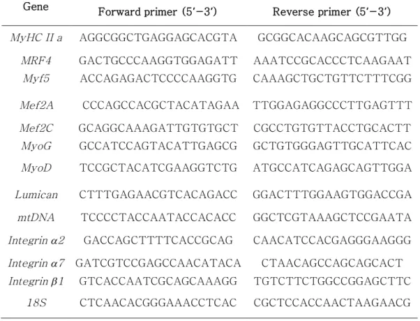
Table 1. Primer sequences
Gene Forward primer (5’-3’) Reverse primer (5’-3’) MyHC II a AGGCGGCTGAGGAGCACGTA GCGGCACAAGCAGCGTTGG
MRF4 GACTGCCCAAGGTGGAGATT AAATCCGCACCCTCAAGAAT Myf5 ACCAGAGACTCCCCAAGGTG CAAAGCTGCTGTTCTTTCGG Mef2A CCCAGCCACGCTACATAGAA TTGGAGAGGCCCTTGAGTTT Mef2C GCAGGCAAAGATTGTGTGCT CGCCTGTGTTACCTGCACTT MyoG GCCATCCAGTACATTGAGCG GCTGTGGGAGTTGCATTCAC MyoD TCCGCTACATCGAAGGTCTG ATGCCATCAGAGCAGTTGGA Lumican CTTTGAGAACGTCACAGACC GGACTTTGGAAGTGGACCGA mtDNA TCCCCTACCAATACCACACC GGCTCGTAAAGCTCCGAATA Integrin a2 GACCAGCTTTTCACCGCAG CAACATCCACGAGGGAAGGG Integrin a7 GATCGTCCGAGCCAACATACA CTAACAGCCAGCAGCACT Integrin b1 GTCACCAATCGCAGCAAAGG TGTCTTCTGGCCGGAGCTTC
18S CTCAACACGGGAAACCTCAC CGCTCCACCAACTAAGAACG
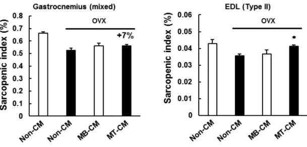
Figure 1. Muscle-derived factors increase muscle mass in mice.
90% confluent C2C12 myoblasts were differentiated into myotubes by culturing the cells in DMEM containing 2% HS for 5 days. Then, 24-hr conditioned media (CM) were collected from C2C12 myoblast (MB) or myotubes (MT). Sham-operated (Sham) or ovariectomized (OVX) mice were intravenously injected with non-CM, MB-CM, or MT-CM for 4 weeks.
At the time of sacrifice, the muscles [gastrocnemius and extensor digitorum longus (EDL)] were removed and weighted. Muscle mass was expressed as a percentage of body weight (sarcopenic index). Each bar represents the mean ± SEM (n = 6). *p< 0.05 as compared with the OVX/non-CM group.
Figure 2. Changes in Lumican expression and secretion during myogenesis.
90% confluent C2C12 myoblasts (MB) were differentiated into myotubes (MT) by culturing the cells in DMEM containing 2% HS for 5 days. (A) Total RNA was isolated and real-time RT-PCR performed. Each bar represents the mean ±SEM (n = 3). *p< 0.05 as compared with the myoblast group. (B) Total cell lysates were subjected to Western blot assay with their relevant antibodies. (C) The 24-hr conditioned media (CM) were collected and subjected to Western blot assay with an anti-Lumican antibody.
Figure 3. Exercise, but not aging nor estrogen-deficiency, stimulates Lumican expression in muscle.
(A, B) Young mouse vs Old mouse. (C, D) Sham-operated mouse (Sham) vs ovariectomized mouse (OVX). (E, F) Unloading mouse vs exercise mouse.
(A, C, D) Muscle mass is expressed as a percentage of body weight (sarcopenic index). (B, D, E) Total RNA was isolated from EDL muscles and real-time RT-PCR was performed. Each bar represents the mean ± SEM (n = 5). *p< 0.05 between the two groups.
Figure 4. Effect of Lumican on in vitro myotube formation.
C2C12 myoblasts were differentiated with various concentrations (0, 1, 10, and 100 nM) of Lumican for 3 days. (A) Myotubes were stained with anti- myosin heavy chain antibody (MyHC, red) and nuclei were counter -stained with DAPI (blue). Representative photographs of immunocytoche- mistry
(ICC) are shown. (B-E) Their quantitative results (B, number of myotubes;
C, myotube area; D, fusion index; E, frequency distribution) from ICC are shown as bar graph. Each bar represents the mean ±SEM (n = 3). *p< 0.05 as compared with the control group.
Figure 5. Effect of Lumican on myoblast’s proliferation.
(A) C2C12 myoblasts were incubated with various concentrations (0, 1, 10, and 100 nM) of Lumican for indicated time periods. Cell viability was assessed using a CCK-8 assay. (B) C2C12 myoblasts were incubated with various concentrations (0, 10, and 100 nM) of Lumican for 24 hr. Cell proliferation was evaluated with a BrdU-based enzyme-linked immunosorbent assay. Each bar represents the mean ±SEM (n = 4).
Figure 6. Effect of Lumican on myoblast’s migration.
The migration of C2C12 myoblasts through Transwell filter was assessed with 10 nM Lumican. Cells were incubated for 4 hr. (A) Photographs of migrated crystal violet-stained cells are shown. (B) Quantitative analysis of the migrated cells. Each bar represents the mean ±SEM (n = 3).
Figure 7. Effects of Lumican on myogenesis-related genes.
C2C12 myoblasts were differentiated with or without 10 nM Lumican for 24 hr. Total RNA was isolated and real-time RT-PCR performed. Each bar represents the mean ± SEM (n = 3). *p< 0.05 as compared with the control group.
Figure 8. Effect of Lumican on mitochondrial DNA content in myoblasts.
C2C12 myoblasts (MB) were differentiated with or without 10 nM Lumican for indicated time periods. Total RNA was isolated and real-time RT-PCR performed. Each bar represents the mean ± SEM (n = 3).
Figure 9. Effects of Lumican on autophagy of myoblasts.
C2C12 myoblasts were incubated with or without 10 nM Lumican for indicated time periods. Total cell lysates were subjected to Western blot assay with their relevant antibodies.
Figure 10. Possible receptors for Lumican in myoblasts.
(A) C2C12 myoblasts were differentiated with or without 10 nM Lumican for indicated time periods. Total RNA was isolated and real-time RT-PCR performed. Each bar represents the mean ± SEM (n = 3). (B, C) C2C12 myoblasts were pretreated with Echistatin (Integrin a5b3 inhibitor) or TC- I15 (Integrin a2b1 inhibitor) for 1 hr. Then cells were differentiated with or without 10 nM Lumican for 48 hr. (D, E) C2C12 myoblasts were pretreated with Integrin a7 blocking antibody for 1 hr. Then cells were differentiated with or without 10 nM Lumican for 48 hr. (F, G) C2C12 myoblasts were transfected with negative control siRNA or Integrin a7 siRNA. Then cells were differentiated with or without 10 nM Lumican for 48 hr. (B, D, F) Myotubes were stained with anti- MyHC (red) and nuclei were counter- stained with DAPI (blue). (C, E, G) The myotube area from ICC is shown.
Each bar represents the mean ± SEM (n = 3).
Figure 11. Possible signalings of Lumican-stimulated myogenesis.
C2C12 myoblasts were incubated with or without 10 nM Lumican for indicated time periods. (A) Total cell lysates were subjected to Western blot assay with their relevant antibodies. (B) p-Akt/Akt expression level. (C) p- mTOR/mTOR expression level. (D, E) C2C12 myoblasts were pretreated with PI3K inhibitor for 1 hr. Then cells were differentiated with or without 10 nM Lumican for 48 hr. (F, G) C2C12 myoblasts were pretreated with Rapamycin (mTOR inhibitor) for 1 hr. Then cells were differentiated with or without 10 nM Lumican for 48 hr. (D, F) Myotubes were stained with anti- MyHC (red) and nuclei were counter-stained with DAPI (blue). (E, G) The myotube area from ICC is shown. Each bar represents the mean ±SEM (n = 3). *p< 0.05 as compared with the control group.
.
Figure 12. Systemic administration of lumican increases muscle mass and strength in mice.
Sham-operated (Sham) or ovariectomized (OVX) mice were injected intravenously with various doses (0, 0.4, or 2 mg) of Lumican (5 days a week) for 4 weeks. (A) At the time of sacrifice, the muscles were removed and weighed. Muscle mass was expressed as a percentage of BW (sarcopenic index). (B) Muscle strength was measured in mice by measuring hang time using Kondziella's inverted screen test. Each bar represents the mean ±SEM (n = 6). a vs b, Duncan’s multiple range test * p < 0.05 as compared with the Sham group; means without a common letter differ among the three OVX group, p< 0.05.
참고 문헌
1. Rosenberg I. Summary comments: epidemiological and meth- odological problems in determining nutritional status of older persons. Am J Clin Nutr. 1989;50:1231-3
2. Rosenberg IH. Sarcopenia: origins and clinical relevance. J Nutr.
1997;127:990S-91S
3. Janssen I, Heymsfield SB, Ross R. Low relative skeletal muscle mass (sarcopenia) in older persons is associated with functional impairment and physical disability. J Am Geriatr Soc. 2002;50:889-96
4. Evans WJ. What is sarcopenia? J Gerontol A Biol Sci Med Sci. 1995; 50.
Spec:5-8
5. Newman AB, Kupelian V, Visser M, Simonsick E, Goodpaster B, Nevitt M, Kritchevsky SB, Tylavsky FA, Rubin SM, Harris TB. Sarcopenia:
alternative definitions and associations with lower extremity function.
AGS. 2003;51:1602–1609
6. Grimby G, Saltin B. The ageing muscle. Clin Physiol 1983;3: 209-18
7. Bijlsma AY, Meskers CG, Westendorp RG, Maier AB. Chronology of age- related disease definitions: osteoporosis and sarcopenia. Ageing Res Rev 2012;11:320-324.
8. Paddon-Jones D, Sheffield-Moore M, Zhang XJ, Volpi E, Wolf SE, Aarsland A, et al. Amino acid ingestion improves muscle protein synthesis in the young and elderly. Am J Physiol Endocrinol Metab 2004;286: E321-8
9. Pourteymour S, Eckardt K, Holen T, Langleite T, Lee S et al., Global mRNA sequencing of human skeletal muscle: Search for novel exercise- regulated myokines. Mol Metab, 2017;6(4): 352-365
10. Vinel C, Lukjanenko L, Batut A, Deleruyelle S, Pradère JP, Le Gonidec S, et al., The exerkine apelin reverses age-associated sarcopenia. Nat Med. 2018;24(9): 1360-1371
11. Blochberger, T.C., J.-P. Vergnes, J. Hempel, and J.R. Hassell. cDNA to chick lumican (corneal keratan sulfate proteoglycan) reveals homology to the small interstitial proteoglycan gene family and expression in muscle and intestine. J. Biol. Chem. 1992;267:347–352
12. Chakravarti, S., R. Stalling, D. Fite, N. SundarRaj, and J.R. Hassell.
Primary structure of human lumican (keratan sulfate proteoglycan) and localization of the gene (LUM) to chromosome 12q21.3-q22. Genomics.
1995;27:481–488
13. Ezura Y, Chakravarti S, Oldberg A, Chervoneva I, Birk DE. Differential expression of lumican and fibromodulin regulate collagen fibrillogenesis in developing mouse tendons. J Cell Biol. 2000;151: 779-788
14. Chakravarti S, Magnuson T, Lass JH, Jepsen KJ, LaMantia C, Carroll H.
Lumican regulates collagen fibril assembly: skin fragility and corneal opacity in the absence of lumican. J Cell Biol. 1998;141: 1277-1286
15. Chakravarti S. Functions of lumican and fibromodulin: lessons from knockout mice. Glycoconj J. 2002;19: 287-293
16. Austin BA, Coulon C, Liu CY, Kao WW, Rada JA. Altered collagen fibril formation in the sclera of lumican-deficient mice. Invest Ophthalmol Vis Sci. 2002;43: 1695-1701
17. Quantock AJ, Meek KM, Chakravarti S. An x-ray diffraction investigation of corneal structure in lumican-deficient mice. Invest Ophthalmol Vis Sci. 2001;42: 1750-1756
18. Chakravarti S, Petroll WM, Hassell JR, Jester JV, Lass JH, Paul J, Birk DE. Corneal opacity in lumican-null mice: defects in collagen fibril
structure and packing in the posterior stroma. Invest Ophthalmol Vis Sci.
2000;41: 3365-3373
19. Saika S, Miyamoto T, Tanaka S, Tanaka T, Ishida I, Ohnishi Y, Ooshima A, Ishiwata T, Asano G, Chikama T, Shiraishi A, Liu CY, Kao CW, Kao WW. Response of lens epithelial cells to injury: role of lumican in
epithelial-mesenchymal transition. Invest Ophthalmol Vis Sci. 2003;44:
2094-2102
20. Ishiwata T, Fujii T, Ishiwata S, Ikegawa S, Naito Z. Effect of morpholino
kidney (HEK) 293 cells. Pathol Int. 2004;54: 77-81
21. Ishiwata T, Yamamoto T, Kawahara K, Kawamoto Y, Matsuda Y, Ishiwata S, Naito Z. Enhanced expression of lumican inhibited the attachment and growth of human embryonic kidney 293 cells. Exp Mol Pathol. 2010;88: 363-370
22. Lu YP, Ishiwata T, Kawahara K, Watanabe M, Naito Z, Moriyama Y, Sugisaki Y, Asano G. Expression of lumican in human colorectal cancer cells. Pathol Int. 2002;52: 519-526
23. Ping Lu Y, Ishiwata T, Asano G. Lumican expression in alpha cells of islets in pancreas and pancreatic cancer cells. J Pathol. 2002;196: 324- 330
24. Grover J, Chen XN, Korenberg JR, Roughley PJ. The human lumican gene. Organization, chromosomal location, and expression in articular cartilage. J Biol Chem. 1995;270: 21942-21949
25. Sztrolovics R, Alini M, Mort JS, Roughley PJ. Age-related changes in fibromodulin and lumican in human intervertebral discs. Spine (Phila Pa 1976) .1999;24: 1765–1771
26. Qin H, Ishiwata T, Asano G. Effects of the extracellular matrix on lumican expression in rat aortic smooth muscle cells in vitro. J Pathol.
2001;195: 604-608
27. Naito Z, Ishiwata T, Kurban G, Teduka K, Kawamoto Y, Kawahara K,
Sugisaki Y. Expression and accumulation of lumican protein in uterine cervical cancer cells at the periphery of cancer nests. Int J Oncol.
2002;20: 943-948
28. Raouf A, Ganss B, McMahon C, Vary C, Roughley PJ, Seth A. Lumican is a major proteoglycan component of the bone matrix. Matrix Biol.
2002;21: 361-367
29. Dolhnikoff M, Morin J, Roughley PJ, Ludwig MS. Expression of lumican in human lungs. Am J Respir Cell Mol Biol. 1998;19: 582-587
30. Schaefer L, Gröne HJ, Raslik I, Robenek H, Ugorcakova J, Budny S, Schaefer RM, Kresse H. Small proteoglycans of normal adult human kidney: distinct expression patterns of decorin, biglycan, fibromodulin, and lumican. Kidney Int. 2000;58: 1557-1568
31. Chakravarti S, Magnuson T, Lass JH, Jepsen KJ, LaMantia C, et al.
Lumican regulates collagen fibril assembly: skin fragility and corneal opacity in the absence of lumican. J Cell Biol. 1998;141: 1277–1286
32. Liu H, Zhang J, Liu CY, Wang IJ, Sieber M, et al. Cell therapy of congenital corneal diseases with umbilical mesenchymal stem cells:
lumican null mice. PLoS ONE. 2010;5: e10707
33. Yeh JT, Yeh LK, Jung SM, Chang TJ, Wu HH, et al. Impaired skin wound healing in lumican-null mice. Br J Dermatol. 2010;163: 1174–1180
formation in the sclera of lumican-deficient mice. IOVS. 2002;43: 1695–
1701
35. Carlson EC, Liu CY, Chikama T, Hayashi Y, Kao CW, et al. Keratocan, a cornea-specific keratan sulfate proteoglycan, is regulated by lumican. J Biol Chem. 2005;280: 25541–25547
36. Carlson EC, Lin M, Liu CY, Kao WW, Perez VL, et al. Keratocan and lumican regulate neutrophil infiltration and corneal clarity in
lipopolysaccharide-induced keratitis by direct interaction with CXCL1. J Biol Chem. 2007;282:35502–35509
37. Carlson EC, Sun Y, Auletta J, Kao WW, Liu CY, et al. Regulation of corneal inflammation by neutrophil-dependent cleavage of keratan sulfate proteoglycans as a model for breakdown of the chemokine gradient. J Leukoc Biol. jlb. 2010;0310134
38. Hayashi Y, Call MK, Chikama T, Liu H, Carlson EC, et al. Lumican is required for neutrophil extravasation following corneal injury and wound healing. J Cell Sci. 2010;123: 2987–2995
39. Saika S, Shiraishi A, Saika S, Liu C-Y, Funderburgh JL, et al. Role of Lumican in the Corneal Epithelium during Wound Healing. J Biol Chem.
2000;275: 2607–2612
40. Meij JT, Carlson EC, Wang L, Liu CY, Jester JV, et al. Targeted expression of a lumican transgene rescues corneal deficiencies in lumican-null mice. Mol Vis. 2007;13: 2012–2018
41. Saika S, Miyamoto T, Tanaka S, Tanaka T, Ishida I, et al. Response of lens epithelial cells to injury: role of lumican in epithelial-mesenchymal transition. IOVS .2003;44: 2094–2102
42. Yeh LK, Chen WL, Li W, Espana EM, Ouyang J, et al. Soluble lumican glycoprotein purified from human amniotic membrane promotes corneal epithelial wound healing. IOVS. 2005;46: 479–486
43. Yeh JT, Yeh LK, Jung SM, Chang TJ, Wu HH, et al. Impaired skin wound healing in lumican-null mice. BJD. 2010;163: 1174–1180
44. Niewiarowska J, Brezillon S, Sacewicz-Hofman I, Bednarek R, Maquart F-X, et al. Lumican inhibits angiogenesis by interfering with alpha 2 beta 1 receptor activity and downregulating MMP-14 expression. Thrombosis research. 2011;128: 452–457
45. Nikitovic, D., Papoutsidakis, A., Karamanos, N. K. & Tzanakakis, G. N.
Lumican affects tumor cell functions, tumor-ECM interactions,
angiogenesis and inflammatory response. Matrix Biol .2014;35, 206–214
46. Deacon RM, Measuring the strength of mice. J Vis Exp. 2013;Jun 2;(76)
47. Levine B, Klionsky DJ. Development by self-digestion: Molecular mechanisms and biological functions of autophagy. Dev Cell. 2004;6:463–
477)
48. Fortini P, Ferretti C, Iorio E, Cagnin M, Garribba L, Pietraforte D, Falchi
metabolism, autophagy and differentiation during in vitro myogenesis.
Cell Death Dis. 2016;7:e2168)
49. Paddon-Jones D, Sheffield-Moore M, Zhang XJ, Volpi E, Wolf SE, Aarsland A, et al. Amino acid ingestion improves muscle protein synthesis in the young and elderly. Am J Physiol Endocrinol Metab.2004;286: E321-8
50. Paddon-Jones D, Rasmussen BB. Dietary protein recommendations and the prevention of sarcopenia. Curr Opin Clin Nutr Metab Care.2009;12:
86-90
51. Walrand S, Short KR, Bigelow ML, Sweatt AJ, Hutson SM, Nair KS.
Functional impact of high protein intake on healthy elderly people. Am J Physiol Endocrinol Metab.2008;295: E921-8
52. Barbosa-Silva MC. Subjective and objective nutritional assessment methods: what do they really assess? Curr Opin Clin Nutr Metab Care.2008;11: 248-54
53. Campbell WW, Trappe TA, Wolfe RR, Evans WJ. The recommended dietary allowance for protein may not be adequate for older people to maintain skeletal muscle. J Gerontol A Biol Sci Med Sci 2001;56: M373- 80
54. Gordon, B. S., Kelleher, A. R. & Kimball, S. R. Regulation of muscle protein synthesis and the effects of catabolic states. Int J Biochem Cell Biol 2013;45, 2147–157
55. Glass, D. J. Skeletal muscle hypertrophy and atrophy signaling pathways. Int J Biochem Cell Biol 37, 2005;1974–984
56. Wolfe, R. R. Regulation of muscle protein by amino acids. JN.2002.
57. Suryawan, A. & Davis, T. A. Regulation of protein degradation pathways by amino acids and insulin in skeletal muscle of neonatal pigs. J Anim Sci Biotechnol .2014;1186/2049
58. Jeyapalan, A. S. et al. Glucose stimulates protein synthesis in skeletal muscle of neonatal pigs through an AMPK- and Mtor independent process. Am J Physiol Endocrinol Metab 2017;293, E595–03
59. Atherton, P. J. & Smith, K. Muscle protein synthesis in response to nutrition and exercise. J Physiol 2012;590, 1049–057
60. Sugden, P. H. & Fuller, S. J. Regulation of protein turnover in skeletal and cardiac muscle. Biochem J 1991;273, 21–.37
61. Iolascon G, Di Pietro G, Gimigliano F, Mauro GL, Moretti A, Giamattei MT, et al., Physical exercise and sarcopenia in older people: position paper of the Italian Society of Orthopaedics and Medicine (OrtoMed), Clin Cases Miner Bone Metab.2014;11(3):215-221
62. Pedersen BK, Febbraio MA, Muscles, exercise and obesity: skeletal muscle as a secretory organ. Nat Rev Endocrino. 2012;8(8): 457-465
63. Yamanak O, Yuan Y, Coulson-Thomas VJ, Gesteira TF, Call MK, Zhang Y, et al., Lumican binds ALK5 to promote epithelium wound healing.
PLOS One.2013;8(12): e82730
64. Chen LK, Lee WJ, Peng LN, Liu LK, Arai H, Akishita M. Recent
Advances in Sarcopenia Research in Asia: 2016 Update From the Asian Working Group for Sarcopenia. J Am Med Dir Assoc. 2016;17: 767.e1-7
65. Deacon RM, Measuring the strength of mice. J Vis Exp 2013;Jun 2;(76)
66. Bentzinger et al., Building Muscle: Molecular regulation of Myogenesis.
Cold Spring Harb Perspect Biol, 2012;4:a008342
67. Holloszy, Biochemical adaptations in muscle. Effects of exercise on mitochondrial oxygen uptake and respiratory enzyme activity in skeletal muscle. J Biol Chem. 1967;242: 2278-2282
68. Goodman CA, et al., Novel insights into the regulation of skeletal muscle protein synthesis as revealed by a new nonradioactive in vivo technique.
FASEB J 2011;25(3): 1028-1039
69. Walter MN, Dehsorkhi A, Hamley IW, Connon CJ. Supra-molecular assembly of a lumican-derived peptide amphiphile enhances its collagenstimulating activity. Biomater Sci 2016;4(2): 346-54
ABSTRACT
Sarcopenia, or muscle loss is an important public health problem but there have been no approved drug for its treatment. It have been well known that exercise increases the amount of muscle mass. Because muscles produce and release the variety of factors (known as muscle-derived factors or myokines), I hypothesized that myokines can increase muscle mass and can be used as therapeutic agents in the treatment of sarcopenia. In this study, Lumican expression is increased in skeletal muscle of exercised mouse and Lumican secretion is increased during myogenesis. Lumican treatment stimulated myogenic differentiation of C2C12 myoblasts and stimulated Akt- mTOR signaling. Lumican-induced myogenesis is blocked by inhibitor of Integrin a2/b1 or Integrin a5/b3. Also, intravenous treatment of Lumican increased skeletal muscle masses and improved muscle strength in ovariectomized mice. The present results indicate that Lumican possesses the pro-myogenic effects and a potential as a therapeutic agent against sarcopenia.
Keywords : Lumican, Sarcopenia, Myokine, Myogenesis, Skeletal muscle