저작자표시-비영리-변경금지 2.0 대한민국 이용자는 아래의 조건을 따르는 경우에 한하여 자유롭게
l 이 저작물을 복제, 배포, 전송, 전시, 공연 및 방송할 수 있습니다. 다음과 같은 조건을 따라야 합니다:
l 귀하는, 이 저작물의 재이용이나 배포의 경우, 이 저작물에 적용된 이용허락조건 을 명확하게 나타내어야 합니다.
l 저작권자로부터 별도의 허가를 받으면 이러한 조건들은 적용되지 않습니다.
저작권법에 따른 이용자의 권리는 위의 내용에 의하여 영향을 받지 않습니다. 이것은 이용허락규약(Legal Code)을 이해하기 쉽게 요약한 것입니다.
Disclaimer
저작자표시. 귀하는 원저작자를 표시하여야 합니다.
비영리. 귀하는 이 저작물을 영리 목적으로 이용할 수 없습니다.
변경금지. 귀하는 이 저작물을 개작, 변형 또는 가공할 수 없습니다.
의학석사 학위논문
Mutation in Retinoic X Receptor-γ is a possible mechanism of ATRA resistance in acute promyelocytic
leukemia: Identifying genetic changes related to drug resistance using whole exome sequencing
ATRA 치료에 내성을 보이는 급성 전골수세포성
백혈병에서 엑솜시퀀싱을 통한 약물내성 기전 탐색:
Retinoic X Receptor-γ 돌연변이의 발견
2016 년 2 월
서울대학교 대학원 의학과 중개의학 전공
육 정 환
ATRA 치료에 내성을 보이는 급성 전골수세포성
백혈병에서 엑솜시퀀싱을 통한 약물내성 기전 탐색:
Retinoic X Receptor-γ 돌연변이의 발견
지도교수 윤 성 수
이 논문을 의학석사 학위논문으로 제출함
2015년 10월
서울대학교 대학원 의학과 중개의학 전공
육 정 환
육정환의 석사학위논문을 인준함
2015년 12월
위 원 장 (인) 부 위 원 장 (인) 위 원 (인)
Mutation in Retinoic X Receptor-γ is a possible mechanism of ATRA resistance in acute promyelocytic
leukemia: Identifying genetic changes related to drug resistance using whole exome sequencing
By Jeonghwan Youk
(Directed by Professor Sung-Soo Yoon, M.D., Ph.D.)
A thesis submitted in partial fulfillment of the requirements for the Degree of Master of Science in Translational Medicine at Seoul
National University College of Medicine
October 2015
Approved by Thesis Committee:
Professor Chairman
Professor Vice chairman
Professor
i
ABSTRACT
Background: Acute promyelocytic leukemia (APL) has the best prognosis among acute myeloid leukemia. However, a subset of APL patients are not cured with all-trans retinoic acid (ATRA) combined with anthracycline-based cytotoxic chemotherapy. Several mechanisms such as increased ATRA metabolism have been suggested to explain acquired resistance to ATRA.
However, genetic mechanisms of ATRA resistance have not been characterized at all. Hence, in this study, I tried to reveal genetic alterations attributable to ATRA resistance.
Methods: Whole exome sequencing (WES) was performed using DNA of three APL patients who showed resistance to ATRA based treatment. These include two patients who failed to achieve complete remission (CR) after induction chemotherapy, and one patient who experienced relapse after CR despite consolidation treatment. DNA extracted from bone marrow at the time of diagnosis was used for analysis, while DNA extracted from saliva at the time of CR was used as germline control. Calling of single nucleotide variants (SNVs) was performed using internal pipeline called Adiscan (http://www.syntekabio.com). Annotation was performed using Polyphen-2.
SNVs found by WES were validated by direct Sanger sequencing. The frequency of those validated SNVs was defined in a separate APL cohort.
Results: I identified 33 somatic non-synonymous SNVs in three patients.
Polyphen-2 algorithm predicted 9 among 33 SNVs to damage protein function.
Sanger sequencing validated 7 from 9 SNVs. These validated SNVs include
ii
RXRG M77R, N6AMT2 A78S, TXNDC15 D198E, B3GALTL A444T, RBBP8NL E182G, BHMT M185I and ADAMTS5 G85D. When these 7 SNVs were genotyped in a separate cohort, none of these SNVs were found in an APL cohort composed of 29 ATRA sensitive patients, suggesting these SNVs would be truly related to ATRA resistance in APL. Especially, when a simulation using amber molecular dynamics, I observed 1) increase in hydrogen bonding, 2) decreased helix folding structure and 3) decreased energy state in RXRG mutant case.
Conclusions: WES identified seven SNVs which were unique in ATRA resistant cases. Among those mutations, RXRG mutation could be a promising genetic mechanism of ATRA resistance.
Keywords: Drug resistance; Leukemia, Promyelocytic, Acute; Retinoid X Receptor gamma; Tretinoin;
Student number: 2014-21164
iii
CONTENTS
Abstract ... i
Contents... iii
List of tables and figures ... iv
List of abbreviation ... v
Introduction ... 1
Methods ... 2
Results ... 5
Discussion ... 9
References ... 11
Abstract in Korean... 13
iv
LIST OF TABLES AND FIGURES
Table 1.
Clinical and laboratory characteristics of three patients ... 3Table 2.
Non-synonymous SNVs in three APL patients ... 6Table 3.
Frameshift-causing indels associated with ATRA resistance ... 7Table 4.
Copy number variations associated with ATRA resistance ... 7Figure 1.
The Amber molecular dynamics predicted structural change of RXRG by M77R mutation. ... 8v
LIST OF ABBREVIATIONS
AA amino acid ALT alternative
AML acute myeloid leukemia APL Acute promyelocytic leukemia ATRA all-trans retinoic acid
CNVs copy number variations CR complete remission CTx chemotherapy
NGS next-generation sequencing RARα retinoic acid receptor α REF reference
RFS relapse free survival RXR retinoid X receptor SNVs single-nucleotide variants WBC white blood count
WES whole-exome sequencing
1
INTRODUCTION
Acute promyelocytic leukemia (APL) accounts for fewer than 10% of acute myeloid leukemia (AML) cases in adults1. Most APL survivors achieve complete remission (CR) after induction chemotherapy, which, prior to arsenical use (i.e., arsenic trioxide), consisted of anthracycline-based
cytotoxic agents and all-trans retinoic acid (ATRA). However, about 10–15%
of such patients are either refractory to the latter, or relapse after CR, likely due to ATRA resistance. Clinically, resistance to ATRA is categorized as either primary or secondary: Primary resistance, equated with failure to achieve CR during the first-round induction chemotherapy, is uncommon; secondary (or delayed) ATRA resistance, marked by APL recurrence after CR, is more common.
The following mechanisms have been suggested to explain ATRA resistance: hypercatabolism of ATRA through extreme cytochrome P450 enzymatic activity2; constitutive expression of cellular retinoic acid-binding proteins by leukemic cells, leading to ATRA sequestration3; and mutation in the ligand-binding domain of the retinoic acid receptor α (RARα) region4. However, this particular mutation has yet to be demonstrated in patients with ATRA resistance, thus questioning its clinical relevance.
At present, next-generation sequencing (NGS) has not been applied for the comprehensive genomic analysis of ATRA resistance. Consequently, whole-exome sequencing (WES) was utilized for this study to explore the genetic variations that contribute to ATRA resistance in patients with APL.
2
METHODS
Patient characteristics
DNA samples of three patients with ATRA-resistant APL, treated at Seoul National University Hospital between 2009 and 2012, were available for NGS.
Written informed consent was obtained from all patients. This study was conducted in accordance with the Declaration of Helsinki provisions and was approved by our Institutional Review Board. The clinical characteristics of these patients are summarized in Table 1. Of note, patient A had low-risk disease but still failed to achieve morphologic CR after induction with idarubicin and ATRA. She eventually achieved molecular CR following induction with arsenic trioxide. Patient B also achieved molecular CR after the second round of induction chemotherapy, having failed during the first attempt. Although ATRA treatment was maintained, his disease relapsed 14 months later and he died during consolidation chemotherapy. Patient C achieved CR after the first-round induction chemotherapy with idarubicin plus ATRA. However, his disease relapsed after 15 months during ATRA maintenance and he died of a related intracranial hemorrhage.
3
Table1. Clinical and laboratory characteristics of three patients
Patient A Patient B Patient C
Sex Female Male Male
Age at diagnosis 38 64 67
Initial Bone Marrow blast 90.3% 89.8% 95.1%
Initial WBC 1100 10750 51730
Initial platelet 78k 93k 32k
Cytogenetics
46,XX,der(15)t(15;17)(q 22;q21),der(17)t(15;17)
i(17)(q10)[20]
46,XY,15pstk+ps+, t(15;17)(q22;q21)[17]/
46,XY,15pstk+ps+[3]
45,X,-Y, t(15;17)(q22;q21)[20]
Induction regimen idarubicin, ATRA idarubicin, ATRA idarubicin, ATRA
Result of induction persistence persistence CR
Number of CTx to get CR 2 2 1
Relapse No Yes Yes
RFS (months) - 15.1 14.7
Last status Alive Dead Dead
Overall survival (months) 37.4 37.2 15.9
WBC, white blood count; CTx, chemotherapy; RFS, relapse free survival;
Whole-exome sequencing
Leukemic cells were obtained from the bone marrow aspirated from each subject at the time of the diagnosis. Saliva collected at the time of CR provided germline controls. Genomic DNA purification kits for blood and saliva (Norgen Biotek Corp., Thorold, ON, Canada) were used to isolate the genomic DNA of leukemic blasts and epithelial cells in saliva. Quality was
4
monitored by the NGS QC Toolkit (National Institute of Plant Genome Research, New Delhi, India).
Genomic DNA was then fragmented for massively parallel sequencing by the HiSeq 2000 system (Illumina Inc., San Diego, CA, USA), according to the manufacturer’s instructions. The SureSelect Human All Exon Kit (Agilent Technologies Inc., Santa Clara, CA, USA) was used for DNA capture. FASTQ files were aligned to the human reference genome (human_g1k_v 37.fasta) using the Burrows-Wheeler Aligner (bwa-0.7.5)5 to generate a sequence alignment/map file. Thereafter, the Genome Analysis Toolkit (Broad Institute, Cambridge, MA, USA) was used for local sequence alignment6. Somatic single-nucleotide variants (SNVs) were identified using the Allelic Depth Imbalance Scanning (ADISCAN;
http://www.syntekabio.com) tool. Briefly, ADISCAN assumes that for a
genome position determined to be heterozygous by NGS, the reference allele frequency should be balanced to 0.5. Based on this hypothesis, ADISCAN uses pair allelic fractions at two counterpart genome positions (e.g., normal vs.
tumor) and returns their adjusted tangent difference and ranking.
Complimentary SNVs were identified using MuTect software (Broad Institute)7. VarScan was utilized to verify indels8. Among a variety of genes, I chose frameshift-causing indels, which were shared among all three patients.
CONTRA freeware was used to determine copy number variations (CNVs)9. A polyphen-2 algorithm10 was applied to predict SNV function. SNVs likely to damage proteins were validated by Sanger sequencing.
5
Validation and screening of SNVs in a separate APL cohort
A separate APL cohort (N=29; male-to-female ratio, 21:8; median age, 48 years), treated at our facility between May 2002 and October 2013, was screened for Sanger-validated SNVs. Nine of these patients had high-risk disease, and all had achieved CR following first-round induction chemotherapy, with no relapses to date.
RESULTS
Possible somatic mutations associated with ATRA resistance identified by WES
WES yielded a total of 412,442,511 mapped nucleotide sequences (blast DNA:
209,631,235; salivary epithelial DNA: 202,811,276). Mean depth of the samples was more than 110-fold. Nonsynonymous SNVs identified by ADISCAN and MuTect were as follows: patient A: 14 and 0; patient B: 8 and 1; and patient C: 11 and 19, respectively. None of three patients showed FLT3 and RARA mutations. The polyphen-2 algorithm marked 9 of the 33 nonsynonymous SNVs as damaging mutations. Of these, seven were verified by Sanger sequencing: RXRG M77R, N6AMT2 A78S, TXNDC15 D198E, B3GALTL A444T, RBBP8NL E182G, BHMT M185I, and ADAMTS5 G85D (Table 2). To date, The Cancer Genome Atlas (http://www.cbioportal.org) has no record of three of the genes (RXRG, N6AMT2, and B3GALTL) as being altered in AML cases, whereas abnormalities of the other four genes have been previously annotated (TXNDC15 deletion; ADAMTS5 amplification;
6
RBBP8NL amplification; and BHMT deletion).
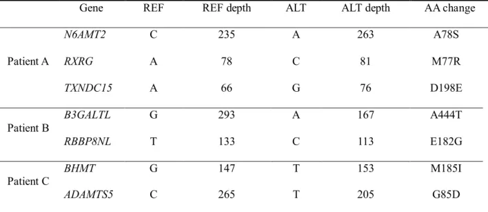
Table 2. Non-synonymous SNVs in three APL patients
Gene REF REF depth ALT ALT depth AA change
Patient A
N6AMT2 C 235 A 263 A78S
RXRG A 78 C 81 M77R
TXNDC15 A 66 G 76 D198E
Patient B
B3GALTL G 293 A 167 A444T
RBBP8NL T 133 C 113 E182G
Patient C
BHMT G 147 T 153 M185I
ADAMTS5 C 265 T 205 G85D
SNV, single nucleotide variant; APL, acute promyelocytic leukemia; REF, reference;
ALT, alternative; AA, amino acid;
In terms of indels, 123, 119 and 127 frameshift-causing insertions or deletions were identified in patient A, B and C, respectively. Among them, 55 genes were repetitively identified in all three patients, although the sites of indels were different (Table 3).
No significant CNVs were identified in patient A. However, Patient B exhibited copy number loss in six genes (HAUS1, PPP1R13L, SPTBN5, RRBP1, FANCA, and COL5A1) and copy number gain in two genes (NSMAF and SLC25A6). In patient C, NKD1 copy number gain was confirmed (Table 3).
7
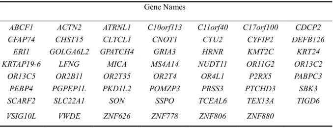
Table 3. Frameshift-causing indels associated with ATRA resistance Gene Names
ABCF1 ACTN2 ATRNL1 C10orf113 C11orf40 C17orf100 CDCP2 CFAP74 CHST15 CLTCL1 CNOT1 CTU2 CYFIP2 DEFB126
ERI1 GOLGA6L2 GPATCH4 GRIA3 HRNR KMT2C KRT24 KRTAP19-6 LFNG MICA MS4A14 NUDT11 OR11G2 OR13C2
OR13C5 OR2B11 OR2T35 OR2T4 OR4L1 P2RX5 PABPC3 PEBP4 PGPEP1L PKD1L2 POMZP3 PRSS3 PTCHD3 SBK3 SCARF2 SLC22A1 SON SSPO TCEAL6 TEX13A TIGD6 VSIG10L VWDE ZNF626 ZNF778 ZNF806 ZNF880
Table 4. Copy number variations associated with ATRA resistance
Patient Gene Locus Mean of log ratio p-value Gain/Loss
B HAUS1 chr18:43684298-43684407 -2.4 <0.001 loss PPP1R13L chr19:45882893-45883486 -2.0 <0.001 loss SPTBN5 chr15:42151088-42151178 -2.0 <0.01 loss RRBP1 chr20:17607993-17608027 -1.9 <0.001 loss FANCA chr16:89846277-89846365 -1.9 <0.001 loss COL5A1 chr9:137710502-137710609 -1.8 <0.001 loss NSMAF chr8:59535779-59535831 1.9 <0.001 gain SLC25A6 chrX:1510792-1511039 1.9 <0.001 gain C NKD1 chr16:50582628-50582660 -1.9 <0.01 loss
ATRA, all-trans retinoic acid;
8
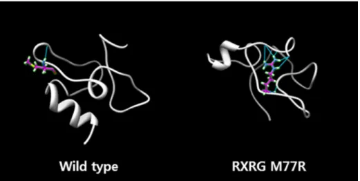
Sanger sequencing of the seven genes mentioned above in the ATRA-sensitive APL cohort confirmed the absence of the expected mutations in all 29 subjects. To predict the functional roles of these mutations, the Amber molecular dynamics simulation (http://www.ambermd.org)11 was applied. Because the retinoid X receptor (RXR) closely interacts with RAR, the chief protein involved in APL pathogenesis, I focused specifically on SNVs in RXRG. Increased hydrogen bonding, diminished helical folding, and lower energy state were all associated with the RXRG M77R mutation (Figure 1).
Figure 1. The Amber molecular dynamics predicted structural change of RXRG by M77R mutation.
9
DISCUSSION
To our knowledge, this is the first report on the use of WES to identify putative genetic mutations related to ATRA resistance. I found seven SNVs unique to ATRA-resistant APL patients, three of which were not previously reported in a public database. Such voids in the published data are understandable, given the low frequency of APL. Moreover, ATRA resistance in low-risk APL is so rare, that a solitary SNV should be viewed as a functional deviation and not dismissed as an incidental finding.
As such, the RXRG M77R I identified may be a bona fide factor in ATRA resistance. In addition to its occurrence in a patient with low-risk, ATRA-resistant APL, significant functional derangement of the RXR protein was predicted for this mutation through simulated dynamics. On a molecular level, RXR-RARα heterodimers form and then bind to retinoic acid response elements. Pharmacological doses of ATRA cause the release of PML-RARα fusion protein co-repressors, enabling PML-RARα and RXR proteins to heterodimerize for the transcription of genes governing normal myeloid differentiation. Furthermore, PML-RARα is degraded by pharmacological doses of ATRA, and wild-type RARα is upregulated, moving into the nucleus along with RXR12. In the event of RXR dysfunction (such as with RXRG M77R), PML-RARα or wild-type RARα proteins are unable to heterodimerize with RXR, potentially blocking myeloid differentiation despite the ATRA exposure.
A biological basis for ATRA resistance may also exist in the other
10
implicated SNVs. In particular, N6AMT2 plays a role in DNA methylation, thereby impacting transcriptional regulation. Methylation of a key gene in cell maturation may block myeloid differentiation, accounting for ATRA resistance in the APL patients. Indeed, N6AMT2 is a frequently mutated gene in various malignancies, including prostatic cancer (http://www.genecard.org).
Conversely, BHMT is indirectly involved in folate metabolism via homocysteine. Considering the important role of folate in DNA synthesis and methylation, genetic variants of BHMT may thus confer ATRA resistance.
Associations between BHMT defects and risks of certain cancers have been previously reported13,14.
In conclusion, this brief study is the first to suggest a genetic mechanism for ATRA resistance in APL patients by WES. A mutation in RXRG has been identified as a putative genetic marker that requires further analysis. Individualized use of ATRA in the therapeutic regimens for APL patients may thus be indicated.
11
REFERENCES
1. Warrell, R.P., Jr., de The, H., Wang, Z.Y. & Degos, L. Acute promyelocytic leukemia. The New England journal of medicine 329, 177-189 (1993).
2. Gallagher, R.E. Retinoic acid resistance in acute promyelocytic leukemia. Leukemia 16, 1940-1958 (2002).
3. Boylan, J.F. & Gudas, L.J. The level of CRABP-I expression influences the amounts and types of all-trans-retinoic acid metabolites in F9 teratocarcinoma stem cells. The Journal of biological chemistry 267, 21486-21491 (1992).
4. Shao, W., Benedetti, L., Lamph, W.W., Nervi, C. & Miller, W.H., Jr. A retinoid-resistant acute promyelocytic leukemia subclone expresses a dominant negative PML-RAR alpha mutation. Blood 89, 4282-4289 (1997).
5. Li, H. & Durbin, R. Fast and accurate short read alignment with Burrows-Wheeler transform. Bioinformatics 25, 1754-1760 (2009).
6. McKenna, A., et al. The Genome Analysis Toolkit: a MapReduce framework for analyzing next-generation DNA sequencing data.
Genome research 20, 1297-1303 (2010).
7. Cibulskis, K., et al. Sensitive detection of somatic point mutations in impure and heterogeneous cancer samples. Nat Biotechnol 31, 213-219 (2013).
8. Koboldt, D.C., et al. VarScan 2: somatic mutation and copy number alteration discovery in cancer by exome sequencing.
Genome research 22, 568-576 (2012).
9. Li, J., et al. CONTRA: copy number analysis for targeted resequencing. Bioinformatics 28, 1307-1313 (2012).
10. Adzhubei, I., Jordan, D.M. & Sunyaev, S.R. Predicting functional effect of human missense mutations using PolyPhen-2. Current protocols in human genetics / editorial board, Jonathan L.
Haines ... [et al.] Chapter 7, Unit7 20 (2013).
11. Beveridge, D.L., Cheatham, T.E., 3rd & Mezei, M. The ABCs of molecular dynamics simulations on B-DNA, circa 2012. Journal of biosciences 37, 379-397 (2012).
12. Melnick, A. & Licht, J.D. Deconstructing a disease: RARalpha, its fusion partners, and their roles in the pathogenesis of acute promyelocytic leukemia. Blood 93, 3167-3215 (1999).
13. Koushik, A., et al. Nonsynonymous polymorphisms in genes in the one-carbon metabolism pathway and associations with colorectal cancer. Cancer epidemiology, biomarkers &
prevention : a publication of the American Association for Cancer Research, cosponsored by the American Society of Preventive Oncology 15, 2408-2417 (2006).
12
14. da Silva, L.M., et al. MTHFD1 G1958A, BHMT G742A, TC2 C776G and TC2 A67G polymorphisms and head and neck squamous cell carcinoma risk. Molecular biology reports 39, 887-893 (2012).
13
국문 초록
서론: 급성 전골수성 백혈병은 급성 골수성 백혈병중에서 가장 예후 가 좋은 아형이다. 하지만 일부 환자들은 ATRA 를 기초로 한 표준 치료로 완치가 되지 않는다. ATRA 대사의 활성화 등 몇몇 기전이 후천적인 ATRA 저항성으로 제시되고 있지만 ATRA 내성의 유전 적인 기전은 잘 알려지지 않았다. 따라서 이번 연구를 통하여 ATRA 내성에 기여하는 유전적 변이를 찾고자 한다.
방법: ATRA 를 포함한 표준치료에 내성을 보인 3 명의 급성 전골 수성 백혈병 환자의 DNA 를 사용하여 전체 엑솜시퀀싱을 시행하였 다. 3 명의 환자중, 2 명은 첫 번째 관해치료에 저항성을 보인 환자 였고, 나머지 한 환자는 첫번째 관해치료에 반응을 보였지만 ATRA 유지 치료 중에 재발을 한 환자였다. DNA 는 환자들의 진단시 검체 에서 획득되었고, 분석을 위하여 관해를 얻었을 때의 구강상피세포 를 대조군으로 사용하였다. 단일염기서열변이는 Adiscan 으로 불리
는 내부 파이프라인을 사용하여 확인하였다
(http://www.syntekabio.com). 전체 엑솜시퀀싱에서 발견된 단일염기서 열변이는 Sanger 시퀀싱을 통해 재확인되었다. 이들 돌연변이의 존 재를 ATRA 에 반응을 보이는 급성 전골수성 백혈병 환자 코호트에 서 추가적으로 확인하였다.
14
결과: 전체 엑솜시퀀싱을 통하여 3 명의 환자에서 33 개의 비동일 단일염기 다형성이 발견되었다. Polyphen-2 알고리즘을 활용하여 단백질 기능에 영향을 미치는 9 개의 비동일 단일염기 다형성을 추 렸고 Sanger 시퀀싱에서 7 개가 증명되었다. 그들은 RXRG M77R, N6AMT2 A78S, TXNDC15 D198E, B3GALTL A444T, RBBP8NL E182G, BHMT M185I and ADAMTS5 G85D 였다. 이 7 개의 비동일 단일염기 다형성은 ATRA 에 반응을 보인 29 명의 급성 전골수성 백혈병환자 코호트에서 단 한 번도 확인되지 않은 염기 다형성이었다. 이는 본 비동일 단일염기 다형성이 ATRA 내성의 기전과 연관되어 있을 가 능성을 시사한다. Amber 분자 역동학 시물레이션을 진행한 결과 1) 수소결합력의 증가, 2) 나선형 분자구조의 감소, 3) RXRG 돌연변이 에서 에너지의 감소가 확인되었다.
결론: 전체 엑솜시퀀싱을 통하여 단백질 기능에 영향을 미치는 7 개 의 비동일 단일염기 다형성을 ATRA 내성 환자들에서 확인하였다.
그 중에서 RXRG 돌연변이가 기전적으로 ATRA 내성에 기여하는 유전적 변이의 가능성이 있을 것이다.
주요어 : 급성 전골수성 백혈병, 약제 내성, 트레티노인, 레티노이트 X 수용체 감마
학 번 : 2014-21164