저작자표시-비영리-변경금지 2.0 대한민국 이용자는 아래의 조건을 따르는 경우에 한하여 자유롭게
l 이 저작물을 복제, 배포, 전송, 전시, 공연 및 방송할 수 있습니다. 다음과 같은 조건을 따라야 합니다:
l 귀하는, 이 저작물의 재이용이나 배포의 경우, 이 저작물에 적용된 이용허락조건 을 명확하게 나타내어야 합니다.
l 저작권자로부터 별도의 허가를 받으면 이러한 조건들은 적용되지 않습니다.
저작권법에 따른 이용자의 권리는 위의 내용에 의하여 영향을 받지 않습니다. 이것은 이용허락규약(Legal Code)을 이해하기 쉽게 요약한 것입니다.
Disclaimer
저작자표시. 귀하는 원저작자를 표시하여야 합니다.
비영리. 귀하는 이 저작물을 영리 목적으로 이용할 수 없습니다.
변경금지. 귀하는 이 저작물을 개작, 변형 또는 가공할 수 없습니다.
의학박사 학위논문
Targeted massive parallel sequencing and comprehensive phenome-genome assessment in neuromuscular patients
without genetic etiology by conventional diagnostic methods
기존 진단 방법으로 원인유전자를 발견하지 못한 신경근육 질환 환자에서 표적 병렬 시퀀싱 및
포괄적 표현형-유전형 분석 연구
2023 년 2 월
서울대학교 대학원 의학과 중개의학전공
조 안 나
기존 진단 방법으로 원인유전자를 발견하지 못한 신경근육 질환 환자에서 표적 병렬 시퀀싱 및 포괄적 표현형-유전형 분석 연구
지도 교수 채 종 희
이 논문을 의학박사 학위논문으로 제출함 2022년 10월
서울대학교 대학원 의학과 중개의학전공
조 안 나
조안나의 의학박사 학위논문을 인준함
2023년 1월
위 원 장 (인)
부위원장 (인)
위 원 (인)
위 원 (인)
위 원 (인)
Targeted massive parallel sequencing and comprehensive phenome-genome assessment in neuromuscular patients
without genetic etiology by conventional diagnostic methods
By
Anna Cho, M.D.
Directed by Jong Hee Chae, M.D., Ph.D.
A thesis submitted in partial fulfillment of the requirements for the Degree of Doctor of Philosophy in Medicine (Major in Translational Medicine) at the Seoul
National University College of Medicine October 2022
Approved by Thesis Committee:
Professor ( 인 ) Chairman Professor ( 인 ) Vice chairman Professor (인)
Professor ( 인 )
Professor ( 인 )
i
Abstract
Targeted massive parallel sequencing and comprehensive phenome-genome assessment in neuromuscular patients without genetic etiology by
conventional diagnostic methods
Anna Cho Translational Medicine The Graduate School Seoul National University
Neuromuscular disorders are clinically, pathologically, and genetically heterogeneous groups of disorders. An accurate genetic diagnosis has often been challenging due to the heterogeneity and complexity of these groups of disorders.
The advent of next generation sequencing (NGS) technologies have started a new era of molecular genetic diagnosis in neuromuscular disorders. It is an effective diagnostic tool for the parallel investigation of a large number of genes and has been increasingly used in the recent clinical and research fields. In this study, NGS-
ii
based targeted sequencing panels were applied to the diagnostic workflow in the genetic characterization of 327 patients who were suspected of having a hereditary neuromuscular disorders but without a definite molecular diagnosis using traditional genetic analysis. Four sets of targeted sequencing panels prepared at different times and were sequentially applied. Fifty eight patients were sequenced for 579 genes, 29 patients for 383 genes, and 150 patients for 436 genes. The last 90 patients were analyzed with tests selected among 6 custom panels for congenital myopathies, congenital muscular dystrophies, limb girdle muscular dystrophies, metabolic myopathies, myofibrillar myopathies, and Charcot-Marie-Tooth diseases. Each panel contains 29 to 79 genes according to the clinical subgroups.
Pathogenic mutations were confirmed in 161 cases (49.2%) out of 327 patient. The diagnostic yield by phenotype groups were 62.7% in congenital muscular dystrophies (n = 47), 45.3% in limb girdle muscular dystrophies (n = 43), 54.3% in congenital myopathies (n = 50), 80% in congenital myasthenic syndromes (n = 4), 14.3% in metabolic myopathies (n = 3), 35.3% in motor neuron disorders (n = 6), 50% in hereditary motor and sensory neuropathies (n = 3), and 31.3% in other neuromuscular disorders (n = 5) respectively. The causative mutations were found in 50 different genes and the six most frequently found genes were COL6A1 (n = 21), RYR1 (n = 18), DMD (n = 10), COL6A2 (n = 9), ACTA1 (n = 7), and LMNA (n
= 7) in order. This study illustrates the clinical utility of targeted NGS as a powerful diagnostic tool in hereditary neuromuscular disorders. Integrated phenome-genome
iii
analysis is very important for improving the diagnostic efficiency of this technique.
It is necessary to establish a comprehensive diagnostic platform for the molecular diagnosis of neuromuscular disorders that integrates single gene testing and NGS based on the phenotype analysis.
Keywords: targeted parallel sequencing, next generation sequencing, molecular
diagnosis, neuromuscular disorders, muscular dystrophy, congenital myopathy Student number: 2015-30012
iv
Contents
Abstract --- ⅰ Contents --- ⅳ List of Tables --- ⅴ List of Figures --- ⅵ List of Abbreviations --- ⅶ
Introduction --- 1
Methods --- 4
Results ---25
Discussion --- 63
References --- 71
국문초록 --- 79
v
List of Tables
Table 1. List of 579 Genes for Targeted NGS Sequencing [Seattle Panel] ---11
Table 2. List of 383 Genes for Targeted NGS Sequencing [NMD-2014] ---15
Table 3. List of 436 Genes for Targeted NGS Sequencing [NMD-2015] ---17
Table 4. Lists of Genes for Targeted NGS Sequencing [SNUH-NMD] --- 20
Table 5. Pathogenic and likely pathogenic variants in 83 muscular dystrophy patients --- 33
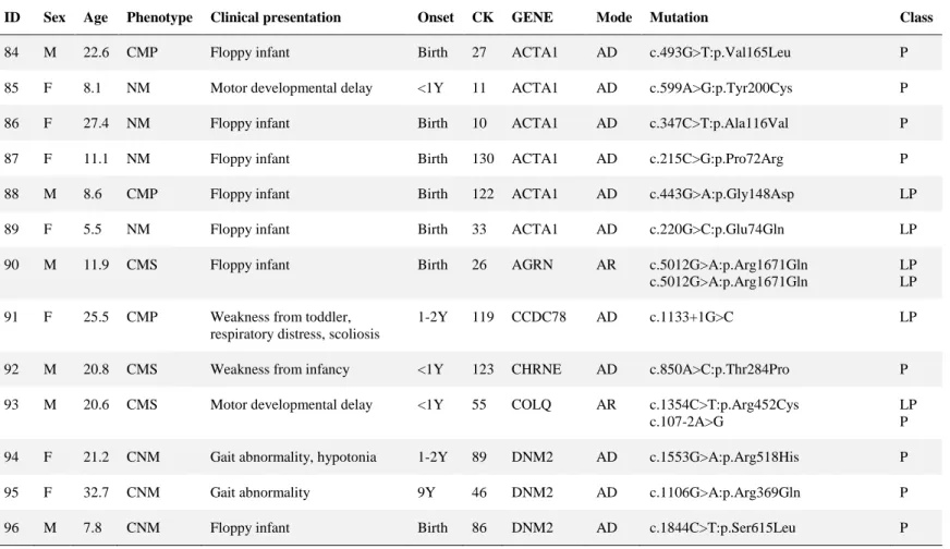
Table 6. Pathogenic and likely pathogenic variants in 55 congenital myopathy and congenital myasthenic syndrome patients --- 41
Table 7. Pathogenic and likely pathogenic variants in 10 motor neuron disease and hereditary motor and sensory neuropathy patients --- 48
Table 8. Pathogenic and likely pathogenic variants in 13 metabolic and other myopathies patients --- 50
Table 9. Variants of uncertain significance in 31 patients --- 54
Table 10. List of diagnoses confirmed by further testing in 28 patients --- 59
vi
List of Figures
Figure 1. Flowchart of the diagnostic workflow with NGS application --- 6
Figure 2. Demographics and phenotype classification --- 27
Figure 3. Serum creatine kinase (CK) by phenotype groups --- 28
Figure 4. Fraction of diagnostic yields by phenotype groups --- 30
Figure 5. Diagnosis frequency in 50 genes with mutations --- 31
Figure 6. Distribution of genotypes in muscular dystrophies and congenital myopathies by NGS analysis --- 39
Figure 7. Muscle histopathology in congenital myopathies --- 46
Figure 8. Muscle histopathology in myofibrillar myopathies --- 52
Figure 9. Summary of the targeted sequencing, phenotypic classification, and diagnostic yield ---62
vii
List of Abbreviations
ACMG: American College of Medical Genetics and Genomics AD: autosomal dominant
AR: autosomal recessive
BMD: Becker muscular dystrophy CCD: central core disease
CFTD: congenital myopathy with fiber type disproportion CMA: chromosome microarray
CMD: congenital muscular dystrophy CMP: congenital myopathy
CMS: congenital myasthenic syndromes CMT: Charcot-Marie-Tooth disease CNM: centronuclear myopathy CNV: copy number variation CK: creatine kinase
DCMP: dilated cardiomyopathy DM: dermatomyositis
viii
DMD: Duchenne muscular dystrophy EMG: electromyography
FSHD: facioscapulohumeral muscular dystrophy HGMD: Human Gene Mutation Database
HMSN: hereditary motor and sensory neuropathies IHC: immunohistochemistry
IMNM: immune mediated necrotizing myopathy LGMD: limb girdle muscular dystrophy
MD: muscular dystrophy
MDCMD: merosin deficient congenital muscular dystrophy MFM: myofibrillar myopathy
MLPA: multiplex ligation dependent probe amplification MM: metabolic myopathy
MMD: multi-minicore disease MND: motor neuron diseases MRI: magnetic resonance imaging MTM: myotubular myopathy MyoD: myotonic dystrophy
ix
NCV: nerve conduction velocity NGS: next generation sequencing NM: nemaline myopathy
NMD: neuromuscular disorders ONM: other neuromuscular disorders P: pathogenic
LP: likely pathogenic
SMA: spinal muscular atrophy
SNUH: Seoul national university hospital UCMD: Ullrich congenital muscular dystrophy VUS: variants of unknown significance
WB: Western blot
WES: whole exome sequencing XD: X linked dominant
XR: X linked recessive
1
Introduction
Neuromuscular disorders (NMD) are clinically, pathologically, and genetically heterogeneous groups of disorders. The clinical presentation shares similar features such as hypotonia, weakness, and motor developmental delay, which make it difficult to reach a specific diagnosis even for the experienced clinicians (1-4).
Traditionally, most of neuromuscular disorders have been categorized and diagnosed according to their clinical phenotypes and histological presentations (5, 6) and genetic diagnosis has not been always possible because of the diversity of disease causing genes.
There are at least three major reasons that many cases with probable hereditary neuromuscular disorders have been genetically undiagnosed. Firstly, it is often difficult to determine the target gene to be analyzed due to genetic complexity and heterogeneity. For example, nemaline myopathy, one of the most common of the congenital myopathies, is caused by at least 12 different genes including ACTA1, NEB, TPM2, TPM3, MYPN, TNNT1, TNNT3, KLHL40, KLHL41, KBTBD13, CFL2, and LMOD3 (7). While ACTA1, a representative causative gene of nemaline myopathy, shares various phenotypes including intra-nuclear rod myopathy, congenital myopathy with fiber type disproportion, and zebra body myopathy.
Secondly, muscle genes are often big in size such as TTN with 362 exons, making it difficult to routinely sequence such genes even though there are known causative
2
genes (8). Thus far, we have often performed routine gene sequencing of small genes such as ACTA1 and MTM1 and large genes such as RYR1 and COL6A1 were occasionally subjected to sequencing analysis only at hot spots. Thirdly, most cases are sporadic without parental consanguinity, which makes us difficult to do linkage study to identify new causative genes.
The advent of next generation sequencing (NGS) technologies have started a new era of molecular genetic diagnosis in neuromuscular disorders (9-12). It is an effective diagnostic tool for the parallel investigation of a large number of genes and has been increasingly used in the recent clinical and research fields (13-16).
Genome-wide approaches such as whole exome sequencing (WES) or whole- genome sequencing (WGS) can be the ultimate goal in genetic testing, in that it allows the screening of numerous known gene mutations and can potentially identify new disease causative genes (12). But selected gene panel tests do have multiple clinical advantages over genome-wide sequencing methods in terms of time and cost effectiveness (17, 18). The important point is to establish a comprehensive diagnostic system in patients with hereditary neuromuscular disease that is clinically very complex and contains a wide range of genes.
In this study, we applied an NGS-based platform to the diagnostic workflow in the genetic characterization of neuromuscular patients who were suspected of having
3
a Mendelian disorder but without a definite molecular diagnosis using the traditional genetic analysis.
4
Methods
Design and subjects
The study enrolled a total of 327 patients with suspected hereditary neuromuscular disorders based on their clinical and/or muscle histopathological analysis but without a definite molecular diagnosis. All cases were ascertained from the Seoul National University Hospital (SNUH) – Neuromuscular Disorders (NMD) database, which includes a cohort of more than 2000 cases from 2000 to 2020. All clinical information and samples used form diagnostic purpose in this study were collected after obtaining written informed consent from.
Before the NGS testing, several genetic testing including SMN1/2 Multiplex ligation dependent probe amplification (MLPA), DMD MLPA, DMPK PCR and Southern blot analysis, and Sanger sequencing targeting entire genes or hot spots of severe genes (ACTA1, CPT2, FKTN, and MTM1) were performed based on the clinical and pathologic findings in selected patients, but were negative. Patients whose molecular diagnosis was confirmed by these conventional genetic analysis methods were excluded from this study.
Four sets of targeted sequencing panels prepared at different times were sequentially applied for the analysis of 327 patients. In 2013, the first 58 patients were analyzed with NGS in a collaborative study with Seattle Children’s Research
5
Institute (10). Then we developed the first in-house panel for the neuromuscular disorders and analyzed 29 patients in 2014. The gene panel library was updated once in 2015 and an additional 150 patients were analyzed. As Korea’s National Health Insurance Service coverage became available for the panel NGS analysis of hereditary diseases in 2017 (Ministry of Health and Welfare Notice No.2017-15), the validated NGS custom panels by the Department of Laboratory Medicine at Seoul National University Hospital has begun. We set up 6 kinds of different targeted panels for the hereditary neuromuscular disorders according to the clinical subgroups: 1) congenital muscular dystrophy (CMD), 2) limb girdle muscular dystrophy (LGMD), 3) congenital myopathy (CMP), 4) metabolic myopathy (MM), 5) Charcot-Marie-Tooth disease (CMT), and 6) myofibrillar myopathy (MFM).
Total 90 patients underwent the SNUH-NGS panel analysis corresponding to the clinical diagnosis. The diagnostic workflow and study design is summarized as a flow chart in Figure 1.
6
Figure 1. Flowchart of the diagnostic workflow with NGS application
SNUH, Seoul National University Hospital; NMD, neuromuscular disorders; CK, creatine kinase; EMG/NCS, electromyography & nerve conduction velocity; MRI, Magnetic Resonance Imaging; IHC & WB, immunohistochemistry & Western blot;
NGS, next generation sequencing; N, numbers; CMD, congenital muscular dystrophy; LGMD, limb girdle muscular dystrophy; CMP, congenital myopathy;
MM, metabolic myopathy; CMT, Charcot-Marie-Tooth disease; MFM, myofibrillar myopathy; CMA, chromosome microarray; PW/AS, Prader- Willi/Angelman Syndrome methylation test; DMD, Duchenne muscular dystrophy;
SMA, spinal muscular atrophy; MyoD, myotonic dystrophy; FSHD, facioscapulohumeral muscular dystrophy
7
Clinical and histological data analysis
Clinical information was reviewed including demographic information, the age of onset, clinical presentation, and serum creatine kinase (CK) levels.
1) Clinical phenotype classification
All patients were classified into 8 diagnostic subgroups based on their clinical and muscle histopathological analyses: congenital muscular dystrophies (CMD), limb girdle muscular dystrophies (LGMD), congenital myopathies (CMP), congenital myasthenic syndromes (CMS), metabolic myopathies (MM), motor neuron diseases (MND), hereditary motor and sensory neuropathies (HMSN), and other neuromuscular disorders (ONM). CMD are categorized based on high CK and/or presence of necrotic and regenerating muscle fibers on muscle pathology with the clinical onset in infancy or before independent gait. LGMD are categorized based on high CK and/or presence of necrotic and regenerating muscle fibers on muscle pathology with the onset of weakness after independent gait. CMP are categorized based on pathological diagnosis and/or characteristic clinical features including hypotonia and facial weakness at birth. CMS are categorized based on clinical diagnosis with characteristics of weakness with diurnal variation, ptosis, and/or positive Jolly test. MM are categorized based on clinical diagnosis with characteristics of exercise intolerance, rhabdomyolysis, and/or muscle weakness
8
with high CK. MND and HMSN are categorized based on electrophysiological findings and/or presence of fibers type grouping (denervation pattern) on muscle pathology. Patients who could not be classified into a specific group were classified as ONM.
2) Muscle pathology analysis
Histopathological phenotype was reviewed in 261 cases that underwent muscle biopsies at Seoul National University Children’s Hospital. Serial frozen sections from each muscle sample were stained using a set of histochemical methods including hematoxylin and eosin (H&E), modified Gomori trichrome, NADH- Tetrazolium Reductase (NADH-TR), succinyl dehydrogenase, ATPase, and immunohistochemistry for dystrophin, merosin and dysferlin. Additional immunohistochemical stainings with mouse monoclonal anti-human collagen VI primary antibodies (Chemicon, MAB1944) or monoclonal antibodies against alpha-dytroglycan with glycoepitope-dependent antibody (VIA4-1) (Merck Millipore, Catalogue No. 05-298) were performed on clinically suspected. Reports or slides of muscle histopathology performed at an external institution in 10 cases were reviewed.
9
Gene selection, sequencing, and variant annotation 1) Seattle Panel
The first NGS series were performed at Seattle Children’s Research Institute, Seattle, Washington, USA (10). A DNA library was prepared for each sample by capturing the exons of the genes of interest using custom made DNA probes (Haloplex Agilent, Santa Clara, CA). The selected 579 genes (Table 1) comprised 10,706 exons and a total of 3.88 Mb. Fifty eight samples were sequenced at 5-8 samples per lane on a GAIIx instrument (Illumina, San Diego, CA) using 2x100 paired-ends reads. Reads were aligned using Burrows-Wheeler Aligner (BWA, version 0.7.3) and data were analyzed with the Genome Analysis Toolkit (GATK, version 2.4.9) (Broad Institute, Cambridge, MA) as previously described (19).
Variants were annotated using SeattleSeq (20) and wAnnovar (21). Variants found within the targeted regions were further evaluated for their possible clinical significance by cross-referencing to the Single Nucleotide Polymorphism Database (dbSNP), the Exome variant server, and the 1000 Genomes browser. An internal database of polymorphisms was also used for quality assurance during this analysis.
PolyPhen-2 and SIFT (sorting intolerant from tolerant) scores were used only for reference but were not used for filtering the variants. For variants in genes with autosomal dominant and X-linked disease inheritance, we used the minor allele frequency (MAF) cut-off of 0.2%, and for variants in genes with autosomal
10
recessive disease inheritance, we used the MAF cut-off of 0.5% (19). Variants that exceeded these frequencies were not considered as potential mutations, even if they were previously reported as such in the Human Gene Mutation Database (HGMD).
Finally, variants were searched in the HGMD by Genometrax (22, 23).
11
Table 1. List of 579 Genes for Targeted NGS Sequencing [Seattle Panel]
Congenital myopathy (30 genes)
ACTA1, ACTG2, ATP2A1, BIN1, CCDC78, CFL2, CLCN1, CNTN1, DNM2, FAM123B, FLNC, GNE, HOXD10, HRAS, KBTBD13, LDB3, MEGF10, MTM1, MTMR14, MYF6, MYH2, MYH7, NEB, TIA1, TNNT1, TPM2, TPM3, TTN, VCP, VMA21
Muscular dystrophy (48 genes)
ANO5, BAG3, CAPN3, CAV3, CHKB, CNBP, COL6A1, COL6A2, COL6A3, CRYAB, DAG1, DES, DMD, DMPK, DNAJB6, DPM2, DPM3, DYSF, EMD, FHL1, FKBP14, FKRP, FKTN, GTDC2, ISPD, ITGA7, ITGA9, KLHL9, LAMA2, LARGE, LMNA, MTAP, MYOT, PABPN1, PLEC, PLOD3, POMGNT1, POMT1, POMT2, SECISBP2, SGCA, SGCB, SGCD, SGCG, SPP1, SYNE2, TCAP, TRIM32
Congenital myopathy or Muscular dystrophy (2 genes)
RYR1, SEPN1
Metabolic myopathy (202 genes)
AARS2, ACACA, ACAD9, ACADM, ACADVL, ACSF3, ACY1, ADCK3, AGK, AGL, AIFM1, ALDH1B1, ALDOA, ALG1, ALG11, ALG12, ALG13, ALG2, ALG3, ALG6, ALG8, ALG9, ALPL, AMACR, AMPD1, ATP5A1, ATP5E, ATP5G3, ATPAF2, B4GALT1, BCS1L, BRP44L, C10orf2, C12orf65, CACNA1S, COA5, COG1, COG4, COG5, COG6, COG7, COG8, COQ2, COQ4, COQ6, COQ9, COX10, COX14, COX15, COX20, COX4I2, COX6B1, COX7B, CPT1A, CPT1B, CPT2, CYC1, CYCS, D2HGDH, DARS2, DGUOK, DMGDH, DNA2, DOLK, DPM1, EARS2, ENO3, ETFA, ETFB, ETFDH, FARS2, FASTKD2, FOXRED1, GAA, GALC, GBE1, GFER, GFM1, GPR172B, GYG1, GYS1, HADH, HADHA, HADHB, HARS2, IARS, ISCU, LAMP2, LARS2, LDHA, LDHB, LIAS, LPIN1, LRPPRC, LYRM4, LYRM7, MARS, MATR3, MGAT2, MGME1, MPV17, MRPL12, MRPL3, MRPL44, MRPS16, MRPS22, MTFMT, MTHFD1, MTHFR, MTO1, NDUFA1, NDUFA10, NDUFA11, NDUFA12, NDUFA2, NDUFA4, NDUFA9, NDUFAF1, NDUFAF2, NDUFAF3, NDUFAF4, NDUFAF5,
12
NDUFAF6, NDUFB3, NDUFB9, NDUFS1, NDUFS2, NDUFS3, NDUFS4, NDUFS6, NDUFS7, NDUFS8, NDUFV1, NDUFV2, NEU1, NFU1, NUBPL, OAT, OPA1, OXCT1, PDSS1, PDSS2, PEX12, PEX14, PEX26, PEX3, PEX5, PEX6, PEX7, PFKM, PGAM2, PGK1, PGM1, PHKA1, PHKA2, PHKB, PHKG2, PHYH, PMM2, PNPLA2, PNPT1, POLG, POLG2, PSAP, PTEN, PTRF, PUS1, PYGL, PYGM, RMND1, RRM2B, RSPH9, SCO1, SCO2, SDHA, SDHAF1, SDHAF2, SDHB, SDHC, SDHD, SLC22A5, SLC25A20, SLC25A3, SLC25A4, SUCLA2, SUCLG1, SURF1, TACO1, TARS2, TAZ, TIMM44, TK2, TMEM165, TMEM70, TSFM, TTC19, TYMP, UQCRB, UQCRC2, UQCRQ, VARS2, YARS2
Motor neuron or peripheral nerve disease (75 genes)
AARS, AFG3L2, APTX, ARHGEF10, ATL1, ATXN2, BSCL2, CCT5, COLQ, DCTN1, DHTKD1, DNAJB2, DNMT1, DYNC1H1, EGR2, ELAVL4, ERLIN2, FAM134B, FGD4, FOXE1, GAN, GARS, GDAP1, GJB1, HARS, HINT1, HOXB1, HSPB1, HSPB3, HSPB8, HSPG2, IGHMBP2, IKBKAP, INF2, KARS, KIF1B, KIF5A, LITAF, MED25, MFN2, MPZ, MTMR2, MYH14, NAGA, NDRG1, NEFL, NGF, NIPA1, PDK3 , PLEKHG5, PLP1, PMP22, PNPLA6, PRX, RAB7A, REEP1, SACS, SBF2, SH3TC2, SLC12A6, SLC5A7, SMN1, SMN2, SNX25, SPTLC1, SPTLC2, SYNE1, TDP1, TFG, TGM6, TRPV4, UBA1, VRK1, WNK1, YARS
Myasthenic syndrome (12 genes)
AGRN, CHAT, CHRNA1, CHRNB1, CHRND, CHRNE, DOK7, DPAGT1, GFPT1, LAMB2, MUSK, SCN4A
Cardiomyopathy (3 genes)
MYBPC3, MYLK2, RYR2
Other candidate genes (207 genes)
ACAD10, ACAD11, AIFM2, ATP5B, ATP5C1, ATP5D, ATP5F1, ATP5G1, ATP5G2, ATP5H, ATP5I, ATP5J, ATP5J2, ATP5L, ATP5O, ATP5S, ATPAF1, ATPIF1, C14orf2, CARS, CARS2, CD36, CHCHD1, CHCHD7, CMC1, CMC2, COA1, COA3, COA6,
13
COQ10A, COQ10B, COQ3, COQ5, COQ7, COX11, COX16, COX17, COX18, COX19, COX4I1, COX5A, COX5B, COX6A1, COX6A2, COX6B2, COX6C, COX7A1, COX7A2, COX7A2L, COX7B2, COX7C, COX8A, COX8C, ECi1, ECSIT, EPRS, FARSA, FARSB, FRG1, FUNDC2, HINT3, IARS2, ICT1, LACTB, LYRM1, MCAT, MDH2, METTL17, MNF1, MRP63, MRPL1, MRPL10, MRPL11, MRPL13, MRPL14, MRPL15, MRPL16, MRPL17, MRPL18, MRPL19, MRPL2, MRPL20, MRPL21, MRPL22, MRPL23, MRPL24, MRPL27, MRPL28, MRPL30, MRPL32, MRPL33, MRPL34, MRPL35, MRPL36, MRPL37, MRPL38, MRPL39, MRPL4, MRPL40, MRPL41, MRPL42, MRPL43, MRPL45, MRPL46, MRPL47, MRPL48, MRPL49, MRPL50, MRPL51, MRPL52, MRPL53, MRPL54, MRPL55, MRPL9, MRPS10, MRPS11, MRPS12, MRPS14, MRPS15, MRPS17, MRPS18A, MRPS18B, MRPS18C, MRPS2, MRPS21, MRPS23, MRPS24, MRPS25, MRPS26, MRPS27, MRPS28, MRPS30, MRPS31, MRPS33, MRPS34, MRPS35, MRPS36, MRPS5, MRPS6, MRPS7, MRPS9, MRS2, MTCH1, MTERF, MTG1, MTHFD1L, MTHFS, MTIF3, MTRF1L, NARS, NARS2, NDUFA3, NDUFA4L2, NDUFA5, NDUFA6, NDUFA7, NDUFA8, NDUFAB1, NDUFB1, NDUFB10, NDUFB11, NDUFB2, NDUFB4, NDUFB5, NDUFB6, NDUFB7, NDUFB8, NDUFC1, NDUFC2, NDUFS5, NDUFV3, NIPSNAP3A, NRF1, OXA1L, OXSM, PARS2, PGAM1, POLRMT, PRELID1, PRELID2, PTCD1, PTCD2, PTCD3, PTRH2, QARS, RARS, SLIRP, SMCR7, SMCR7L, SUCLG2, TARS, TARSL2, TIMM21, TIMM23, TOMM70A, TOP1MT, TYMS, UQCR10, UQCR11, UQCRC1, UQCRFS1, UQCRH, USMG5, VARS, WARS, WARS2, YME1L1
This table was reproduced from [Supplement Table 1] in “Utility of next generation sequencing in genetic diagnosis of early onset neuromuscular disorders” J Med Genet 52(3):208-16 (10).
14
2) NMD-2014 and NMD-2015 Panels
The custom-designed SureSelect Target Enrichment System Kit (Agilent Technologies, USA) was used to assess hereditary neuromuscular genes at SNUH.
The targeted genes were selected using the 2014 and 2015 version of the gene table of monogenic neuromuscular disorders (24, 25) and some of the genes were added based on a literature search. The capture kits were updated once to include newly identified genes. Twenty nine patients were sequenced with the first kit (NMD- 2014 including 383 genes, Table 2) and 150 with the second kit (NMD-2015 including 436 genes, Table 3). Library preparation was completed according to the manufacturer’s instructions (Agilent Technologies). The library was paired-end sequenced on an Illumina HiSeq 2500 sequencing system.
Paired-end sequencing reads with a read length of 101 bp were aligned to Genome Reference Consortium Human Genome build 37 (patch release 13) using BWA-0.7 (26). The Picard software (v.2.1.1), SAMtools (v.1.3.1) (27), and Genome Analysis Toolkit (v.3.8) (28) best-practice pipelines were used in data analyses. Variant calling was performed using HaplotypeCaller and variant annotation using ANNOVAR (29). mTect2 (30) was used for low-frequency variant detection to search for variants with a variant allele frequency from 0.05 to 0.25. Only low- frequency variants with a variant allele count ≥ 30 were selected.
15
Table 2. List of 383 Genes for Targeted NGS Sequencing [NMD-2014]
The 2014 version of the gene table of monogenic neuromuscular disorders (361 genes)
AARS, ABCC9, ABHD5, ACADVL, ACTA1, ACTC1, ACTN2, ACVR1, AFG3L2, AGL, AGRN, AIFM1, AKAP9, ALDH3A2, ALG13, ALG14, ALG2, ALS2, ANG, ANK2, ANKRD1, ANO5, AP4E1, AP4M1, AP5Z1, APTX, AR, ARHGEF10, ASAH1, ATL1, ATM, ATP2A1, ATP7A, ATXN1, ATXN10, ATXN2, ATXN3, ATXN7, ATXN8OS, B3GALNT2, B3GNT1, BAG3, BEAN, BICD2, BIN1, BSCL2, C9orf72, CABC1, CACNA1A, CACNA1C, CACNA1S, CACNB2, CACNB4, CAPN3, CASQ2, CAV3, CFL2, CHAT, CHKB, CHMP2B, CHRNA1, CHRNB1, CHRND, CHRNE, CHRNG, CLCN1, CNTN1, COL6A1, COL6A2, COL6A3, COLQ, COX15, CPT2, CRYAB, CSRP3, CTDP1, CYP7B1, DAG1, DCTN1, DES, DMD, DMPK, DNAJB2, DNAJB6, DNM2, DNMT1, DOK7, DOLK, DPAGT1, DPM1, DPM2, DPM3, DSC2, DSG2, DSP, DTNA, DUX4, DYNC1H1, DYSF, EGR2, EMD, ENO3, ERBB3, ETFA, ETFB, ETFDH, EXOSC3, EYA4, FA2H, FBLN5, FGD4, FGF14, FHL1, FIG4, FKRP, FKTN, FLNA, FLNC, FUS, FXN, GAA, GAN, GARS, GATAD1, GBE1, GDAP1, GDF8, GFPT1, GJA5, GJB1, GLE1, GMPPB, GNB4, GNE, GPD1L, GTDC2, GYG1, GYS1, HCN4, HEXB, HINT1, HK1, HOXD10, HSPB1, HSPB3, HSPB8, HSPD1, HSPG2, ICSU, IFRD1, IGHMBP2, IKBKAP, ILK, INF2, ISPD, ITGA7, ITPR1, JPH2, JUP, KARS, KBTBD13, KCNA1, KCNA5, KCNC3, KCNE1, KCNE2, KCNE3, KCNH2, KCNJ18, KCNJ2, KCNJ5, KCNQ1, KIAA0196, KIF1A, KIF1B, KIF21A, KIF5A, KLHL40, KLHL9, L1CAM, LAMA2, LAMA4, LAMB2, LAMP2, LARGE, LDB3, LDHA, LITAF, LMNA, LPIN1, LRSAM1, MARS, MATR3, MED25, MEGF10, MFN2, MPZ, MRE11A, MRPL3, MTM1, MTMR2, MTPAP, MURC, MUSK, MYBPC3, MYH2, MYH3, MYH6, MYH7, MYH8, MYL2, MYL3, MYLK2, MYOT, MYOZ2, MYPN, NDRG1, NDUFAF1, NEB, NEFH, NEFL, NEXN, NGFB, NIPA1, NOP56, NPPA, OPA1, OPTN, PABPN1, PDK3, PEO1, PEX7, PFKM, PFN1, PGAM2, PGK1, PGM1, PHKA1, PHOX2A, PHYH, PIP5K1C, PKP2, PLEC1, PLEKHG5, PLN, PLP1, PMP22, PNPLA2 ,PNPLA6, POLG, POLG2, POMGNT1, POMK, POMT1, POMT2, PPP2R2B, PRKAG2, PRKCG, PRPH,
16
PRPS1, PRX, PSEN2, PTPLA, PTRF, PYGM, RAB7, RAPSN, RBCK1, RBM20, REEP1, RRM2B, RTN2, RYR1, RYR2, SACS, SBF1, SBF2, SCN1B, SCN3B, SCN4A, SCN4B, SCN5A, SDHA, SEPN1, SEPTIN9, SETX, SGCA, SGCB, SGCD, SGCE, SGCG, SH3TC2, SIGMAR1, SIL1, SLC12A6, SLC1A3, SLC22A5, SLC25A20, SLC25A4, SLC33A1, SLC52A2, SLC52A3, SLC5A7, SMCHD1, SMN1, SNTA1, SOD1, SPAST, SPG11, SPG20, SPG21, SPG3A, SPG7, SPTBN2, SPTLC1, SPTLC2, SUCLA2, SYNE1, SYNE2, TARDBP, TAZ, TBP, TCAP, TDP1, TFG, TGFB3, TIA1, TK2, TMEM43, TMEM5, TMPO, TNNC1, TNNI2, TNNI3, TNNT1, TNNT2, TNNT3, TNPO3, TOR1A, TPM1, TPM2, TPM3, TRAPPC11, TRIM32, TRPV4, TTBK2, TTN, TTPA, TTR, TUBB3, UBA1, UBQLN2, VAPB, VCL, VCP, VRK1, WNK1, YARS, ZFYVE26, ZFYVE27, ZNF9
Genes added after the literature review (22 genes)
ANKB, CACNA2D1, CACNB2b, CTF1, DNAJC19, FHL2, FOXD4, GATA4, GATA5, GATA6, GLA, GREM2, KCND3, KCNE5, KCNH2, KCNJ8, MOG1, NKX2.5, NUP155, PITX2c, PSEN1, SCN2B
17
Table 3. List of 436 Genes for Targeted NGS Sequencing [NMD-2015]
The 2015 version of the gene table of monogenic neuromuscular disorders (406 genes)
AARS, AARS2, ABCC9, ABHD5, ACADVL, ACTA1, ACTC1, ACTN2, ACVR1, AFG3L2, AGL, AGRN, AIFM1, AKAP9, ALDH3A2, ALG13, ALG14, ALG2, ALS2, AMPD2, ANG, ANK2, ANKRD1, ANO5, AP4B1, AP4E1, AP4M1, AP4S1, AP5Z1, APTX, AR, ARHGEF10, ARL6IP1, ASAH1, ATL1, ATM, ATP2A1, ATP7A, ATXN , ATXN10, ATXN2, ATXN3 ,ATXN7, ATXN8OS, B3GALNT2, B3GNT1, B4GALNT1, BAG3, BEAN, BICD2, BIN1, BSCL2, C12orf65, C19orf12, C9orf72, CABC1, CACNA1A, CACNA1C, CACNA1S, CACNB2, CACNB4, CAPN3, CASQ1 CASQ2, CAV3, CCDC88,. CFL2, CHAT, CHCHD10, CHKB, CHMP2B, CHRNA1, CHRNB1, CHRND, CHRNE, CHRNG, CLCN1, CNTN1, COL6A1, COL6A2, COL6A3, COLQ, COX15, COX6A1, CPT2, CRYAB, CSRP3, CTDP1, CYP2U1, CYP7B1, DAG1, DCTN1, DDHD1, DDHD2, DES, DMD, DMPK, DNAJB2, DNAJB6, DNM2, DNMT1, DOK7, DOLK, DPAGT1, DPM1, DPM2, DPM3, DSC2, DSG2, DSP, DTNA, DUX4, DYNC1H1, DYSF, EEF2, EGR2, ELOVL4, ELOVL5, EMD, ENO3, ENTPD1, ERBB3, ERLIN2, ETFA, ETFB, ETFDH, EXOSC3, EXOSC8, EYA4, FA2H, FBLN5, FBXO38, FGD4, FGF14, FHL1, FIG4, FKRP, FKTN, FLNA, FLNC, FUS, FXN, GAA, GAN, GARS, GATAD1, GBA2, GBE1, GDAP1, GDF8, GFPT1, GJA5, GJB1, GJC2, GLE1, GMPPB, GNB4, GNE, GPD1L, GTDC2, GYG1, GYS1, HCN4, HEXB, HINT1, HK1, HNRPDL, HOXD10, HSPB1, HSPB3, HSPB8, HSPD1, HSPG2, ICSU, IFRD1, IGHMBP2, IKBKAP, ILK, INF2, ISPD, ITGA7, ITPR1, JPH2, JUP, KARS, KBTBD13, KCNA1, KCNA5, KCNC3, KCND3, KCNE1, KCNE2, KCNE3, KCNH2, KCNJ18, KCNJ2, KCNJ5, KCNQ1, KIAA0196, KIF1A, KIF1B, KIF21A, KIF5A, KLHL40, KLHL41, KLHL9,L1CAM, LAMA2, LAMA4, LAMB2, LAMP2, LARGE, LDB3, LDHA, LITAF, LMNA, LPIN1, LRSAM1, MARS, MATR3, MED25, MEGF10, MFN2, MPZ, MRE11A, MRPL3, MRPL44, MTM1, MTMR2, MTO1, MTPAP, MURC, MUSK, MYBPC3, MYH2, MYH3, MYH6, MYH7, MYH8, MYL2, MYL3, MYLK2, MYOT, MYOZ2, MYPN, NDRG1, NDUFAF1, NEB, NEFH, NEFL, NEXN, NGFB, NIPA1, NOP56, NPPA, NT5C2,
18
NUP155, OPA1, OPTN, ORAI1, PABPN1, PDK3, PDYN, PEO1, PEX7, PFKM, PFN1, PGAM2, PGK1, PGM1, PHKA1, PHOX2A, PHYH, PIP5K1C, PKP2, PLEC1, PLEKHG5, PLN, PLP1, PMP22, PNPLA2, PNPLA6, POLG, POLG2, POMGNT1, POMK, POMT1, POMT2, PPP2R2B, PRKAG2, PRKCG, PRPH, PRPS1, PRX, PSEN2, PTPLA, PTRF, PUS1, PYGM, RAB7, RAPSN, RBCK1, RBM20, REEP1, RNF216, RRM2B, RTN2, RYR1, RYR2, SACS, SBF1, SBF2, SCN1B, SCN2B, SCN3B, SCN4A, SCN4B, SCN5A, SDHA, SEPN1, SEPTIN9, SETX, SGCA, SGCB, SGCD, SGCE, SGCG, SH3TC2, SIGMAR1, SIL1, SLC12A6, SLC1A3, SLC22A5, SLC25A20, SLC25A4, SLC33A1, SLC52A2, SLC52A3, SLC5A7, SMCHD1, SMN1, SNTA1, SOD1, SPAST, SPEG, SPG11, SPG20, SPG21, SPG3A, SPG7, SPTBN2, SPTLC1, SPTLC2, STIM1, SUCLA2, SYNE1, SYNE2, SYT2, TARDBP, TAZ, TBP, TCAP, TDP1, TECPR2, TFG, TGFB3, TGM6, TIA1, TK2, TMEM43, TMEM5, TMPO, TNNC1, TNNI2, TNNI3, TNNT1, TNNT2, TNNT3, TNPO3, TOR1A, TOR1AIP1, TPM1, TPM2, TPM3, TRAPPC11, TRIM32, TRPV4, TSFM, TTBK2, TTN, TTPA, TTR, TUBB3, UBA1, UBQLN2, VAPB, VCL, VCP, VMA21, VRK1, WNK1, YARS, YARS2, ZFYVE26, ZFYVE27, ZNF9
Genes added after the literature review (30 genes)
ACADL, ACADM, ACADS, ALDOA, AMPD1, CACNA2D1, CTF1, DGUOK, DNAJC19, FDX1L, FHL2, FOXD4, GATA4, GATA5, GATA6, GLA, GREM2, HADHA, HADHB, ISCU, KCNE5, KCNH2, KCNJ8, NKX2.5, PHKB, PITX2c, PSEN1, RANGRF, SIL1, TSEN54
19
3) SNUH-NMD Panel
The library preparation was performed according to SureSelectXT Target Enrichment protocol (Agilent, Santa Clara, CA, USA). The target genes were selected for 6 custom SNUH-NMD panels (Table 4) including congenital muscular dystrophy panel (29 genes), limb girdle muscular dystrophy panel (42 genes), congenital myopathy panel (29 genes), metabolic myopathy (29 genes), Charcot- Marie-Tooth disease panel (73 genes), and myofibrillar myopathy panel (30 genes).
Paired-end 150-bp sequencing was performed using the MiSeq platform (Illumina, San Diego, CA, USA). The raw data of targeted sequencing was obtained in the FASTQ format. The sequencing data were mapped to the human reference genome sequence (GRCh37/hg19), and per-base coverage was calculated using the NextGENe software v2.4.0.1. (SoftGenetics).
20
Table 4. List of Genes for Targeted NGS Sequencing [SNUH-NMD]
Congenital muscular dystrophy (29 genes)
ACTA1, ALG13, B3GALNT2, CHKB, COL6A1, COL6A2, COL6A3, CRPPA, DNM2, DPM1, DPM2, FHL1, FKRP, FKTN, GAA, GMPPB, ITGA7, LAMA2, LARGE1, LMNA, POMGNT1, POMGNT2, POMK, POMT1, POMT2, RXYLT1, SELENON, TCAP, TRAPPC11
Limb girdle muscular dystrophy (42 genes)
ANO5, CAPN3, CAV3, CAVIN1, CRPPA, DAG1, DES, DMD, DNAJB6, DPM3, DYSF, EMD, FHL1, FKRP, FKTN, GAA, GMPPB, GNE, HNRNPDL, ITGA7, LIMS2, LMNA, MYOT, PLEC, POMGNT1, POMT1, POMT2, SGCA, SGCB, SGCD, SGCG, SMCHD1, SYNE1, SYNE2, TCAP, TMEM43, TNPO3, TOR1AIP1, TRAPPC11, TRIM32, TTN, VCP Congenital myopathy (29 genes)
ACTA1, AGRN, BIN1, CFL2, CHAT, CHRNA1, CHRNB1, CHRND, CHRNE, COLQ, DNM2, DOK7, GFPT1, IGHMBP2, KBTBD13, KLHL40, LAMB2, MTM1, MUSK, MYH7, NEB, RAPSN, RYR1, SEPN1, SLC5A7, TNNT1, TPM2, TPM3, TTN
Metabolic myopathy (29 genes)
ACTB, ACTG1, AKT3, ARFGEF2, ARX, CDK5, COL4A1, DCX, DYNC1H1, EMX2, FLNA, KIF2A, KIF5C, MCPH1, MTOR, NDE1, PAFAH1B1, PIK3CA, RELN, TUBA1A, TUBA8, TUBB, TUBB2A, TUBB2B, TUBB3, TUBG1, VLDLR, WDR62
Charcot-Marie-Tooth disease (73 genes)
AARS, ABHD12, AIFM1, ARHGEF10, ATP1A1, ATP7A, BAG3, BSCL2, CNTNAP1, COA7, DCTN1, DCTN2, DGAT2, DHTKD1, DNAJB2, DNM2, DNMT1, DRP2, DYNC1H1, EGR2, FGD4, FIG4, GARS, GDAP1, GJB1, GNB4, HARS1, HINT1, HSPB1, HSPB3, HSPB8, IGHMBP2, INF2, KIF1B, KIF5A, LITAF, LMNA, LRSAM1, MARS, MCM3AP, MED25, MFN2, MME, MORC2, MPV17, MPZ, MTMR2, NAGLU, NDRG1, NEFH, NEFL, PDK3, PLEKHG5, PMP2, PMP22, PRPS1, PRX, PTRH2, RAB7A, SBF1, SBF2, SCO2, SETX, SGPL1, SH3TC2, SIGMAR1, SPG11, SPTLC1, TRIM2, TRPV4, VCP, WARS, YARS
21 Myofibrillar myopathy (30 genes)
ACTA1, BAG3, CFL2, CRYAB, DES, DNAJB6, FHL1, FLNC, GNE, KBTBD13, KLHL40, LAMP2, LDB3, MATR3, MEGF10, MYH2, MYOT, NEB, ORAI1, PABPN1, PLEC, SELENON, SIL1, STIM1, TCAP, TIA1, TRIM32, TTN, VCP, VMA21
22
Variant interpretation and validation
All selected sequence variants were further confirmed with Sanger sequencing, which was also conducted for available family members. Sequence variants were classified according to the international guidelines of the American College of Medical Genetics and Genomics (ACMG) (31); those classified as ‘pathogenic’ or
‘likely pathogenic’ were considered causative for the phenotype. Low-frequency variants were further validated with amplicon sequencing in which six nucleotide barcode sequences unique to each sample as well as adaptor sequences (AGAT) were added to forward PCR primer to identify individual samples. The same amounts of PCR products for each sample were then pooled using an Illumina dual- indexed PCR free library preparation kit and sequenced on the Illumina HiSeq 2500 sequencing system. During sequence analysis, each paired-end read was assigned to an individual by barcode sequences, and read numbers with or without the variant for each sample were counted.
23
Comprehensive phenome-genome analysis after the NGS application
All patients underwent a clinical follow-up period of at least 1 year after the initial panel analysis. The patients whose final genetic diagnosis was confirmed through NGS analysis were reclassified into the 8 diagnostic subgroups through integrated phenome-genome analysis. CMD and LGMD are reclassified as cases in which genetic mutations reasonable for MD were found in addition to the clinical findings.
CMPs are reclassified as cases in which compatible genetic variants were confirmed based on clinical and pathological findings. In the CMP patients who underwent muscle tissue examination, it was confirmed whether the results of the genetic test and the pathological test are consistent. All patients in whom the pathogenic mutations of CMS were found are categorized into the CMS group.
Patients with confirmed causal variants of MM that were compatible with their clinical findings are finally classified into the MM group. MND and HMSN patients were also reclassified based on the confirmed genotypes and comprehensive phenotypes including clinical, histopathological, and/or electrophysiological findings. Patients who could not be classified into these 7 groups were classified as ONM.
For patients with negative results, further medical record reviews were performed to check for additional diagnoses through other tests including single genetic tests,
24
WES, WGS, RNA sequencing, etc. After reassessing the clinical phenotype, the autoantibodies were analyzed through a commercial line immunoblot assay (EUROLine, EUROIMMUN) on selected patients.
25
Result
Clinical spectrum
A total of 327 patients (211 males and 116 females) were recruited (Figure 2).
Koreans accounted for the majority with 322 unrelated patients and two Emirates, two Mongolians, and one Indian were included. The mean age was 17.6 (± 10.5) years and patients aged 5 to 20 years were included the most in this study (64.5%, n = 211). The youngest subject was 1.2 years old and the oldest subject was 68.1 years old. MD were the most common clinical diagnosis with 170 patients, among which 75 patients (22.9%) were classified as CMD with the clinical onset in infancy or before independent gait. And the remaining 95 patients (29.1%) were classified as LGMD with the onset of weakness after independent gait. Ninety two patients (28.1%) were classified as CMP and 5 patients (1.5%) were classified as CMS. Twenty one patients were MM (6.4%). Seventeen patients (5.2%) with MND and six patients (1.8%) with HMSN were included. Sixteen patients (4.9%) were grouped as ONM because they did not belong to any of the above 7 clinical subgroups. Serum CK was elevated in CMD, LGMD, and MM groups with the mean values of 1231 (± 1803) IU/L, 3599 (± 5077) IU/L, and 27226 (± 73228) IU/L respectively (Figure 3). On the other hand, the mean CK of patients with CMP, CMS, MND, HMSN, and ONM were all within the normal range, in order of 89.6 (± 59.1) IU/L, 88.5 (± 30.0) IU/L, 121.3 (± 58.7) IU/L, 124.0 (± 15.6) IU/L, and
26
129.5 (± 108.6) IU/L. This is the result of reflecting the clinical classification and characteristics of each subgroup.
27
Figure 2. Demographics and phenotype classification
28
Figure 3. Serum creatine kinase (CK) by phenotype groups
Dotted horizontal line = upper limit of normal CK (270 IU/L)
CMD, congenital muscular dystrophies; LGMD, limb girdle muscular dystrophies;
CMP, congenital myopathies; CMS, congenital myasthenic syndromes; MM, metabolic myopathies; MND, motor neuron diseases; HMSN, hereditary motor and sensory neuropathies; ONM, other neuromuscular disorders
29
Identification of pathogenic variants with phenotypic characteristics
Pathogenic mutations were confirmed in 161 cases (49.2%) out of 327 patient. The diagnostic yield by panel sets were 53.4% (n = 31/58) in Seattle Panel, 51.7% (n = 15/29) in NMD-2014 Panel, 46% (n = 69/150) in NMD-2015 Panel, and 51.1% (n
= 46/90) in SNUH-NMD Panels.
The diagnostic yield by phenotype subgroups were 62.7% (n = 47/75) in CMD, 45.3% (n = 43/95) in LGMD, 54.3% (50/92) in CMP, 80% (n = 4/5) in CMS, 14.3%
(n = 3/21) in MM, 35.3% (n = 6/17) in MND, 50% (n = 3/6) in HMSN, and 31.3%
(5/16) in ONM respectively (Figure 4).
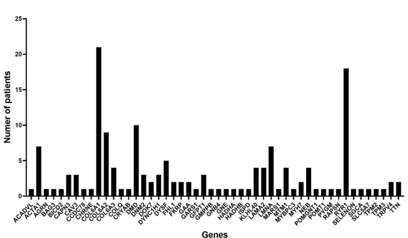
The causative mutations were found in 50 different genes (Figure 5) including ACADVL, ACTA1, AGRN, BAG3, BICD2, CAPN3, CAV3, CCDC78, CHRNE, COL6A1, COL6A2, COL6A3, COLQ, CRYAB, DMD, DNM2, DOK7, DYNC1H1, DYSF, FHL1, FKRP, GAA, GARS1, GFPT1, GMPPB, GNB4, GNE, HADHA, HADHB, ISPD, KLHL40, LAMA2, LMNA, MARS1, MTM1, MYBPC3, MYH7, NEB, POMGNT1, POMT1, PYGM, RAPSN, RYR1, SELENON, SGCA, SLC5A7, TPM2, TPM3, TRPV4, TTN. Only 24 genes were identified in two or more cases, of which 6 most frequently found genes were COL6A1 (n = 21), RYR1 (n = 18), DMD (n = 10), COL6A2 (n = 9), ACTA1 (n = 7), and LMNA (n = 7) in that order.
30
Figure 4. Fraction of diagnostic yields by phenotype groups
The total diagnostic yield was 49.2%. The diagnostic yield in CMD was 62.7%, LGMD was 45.3%, CMP was 54.3%, CMS was 80%, MM was 14.3%, MND was 35.3%, CMT was 50%, and OMN was 31.3%.
CMD, congenital muscular dystrophies; LGMD, limb girdle muscular dystrophies;
CMP, congenital myopathies; CMS, congenital myasthenic syndromes; MM, metabolic myopathies; MND, motor neuron disease; CMT, Charcot-Marie-Tooth disease; ONM, other neuromuscular disorders; P/LP, pathogenic and likely pathogenic variants identified; VUS, variants of uncertain significance
31
Figure 5. Diagnosis frequency in 50 genes with mutations
32
Genetically confirmed 161 patients were reclassified by integrating genotypes and phenotypes.
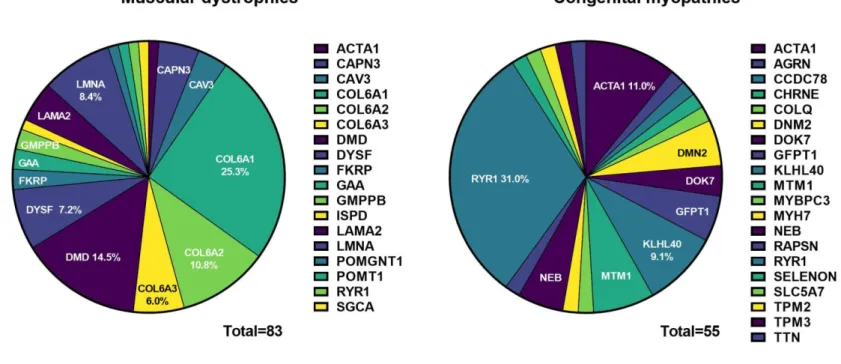
Muscular dystrophies
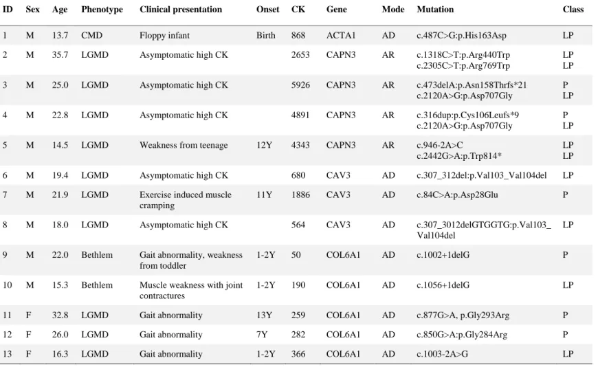
Eighty three patients were diagnosed with MD (Table 5). Pathogenic and likely pathogenic variants were identified in ACTA1 (n = 1), CAPN3 (n = 4), CAV3 (n = 3), COL6A1 (n = 21), COL6A2 (n = 9), COL6A3 (n = 5), DMD (n = 12), DYSF (n
= 6), FKRP (n = 2), GAA (n = 2), GMPPB (n = 2), ISPD (n = 1), LAMA2 (n = 4), LMNA (n = 7), POMGNT1 (n = 1), POMT1 (n = 1), RYR1 (n = 1), and SGCA (n = 1). Clinical features of all the patients were consistent with the detected genotypes.
Type 6 collagenopathies (COL6A1, 2, 3) and dystrophinopathies (DMD) are the most commonly diagnosed genes in MD (Figure 6).
33
Table 5. Pathogenic and likely pathogenic variants in 83 muscular dystrophy patients
ID Sex Age Phenotype Clinical presentation Onset CK Gene Mode Mutation Class
1 M 13.7 CMD Floppy infant Birth 868 ACTA1 AD c.487C>G:p.His163Asp LP
2 M 35.7 LGMD Asymptomatic high CK 2653 CAPN3 AR c.1318C>T:p.Arg440Trp c.2305C>T:p.Arg769Trp
LP LP 3 M 25.0 LGMD Asymptomatic high CK 5926 CAPN3 AR c.473delA:p.Asn158Thrfs*21
c.2120A>G:p.Asp707Gly
P LP
4 M 22.8 LGMD Asymptomatic high CK 4891 CAPN3 AR c.316dup:p.Cys106Leufs*9
c.2120A>G:p.Asp707Gly
P LP 5 M 14.5 LGMD Weakness from teenage 12Y 4343 CAPN3 AR c.946-2A>C
c.2442G>A:p.Trp814*
LP LP 6 M 19.4 LGMD Asymptomatic high CK 680 CAV3 AD c.307_312del:p.Val103_Val104del LP 7 M 21.9 LGMD Exercise induced muscle
cramping
11Y 1886 CAV3 AD c.84C>A:p.Asp28Glu P
8 M 18.0 LGMD Asymptomatic high CK 564 CAV3 AD c.307_3012delGTGGTG:p.Val103_
Val104del
LP
9 M 22.0 Bethlem Gait abnormality, weakness from toddler
1-2Y 50 COL6A1 AD c.1002+1delG P
10 M 15.3 Bethlem Muscle weakness with joint contractures
1-2Y 190 COL6A1 AD c.1056+1delG LP
11 F 32.8 LGMD Gait abnormality 13Y 259 COL6A1 AD c.877G>A, p.Gly293Arg P
12 F 26.0 LGMD Gait abnormality 7Y 282 COL6A1 AD c.850G>A:p.Gly284Arg P
13 F 16.3 LGMD Gait abnormality 1-2Y 366 COL6A1 AD c.1003-2A>G LP
34
14 M 19.7 UCMD Motor developmental delay <1Y 296 COL6A1 AD c.G868A:p.G290R P 15 M 22.1 UCMD Weakness with joint laxity <1Y 116 COL6A1 AD c.958-2A>G P 16 M 17.9 UCMD Weakness with joint laxity Birth 626 COL6A1 AD c.850G>A:p.Gly284Arg P 17 F 16.9 UCMD Gait abnormality, weakness
from toddler
1-2Y 350 COL6A1 AD c.877G>A:p.Gly293Arg P
18 M 20.8 UCMD Floppy infant <1Y 259 COL6A1 AD c.958-2A>G P
19 M 18.6 UCMD Gait abnormality, weakness from toddler
1-2Y 161 COL6A1 AD c.G814A:p.Gly272Ser LP
20 F 15.5 UCMD Hypotonia with joint laxity Birth 119 COL6A1 AD c.1461+3G>C LP
21 M 17.7 UCMD Floppy infant Birth 144 COL6A1 AD c.859-2A>G P
22 M 20.4 UCMD Floppy infant Birth 232 COL6A1 AD c.850G>A:p.Gly284Arg P
23 F 9.3 UCMD Floppy infant <1Y 332 COL6A1 AD c.850G>A:p.Gly284Arg P
24 F 10.6 UCMD Motor developmental delay <1Y 133 COL6A1 AD c.850G>A:p.Gly284Arg P 25 F 8.9 UCMD Motor developmental delay <1Y 297 COL6A1 AD c.850G>A:p.Gly284Arg P 26 M 13.1 UCMD Motor developmental delay <1Y 262 COL6A1 AD c.850G>A:p.Gly284Arg P 27 F 8.4 UCMD Motor developmental delay <1Y 286 COL6A1 AD c.877G>A, p.Gly293Arg P 28 F 14.9 UCMD Weakness with joint laxity 1-2Y 163 COL6A1 AD c.868G>A:p.Gly290Arg P 29 F 14.1 UCMD Weakness with joint laxity <1Y 223 COL6A1 AD c.833G>T:p.Gly278Val LP 30 M 33.1 LGMD Gait abnormality 13Y 287 COL6A2 AD c.349_350delinsAA:p.Ser117Asn LP 31 M 37.6 LGMD Gait abnormality 1-2Y 226 COL6A2 AD c.883G>A:p.Gly295Arg LP
32 M 14.0 LGMD Gait abnormality 6Y 358 COL6A2 AD c.736-2A>G LP
35
33 M 7.7 UCMD Floppy infant <1Y 173 COL6A2 AD c.1615C>T:p.Arg539Ter P
34 M 17.7 UCMD Floppy infant <1Y 247 COL6A2 AD c.875G>A:p.Gly292ASP
35 M 5.1 UCMD Gait abnormality 1-2Y 166 COL6A2 AD c.801+1G>A P
36 F 36.0 UCMD Motor developmental delay <1Y 62 COL6A2 AD c.801+1G>A P
37 F 36.4 LGMD Gait abnormality 12Y 678 COL6A2 AD /
AR
c.1189_1196del:p.G397fs c.2843C>G:p.Thr948Arg
LP VUS 38 M 18.8 UCMD Floppy infant <1Y 228 COL6A2 AR c.2386A>T:p.Lys796X
c.1816+1G>T
P LP 39 M 12.0 Bethlem Gait abnormality, distal
joint contractures.
1-2Y 142 COL6A3 AD c.4367A>G:p.Tyr1456Cys LP
40 F 14.1 UCMD Gait abnormality, weakness from toddler
1-2Y 377 COL6A3 AD c.6282+1G>C P
41 M 13.7 UCMD Motor developmental delay <1Y 308 COL6A3 AD c.6210+1G>A P 42 M 9.1 UCMD Motor developmental delay <1Y 88 COL6A3 AD c.4389+1G>A P 43 M 38.9 Bethlem Gait abnormality, high
arched palate, scoliosis.
2-3Y 164 COL6A3 AR c.1825C>T, p.Arg609*
c.6690+1G>A
P LP 44 M 25.3 BMD Exercise induced muscle
cramping
5Y 4866 DMD XR c.48G>A:p.Trp16Ter P
45 M 13.2 BMD Asymptomatic high CK 4985 DMD XR c.31+36947G>A P
46 M 11.6 BMD Asymptomatic high CK 7892 DMD XR exons 30 - 42 deletion P
47 M 8.2 BMD Asymptomatic high CK 3470 DMD XR c.23A>G:p.Glu8Gly LP
48 M 6.3 BMD Asymptomatic high CK 12708 DMD XR c.9959C>T:p.Pro3320Leu LP
49 M 10.8 DMD Gait abnormality 3Y 8900 DMD XR c.984C>T:p.Gln984Ter P
36
50 M 15.3 DMD Gait abnormality 6Y 5769 DMD XR c.2739G>T:p.Lys913Asn LP
51 M 9.9 DMD Motor developmental delay 1-2Y 7800 DMD XR c.9640A>T:p.Lys3214* LP
52 F 30.7 Carrier Asymptomatic high CK 2525 DMD XR c.5921+2T>A P
53 F 10.6 Carrier Asymptomatic high CK 7310 DMD XR c.433C>T:p.Arg145Ter P
54 F 12.0 Carrier Asymptomatic high CK 3372 DMD XR c.4001dup:p.Val1335Serfs*4 LP 55 F 13.2 Carrier Asymptomatic high CK 2536 DMD XR c.9450_9453del:p.Asn3152Valfs*2 LP
56 F 45.6 LGMD Gait abnormality 22Y 8482 DYSF AR c.663+1G>C
c.2997G>T:p.Trp999Cys
P LP
57 F 45.0 LGMD Gait abnormality 27Y 3246 DYSF AR c.1284+2T>C
c.2974T>C:p.Trp992Arg
P P
58 F 42.7 LGMD Gait abnormality 18Y 2033 DYSF AR c.663+1G>C
c.6057-2A>C
P P
59 M 20.3 LGMD Asymptomatic high CK 7888 DYSF AR c.663+1G>C
c.2997G>T:p.Trp999Cys
P LP 60 M 58.0 LGMD Gait abnormality 20Y NA DYSF AR c.2548C>T:p.Gln850Ter
c.3051G>T:p.Trp1017Cys
P LP 61 M 19.9 LGMD Gait abnormality 15Y 7407 DYSF AR c.2494C>T:p.Gln832*
c.2494C>T:p.Gln832*
P P
62 M 3.9 CMD Floppy infant Birth 7529 FKRP AR c.1170_1171delCG,
p.Gly391Leufs*72 c.1136G>C, p.Arg379Pro
P LP 63 F 5.7 LGMD Gait abnormality, weakness
from toddler
1-2Y 9546 FKRP AR c.501_502delinsCC:p.Arg167_Cys 168delinsSerArg
c.1176C>G:p.Phe392Leu
LP LP
37
64 M 26.4 LGMD Gait abnormality 8Y 661 GAA AR c.1316T>A:p.Met439Lys c.2238G>C:p.Trp746Cys
LP LP 65 M 19.3 LGMD Asymptomatic high CK 1920 GAA AR c.2015G>A:p.Arg672Gln
c.546G>T:p.Thr182=
P P 66 M 12.9 CMD Gait abnormality, weakness
from toddler
1-2Y 817 GMPPB AR c.391G>T:p.Gly131Cys c.391G>T:p.Gly131Cys
LP LP 67 M 4.5 CMD Floppy infant Birth 1014 GMPPB AR c.343T>C, p.Phe115Leu
c.787G>A, p.Gly263Ser
LP LP 68 M 18.1 CMD Floppy infant <1Y 4710 ISPD AR c.894delT:p.Phe298fs*7
c.964_966del:p.Val322del
P LP 69 M 27.5 CMD Motor developmental delay <1Y 845 LAMA2 AR c.595T>C:p.Cys199Arg
c.7605delT:p.Pro2535fs
LP P 70 F 10.8 CMD Floppy infant Birth 5307 LAMA2 AR c.4987C>T:p.Gln1663*
c.5974G>T:p.Glu1992*
P P 71 M 9.2 CMD Motor developmental delay <1Y 2951 LAMA2 AR c.648delC, p.Ser217Leufs
c.7156-4A>G
P LP 72 F 14.9 CMD Floppy infant <1Y 2623 LAMA2 AR c.5866-2A>G
c.8952delG:p.Met2984*
P LP 73 M 18.1 CMD Weakness with ankle
contractures, neck muscle weakness
1-2Y 1071 LMNA AD c.1406T>C:p.Ile469Thr P
74 F 17.3 CMD Gait abnormality 1-2Y 1081 LMNA AD c.149G>C:p.Arg50Pro P
75 F 13.2 CMD Weakness with ankle contractures
<1Y 975 LMNA AD c.745C>T:p.Arg249Trp P
76 F 10.7 CMD Motor developmental delay <1Y 1651 LMNA AD c.1156A>T:p.Arg386Trp LP
38 77 M 6.7 CMD Gait abnormality, weakness <