저작자표시-비영리-변경금지 2.0 대한민국 이용자는 아래의 조건을 따르는 경우에 한하여 자유롭게
l 이 저작물을 복제, 배포, 전송, 전시, 공연 및 방송할 수 있습니다. 다음과 같은 조건을 따라야 합니다:
l 귀하는, 이 저작물의 재이용이나 배포의 경우, 이 저작물에 적용된 이용허락조건 을 명확하게 나타내어야 합니다.
l 저작권자로부터 별도의 허가를 받으면 이러한 조건들은 적용되지 않습니다.
저작권법에 따른 이용자의 권리는 위의 내용에 의하여 영향을 받지 않습니다. 이것은 이용허락규약(Legal Code)을 이해하기 쉽게 요약한 것입니다.
Disclaimer
저작자표시. 귀하는 원저작자를 표시하여야 합니다.
비영리. 귀하는 이 저작물을 영리 목적으로 이용할 수 없습니다.
변경금지. 귀하는 이 저작물을 개작, 변형 또는 가공할 수 없습니다.
약학박사학위논문
Role of SAMHD1 Acetylation in Its dNTPase Activity and Cancer Cell Proliferation
SAMHD1 아세틸화가 뉴클레오티드 3 인산 분해능과 암세포 분열에 미치는 영향 연구
2017년 8월
서울대학교 대학원
ABSTRACT
Role of SAMHD1 acetylation in its dNTPase activity and cancer cell proliferation
Eun ji Lee Division of Pharmaceutical Bioscience College of Pharmacy The Graduate School Seoul National University
SAMHD1 is a deoxynucleotide triphosphohydrolase (dNTPase) which inhibits retroviruses by depleting intracellular deoxynucleotide triphosphates (dNTPs) in noncycling myeloid cells. Although SAMHD1 is expressed ubiquitously throughout the human body, the molecular mechanism to directly regulate its enzymatic activity and its function in non-immune cells are relatively unexplored. Here, I demonstrate that the dNTPase activity of
SAMHD1 is regulated by acetylation, which promotes cell cycle progression in cancer cells. SAMHD1 was acetylated at residue K405 by ARD1, an acetyltransferase, which directly enhanced its dNTPase activity in vitro.
When SAMHD1 wildtype and non-acetylated K405R mutant overexpressing stable cells were constructed in cancer cells, K405R mutant expressing cells showed decreased G1/S transition and slower proliferation over wildtype cells. SAMHD1 acetylation level was strongest during the G1 phase, implicating its role during G1 phase. Collectively, these findings suggest that SAMHD1 acetylation enhances its dNTPase activity and promotes cancer cell proliferation.
Keywords:SAMHD1/ acetylation/ dNTPase/ ARD1/ cell cycle/ cancer
Student number: 2013-30513
TABLE OF CONTENTS
ABSTRACT……… i
TABLE OF CONTENTS………….……..……… iii LIST OF FIGURES……… vii LIST OF TABLES……….. xii
LIST OF ABBREVIATIONS……… xiii
INTRODUCTION……….. 1
I. dNTPs……….. 1
II. SAMHD1………. 6
1. SAMHD1 is a dNTPase….……… 6
2. SAMHD1 and innate immunity………..……….. 10
3. SAMHD1 and cell growth ………..………. 13
4. Posttranslational modifications of SAMHD1…….……….. 15
III. ARD1………... 17
1. Protein
acetylation……….…… 17
2. ARD1 is an acetyltransferase………...……… 21 3. ARD1 is oncogenic………... 23
PURPOSE OF THIS
STUDY……… 25
MATERIALS AND METHODS………... 27
1.
Human liver tissue
samples……….. 27
2.
Cell Culture and
synchronization……….. 27
3.
in vitro acetylation
assay……… 28
4. Mass spectrometric analysis……… 28 5.
in vitro dNTPase
29
7. Transfection and stable cell line construction……… 31
8. Immunoblotting and immunoprecipitation……….. 32
9. Screening of binding proteins………. 32
10. Recombinant protein preparation……….. 33
11. Protein sequence alignment……… 34
12. Flow cytometry analysis……… 34
13. Cell proliferation assay……… 34
14. Colony formation assay……… 35
15. Fluorescence microscopy……… 35
16. Statistical analysis……… 36
RESULTS………... 37
1. S A M H D i s a n o v e l a c e t y l a t i o n s u b s t r a t e o f A R D 1 … … … … … … 37 2. K 4 0 5 r e s i d u e o f S A M H D 1 i s a c e t y l a t e d b y A R D 1 … … … … … … 45 3. ARD1-mediated SAMHD1 acetylation enhances its dNTPase activity……… 56 4. SAMHD1 acetylation is important for cell proliferation in
v a r i o u s c a n c e r c e l l s
… … … … 64
5. SAMHD1 acetylation promotes G1/S transition in cancer
cells……… 77
6. SAMHD1 acetylation may play other undiscovered roles in v a r i o u s c e l l l i n e s
… … … … 94
DISCUSSION ……… 103
CONCLUSION
………….……..………...
108
REFERENCES ……….. 110
ABSTRACT IN KOREAN (국문초록)
………
119
LIST OF FIGURES
Figure 1. Structural elements of three
nucleotides……… 3
Figure 2. Schematic representation of dNTP levels throughout cell cycle
progression……… 4
Figure 3. RNR and de novo dNTP
biosynthesis……… 5
Figure 4. Representative schematic or the important domains of
SAMHD1……… 8
Figure 5. Schematic illustration of the mechanism of SAMHD1 activation by dGTP-induced tetramerization……… 9 Figure 6. Effect of SAMHD1 on HIV-1 replication……… 11 Figure 7. SAMHD1 expression change during cell proliferation……… 14
Figure 8. Model for the regulation of SAMHD1 restriction activity by phosphorylation at the Thr592 residue……… 16 Figure 9. Comparison of two types of protein
acetylation……… 20
Figure 10. Schematics for functional switch of Hsp70 regulated by ARD1- mediated acetylation……… 2 Figure 11. Schematic diagram showing the contribution of ARD1 in cell
cycle progression……… 24 Figure 12. LC-MS/MS analysis reveals SAMHD1 as a novel binding
partner of ARD1……… 40 Figure 13. Co-immunoprecipitation of endogenous SAMHD1 and
FLAG-ARD1……… 41
Figure 14. Recombinant SAMHD1 protein is acetylated by ARD1 in
vitro……… 42
Figure 15. Endogenous SAMHD1 protein is acetylated by ARD1 in vivo 43 Figure 16. Endogenous SAMHD1 protein is less acetylated by ARD1
mutants in vivo……… 44 Figure 17. Construction of SAMHD1 deletion mutants……… 48 Figure 18. Recombinant CTD protein of SAMHD1 was mainly
acetylated by ARD1 in vitro……… 49
Figure 20. K405 is the major site of ARD1-mediated acetylation of
SAMHD1 in vitro……… 52
Figure 21. K405 is the major site of ARD1-mediated acetylation of
SAMHD in vivo……… 53
Figure 22. Sequence alignment of SAMHD1 K405 residue in various
species……… 54
Figure 23. Location of K405 residue in SAMHD1 structure……… 55 Figure 24. Schematic representation of in vitrodNTPase assay………… 58 Figure 25. SAMHD1 acetylation upregulates its dNTPase activity in
vitro 59
Figure 26. SAMHD1 K405R mutant does not show upregulated dNTPase activity in the presence of ARD1 in
vitro……… 60
Figure 27. SAMHD1 WT shows stronger dNTPase activity of K405R mutant in vivo……… 61 Figure 28. SAMHD1 WT shows stronger dNTPase activity in the
presence of ARD1 in
vivo……… 62
Figure 29. Expression of SAMHD1 in hepatocarcinoma tissues and
surrounding normal
tissues……… 67
Figure 30. Establishment of SAMHD1-downregulated cancer cells…… 68 Figure 31. Relative cell growth of SAMHD1-downregulated HeLa cells 69
Figure 32. SAMHD1-downregulated MCF7 cells show decreased anchorage-dependent colony formation……… 70 Figure 33. Establishment of FLAG-SAMHD1 WT or K405R
overexpressing stable cancer cell lines……… 71 Figure 34. Relative cell growth of FLAG-SAMHD1 WT or K405R
overexpressing HeLa cells……… 72 Figure 35. Anchorage-dependent colony formation of FLAG-SAMHD1
expressing stable MCF7
cells……… 73
Figure 36. Anchorage-dependent colony formation of FLAG-SAMHD1 expressing stable A549 cells……… 74 Figure 37. Anchorage-dependent colony formation of FLAG-SAMHD1
expressing stable Hep3B cells……… 75 Figure 38. Anchorage-independent colony formation of FLAG-
SAMHD1 expressing stable HeLa cells……… 76 Figure 39. Cell cycle profiles of SAMHD1-downregulated HeLa cells… 81 Figure 40. Expression levels of cyclins in SAMHD1-downregulated
HeLa cells……… 82
Figure 41. Cell cycle profiles of FLAG-SAMHD1 expressing stable
A549 cells……… 83
Figure 42. Expression levels of cyclins in FLAG-SAMHD1
Figure 43. PCNA expression of FLAG-SAMHD1 overexpressing stable
Hep3B cells……… 85
Figure 44. SAMHD1 acetylation levels during cell cycle progression of FLAG-SAMHD1 overexpressing stable A549 cells………… 87 Figure 45. SAMHD1 acetylation levels during cell cycle progression of
FLAG-SAMHD1 overexpressing stable HeLa cells………… 88 Figure 46. Expression levels of endogenous SAMHD1 during cell cycle
progression of HeLa cells……… 89 Figure 47. Expression levels of endogenous SAMHD1 during cell cycle
progression of Hep3B
cells……… 90
Figure 48. Expression levels of endogenous SAMHD1 during cell cycle progression of A549 cells……… 91 Figure 49. Expression levels of endogenous SAMHD1 during cell cycle
progression of MCF7 cells……… 92 Figure 50. SAMHD1 acetylation does not alter SAMHD1-cyclin A2
binding……… 93
Figure 51. Physiological conditions of SAMHD1 acetylation in THP-1
cells……… 96
Figure 52. Relationship between SAMHD1 and autophagy……… 99 Figure 53. SAMHD1 acetylation alters the expression of junction
proteins in various cancer cells……… 102
Figure 54. Schematic for the mechanism of ARD1-mediated SAMHD1 acetylation in cancer cell proliferation……… 107
LIST OF TABLES
Table 1. Clinicopathological characteristics of 7 hepatocarcinoma
patients ……… 66
LIST OF ABBRIVIATIONS
AGS Aicardi-Goutières syndrome AR Androgen receptor
ARD1 Arrest defective 1
CDK1 Cyclin-dependent kinase 1 CTD C-terminal domain
dATP Deoxyadenosine triphosphate
DCIP Dendritic cell-derived IFN-γ induced protein dCTP Deoxycytidine triphosphate
dGTP Deoxyguanine triphosphate dNTP Deoxyribonucleotide triphosphate dNTPase Deoxynucleotide triphosphohydrolase dTTP Deoxyribonucleotide triphosphate HD Histidine-aspartic
HIV-1 human immunodeficiency virus-1 HSP70 Heat shock protein 70
IFNβ-2a Interferon β-2a
NAT Nα-terminal acetyltransferases PCNA proliferating cell nuclear antigen
RNR Ribonucleotide reductases
Runx2 Runt-related transcription factor 2 SAM Sterile alpha motif
SAMHD1 SAM domain and HD domain containing protein 1
INTRODUCTION
I. dNTPs
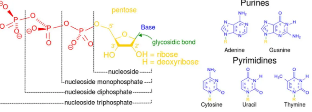
Deoxyribonucleotide triphosphate (dNTP) is a molecule which contains a phosphate group, a deoxyribose sugar and a nitrogenous base (Figure 1). 4 types of dNTPs exist depending on the nitrogenous base they contain; dATP, (deoxyadenosine triphosphate), and dGTP, (deoxyguanine triphosphate), dTTP, (deoxythymidine triphosphate), and dCTP, (deoxycytidine triphosphate). They are necessary for life, as they are the precursors of DNA synthesis. According to base pairing rules (A with T, and C with G), the nitrogenous bases of the two separate dNTPs are bound together with hydrogen bonds to build double-stranded DNA.
A strict balance in dNTP pool sizes is critical for proper DNA replication
Stillman, 2007) (Figure 2). This fluctuation needs to be within a range optimal for chromosomal replication, as imbalances can impede DNA replication and repair resulting in DNA damage, cell cycle arrest or cell death (Mathews, 2006). Thus eukaryotes use diverse mechanisms to strictly regulate the supply of dNTPs (Pai and Kearsey, 2017). Ribonucleotide reductases (RNR) are the major contributors to the pool as they synthesize the dNTPs from ribonucleoside diphosphates (Figure 3) (Nordlund and Reichard, 2006). RNRs have long been recognized as an attractive target for cancer treatment, for their dysregulated activity is associated with genomic instability, malignant transformation and cancer development (Mathews, 2015).
Figure 1. Structural elements of three nucleotides. Each in turn is attached to the nucleoside (in yellow, blue, green) at center: 1st, the nucleotide termed as anucleoside monophosphate nucleotideis formed by adding a phosphate group (in red); 2nd, adding a second phosphate group forms anucleoside diphosphate nucleotide; 3rd, adding a third phosphate group results in anucleoside triphosphate nucleotide. + The nitrogenous base (nucleobase) is indicated by "Base" and "glycosidic bond" (sugar bond). All five primary, or canonical, bases—the purines and pyrimidines—are sketched at right (in blue).
Figure 2. Schematic representation of dNTP levels throughout cell cycle progression. Highest dNTP levels are seen during S phase wheras lowest are during G1 phase. The Low G1-phase dNTP levels contribute to the G1 checkpoint. (Franzolin et al., 2013)
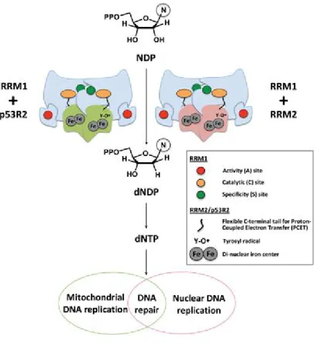
Figure 3. RNR and de novo dNTP biosynthesis. Mammalian RNR enzymes function to reduce the 2' carbon of NDPs to generate dNDPs that are subsequently phosphorylated by nucleoside diphosphate kinase, yielding
II. SAMHD1
1. SAMHD1 is a dNTPase
An additional mode of dNTP pool control was discovered in 2011 as two groups reported a novel enzyme SAMHD1, Sterile alpha motif (SAM) domain and Histidine-aspartic (HD) domain containing protein 1. It is expressed ubiquitously throughout human body, and mainly in the nucleus at cell level. It is the first deoxynucleotide triphosphohydrolase (dNTPase) discovered in mammalian cells, as it depletes the cellular dNTP levels by cleaving them to respective deoxyribonucleosides plus tripolyphosphate (Figure 4) (Goldstone et al., 2011, Powell et al., 2011). This activity of SAMHD1 resides in its histidine-aspartate (HD) domain, while the N- terminal sterile alpha motif (SAM) domain exhibit other activities including substrate-binding. dGTP is reported to be its cofactor, as it binds to the allosteric sites of SAMHD1 and promotes tetramerization (Figure 5). Unlike inactive monomers or dimers, SAMHD1 tetramers show high enzymatic activities and facilitate various biological activities. In addition to the
dNTPase activity, SAMHD1 is reported to have RNase and nuclease activities, although further validations are required.
Figure 4. Representative schematic of the important domains of SAMHD1. The enzymatic activity of SAMHD1 is promoted by the HD domain. SAMHD1 degrades dNTPs into deoxynucleosides (dN) and inorganic triphosphates (PPP). (Rossi, 2014)
Figure 5. Schematic illustration of the mechanism of SAMHD1 activation by allosteric dGTP-induced tetramerization. Subunits of SAMHD1 are labeled A, B, C and D, and the catalytically inactive monomers and dimers are shown with empty substrate-binding pockets.
Binding of four dGTP pairs at the allosteric sites promotes tetramerization and induces large conformational changes at the tetramer interface (AC and
2. SAMHD1 and innate immunity
Originally discovered as a component of the human innate immune system, SAMHD1 was first named as dendritic cell-derived IFN-γ induced protein (DCIP) as its levels were increased in response to IFN-γ treatment in dendritic cells (Li et al., 2000). Later studies revealed that mutations in SAMHD1 causes Aicardi-Goutières syndrome (AGS), a genetic encephalopathy whose symptoms mimic congenital infection (Rice et al., 2009). However, the most widely-known function of SAMHD1 is its anti- HIV-1 restriction activity. Observed in myeloid cells as well as in quiescent CD4+ T cells, SAMHD1 depletes the level of cellular dNTPs to the levels insufficient for reverse transcription, therefore results in blockage of early- stage retroviral replication (Figure 6) (Laguette et al., 2011, Hrecka et al., 2011, Baldauf et al., 2012). Together, these findings suggest new ways to interfere with the immune evasion and immune cell pathology of pandemic HIV-1.
Figure 6. Effect of SAMHD1 on HIV-1 replication. After entry into the target host cell, viral RNA (vRNA) is converted into viral DNA (vDNA) by the viral reverse transcriptase, and the resulting provirus is integrated into the host nucleus. HIV-1 relies on cellular dNTPs for reverse transcription.
Various cellular enzymes that target nucleic acids either positively or negatively affect the early steps of viral replication. SAMHD1, which is located in the nucleus, hydrolyses dNTPs in non-cycling cells, thereby limiting HIV-1 replication. In the presence of SAMHD1, HIV-1 reverse transcription is severely impaired and levels of replication are very low.
(Ayinde et al., 2012)
3. SAMHD1 and cell growth
Like RNR, which controls cell cycle progression by contributing to dNTP pools, SAMHD1 also plays role in cell cycle progression. SAMHD1 stimulates cell proliferation in lung fibroblasts and THP-1 cells (Franzolin et al., 2013, Bonifati et al., 2016). SAMHD1 was variably expressed during the cell cycle, maximally during quiescence and minimally during S phase (Figure 7). Low dNTP levels during G1 phase caused by high levels of SAMHD1 facilitated G1/S transition and resulted in increased cell proliferation in fibroblasts.
Overall, as SAMHD1 controls the size of dNTP pool and cell cycle progression, there is a high possibility of its connection to cancer, similarly to RNRs. Indeed, SAMHD1 is reported to be frequently mutated in chronic lymphocytic leukemia and colon cancers (Clifford et al., 2014, Rentoft et al.,
Figure 7. SAMHD1 expression change during cell proliferation.
Abundance of SAMHD1 (x), and two subunits of RNR, R2 (filled square), and p53R2 (filled triangle) at the indicated days of culture without siRNA.
Left reports in aribitrary units the quantification of the immunoblots on the right. Notice the parallelism between R2 expression and frequency of S- phase cells (open circle) in the cultures. (Franzolin et al., 2013)
4. Posttranslational modifications of SAMHD1
Posttranslational modifications (PTMs) are the chemical modifications of a protein after its translation, which highly increases the functional diversity of the protein (Khoury et al., 2011). A variety of PTMs exist in mammalian systems, including phosphorylation, glycosylation and acetylation. In 2013, two groups independently reported the phosphorylation of SAMHD1. In cycling immune cells, CDK1 (cyclin-dependent kinase 1) and cyclin A2 phosphorylates SAMHD1 at its T592 residue, which keeps SAMHD1 inactive (Cribier et al., 2013, White et al., 2013). However, in non-cycling cells, SAMHD1 is dephosphorylated and shows anti-viral restriction activity towards HIV-1 virus (Figure 8). However, this phosphorylation had no direct influence on the ability of SAMHD1 to decrease dNTP levelsin vitro. These results raises the possibility that unknown forms of PTMs or non-cycling
Figure 8. Model for the regulation of SAMHD1 restriction activity by phosphorylation at the Thr592 residue. SAMHD1 phosphorylation occurs at T592 residue by CDK1 and cyclin A2 in proliferative T-cells.
Phosphorylated SAMHD1 shows no anti-HIV-1 activity (Cribier et al., 2013).
III. ARD1
1. Protein acetylation
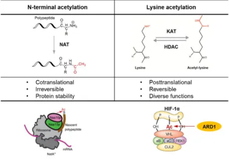
Protein acetylation is one of the major PTMs in cell signaling and metabolism in eukaryotes as its regulation is crucial for various important cellular processes (Drazic et al., 2016, Choudhary et al., 2009, Zhao et al., 2010). It is catalyzed by acetyltransferases, which transfer acetyl groups from the donor acetyl-CoA to the target proteins. Two types of acetylation have been reported, Nα-terminal acetylation and Nε-lysine acetylation (Figure 9). Nα-terminal acetylation is mediated by NATs (Nα-terminal acetyltransferases), as they acetylate the α-amino group of N-terminal residues of proteins. This is one of the most common protein modifications
Lee et al., 1989). Nα-terminal acetylation is known to be co-translational and irreversible, contributing to protein stability and localization. NATs associated to the ribosome acetylates the N-terminal α-amino group of the nascent polypeptide newly synthesized and extruded from the ribosome.
(Driessen et al., 1985).
Nε-lysine acetylation, on the other hand, is less common, but a very important form of protein acetylation which occurs post-translationally on the ε-amino group of lysines. When an acetyl group is added onto lysines, the positive charges on its amino group is neutralized, which results in a significant change in the electrostatic properties of the protein. This type of acetylation is crucial for regulating diverse functions of proteins, such as protein-protein interaction or protein stability (Glozak et al., 2005). Unlike the Nα-terminal acetylation, this form of acetylation is reversible; therefore the acetylation/ deacetylation of a specific residues are reported to act as important switches in the regulation of gene transcription, cellular growth and differentiation.
Many acetyltransferases can regulate their catalytic activity by acetylating themselves (auto-acetylation). For example, p300, an acetyltransferase, is highly auto-acetylated in its activation loop, which results in its enhanced transcriptional activity (Karanam et al., 2006).
Figure 9. Comparison of two types of protein acetylation. N-terminal acetylation and lysine acetylation are compared. ARD1 shows both of the acetylation activities. (Jeong et al., 2002; Starheimm KK et al., 2012; Sharp FR et al., 2004)
2. ARD1 is an acetyltransferase
Arrest defective 1 (ARD1) is an acetyltransferase. Interestingly, it is known to have both Nα-terminal acetylation and Lysine-acetylation activities (ref) (Figure 9). First discovered in Saccharomyces cerevisiae (Lee et al, 1989;
Park and Szostak et al, 1990), ARD1 associates with NAT1 to form NAT complex, and acts as a catalytic component in yeast (Park and Szostak et al, 1992).
Later studies have discovered various substrates of ARD1. Runt-related transcription factor 2 (Runx2) is known to transactivate many genes required for osteoblast differentiation. ARD1 acetylates this protein to block this transactivation, controlling balanced osteogenesis (Yoon et al., 2014). Heat shock protein 70 (HSP70) is also a recently-discovered substrate of ARD1.
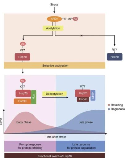
HSP70 acetylation acts as a functional switch to alter its activity from protein refolding to degradation (Figure 10) (Seo et al., 2016). Moreover, ARD1 is also auto-acetylated, which promotes the acetyltransferase activity of itself (Seo et al., 2010). Altogether, these reports demonstrate that ARD1-mediated acetylation acts as an important switch of various substrates to alter their
modes of action.
Figure 10. Schematics for functional switch of Hsp70 regulated by ARD1-mediated acetylation. After stress, ARD1 and Hsp70 are sequentially activated by an acetylation cascade. ARD1 selectively acetylates
deacetylated Hsp70 contributes to protein degradation. (Seo et al., 2016) 3. ARD1 is oncogenic
When initially identified in yeast, ARD1 was reported as a mating-type switch that controls the mitotic cell cycle and alternative development (Whiteway and Szostak, 1985; Whiteway et al., 1987). Later, many studies have reported ARD1 to be oncogenic. It is highly expressed in several types of cancers including breast (Yu et al., 2009, Wang et al., 2011), prostate (Wang et al., 2012), lung and colorectal (Xu et al., 2012). Moreover, various molecular mechanisms of ARD1 were discovered to promote tumorigenesis.
For example, ARD1 is proposed to promote the lung cancer cell proliferation by acetylating b-catenin (Lim et al., 2006; Lim et al, 2008). In prostate cancer, ARD1 activates androgen receptor (AR) by acetylation to promote prostate tumorigenesis (Wang et al., 2012).
Recent study has demonstrated that the nuclear translocation of ARD1 is required for cell cycle progression (Figure 11) (Park et al., 2014). In proliferating cells, ARD1 is imported to the nuclei especially during S phase, contributing to proper cell proliferation in HeLa and A549 cancer cell lines.
Altogether, these growing evidence suggest that ARD1 contributes to cancer proliferation.
Figure 11. Schematic diagram showing the contribution of ARD1 in cell cycle progression.(Park et al., 2014)
PURPOSE OF THIS STUDY
SAMHD1 is the first dNTPase discovered in human cells which contributes to anti-retroviral defense by lowering the cellular dNTP pool levels in non- cycling myeloid cells. This restrictive activity is not shown in cycling cells, implying the existence of posttranslational modifications or cofactors specifically expressed in non-cycling cells. However, molecular mechanisms that directly regulates its enzymatic activity has not been reported, as its phosphorylation did not alter its dNTPase activity in vitro. Moreover, previous studies were mainly limited to its functions in immune cells, despite its ubiquitous expression throughout the body. SAMHD1 was reported to facilitate G1-to-S phase progression in monocytes and fibroblasts, yet further studies on functions of SAMHD1 in other cell types are required.
In this study, I present SAMHD1 as a novel acetylation substrate of ARD1.
As protein acetylation can modulate the activities of the substrate proteins, I hypothesized that ARD1 may acetylate SAMHD1 to alter its dNTPase activity. Moreover, as ARD1 is reported to be oncogenic and the enzymatic activity of SAMHD1 is relevant to the cell cycle regulation, I investigated
whether the ARD1-mediated SAMHD1 acetylation contribute to cancer cell proliferation. By identifying the specific acetylation site of SAMHD1 and mutating it to block its acetylation, I aimed to discover a novel function of SAMHD1 acetylation in cancer cell growth.
MATERIALS AND METHODS
1. Human liver tissue samples
Hepatocarcinoma tissues and surrounding non-tumorous liver samples were obtained from patients at Asan Hospital (Seoul, Republic of Korea).
Clinicopathological characteristics of hepatocarcinoma patients are summarized in Table S1. This study was approved by Institutional Review Board of Asan Hospital, and written informed consent was obtained from all the patients.
2. Cell culture and synchronization
HeLa, A549, Hep3B, MCF-7 and HEK293T cells were grown in DMEM supplemented with 10% fetal bovine serum (FBS) and 1%
penicillin/streptomycin in 5% CO2 humidified atmosphere at 37°C. For serum starvation, cells were incubated in DMEM without serum for 48 h and
was performed to synchronize cell populations in S phase. Cells were treated with 2 mM thymidine (Sigma-Aldrich) for 18 h, cultured for 9 h in normal growth medium, and then retreated with thymidine for another 16 h. After removing the thymidine, cells were released to normal medium and harvested at the indicated times.
3. in vitroacetylation assay
Acetylation assay was performed as described previously [19]. Briefly, 1 μg of purified recombinants or precipitated cellular proteins were incubated in the reaction mixture containing 50 mM Tris–HCl (pH 8), 0.1 mM EDTA, 1 mM DTT, 10% glycerol and 10 mM acetyl-CoA at 37 °C for 1.5 h.
Reaction products were separated by SDS–PAGE and were analyzed by western blot using anti-Lys-Ac antibody. Input proteins were visually quantified using Coomassie brilliant blue staining or Ponceau S staining.
4. Mass spectrometric analysis
used in the in vitro acetylation assay (incubated with ±ARD1) were immediately denatured using 6 M GuHCl and were reduced with 5 mM DTT for 30 min at room temperature, then alkylated with 25 mM iodoacetamide for 30 min. Glu-C digestions were carried out for 6 h at 25 °C then were quenched by 5% formic acid. Micro RPLC–MS/MS analysis was performed as previously described [22].
5. in vitrodNTPase assay
An enzymatic assay based on thin-layer chromatography was performed as described previously [9]. The purified recombinant or immunoprecipated cellular protein (1 μg) was incubated in reaction mixture composed of 50 mM Tris-Cl (pH 8.0), 20 mM KCl, 5 mM MgCl2, 0.1 μCi [α-32P]dTTP, and 200 μM ice-cold dGTP for 3 h at 37 °C. The reactions were stopped by heat inactivation at 70 °C for 10 min. Mixtures were blotted onto polyethyleneimine (PEI)-cellulose plates (Sigma-Aldrich) and subsequently separated using a mobile phase of 1.2 M LiCl. After separation, the α-32P- labeled reaction products were visualized using a phosphorimager.
6. siRNA and plasmid construction
The siRNA sequence targeting human SAMHD1 corresponds to GAUUCAUUGUGGCCAUAUA as previously described [9]. Full-length of cDNAs for human SAMHD1 (Genbank: NM_015474) and human ARD1 (Genbank: NM_003491.3) were obtained from PCR and subcloned into pCDNA3.1 (FLAG-SAMHD1), pCMV-tag2b (FLAG-ARD1), pEGFP-C3 (GFP-ARD1) and pCS2+ (MYC-ARD1) vectors for cellular expression or pGEX-4T (GST-SAMHD1) and pET28a (His-ARD1) vectors for the bacterial induction of recombinant proteins. Deletion mutants of GST- SAMHD1 were constructed from pGEX-4T-SAMHD1 plasmid. cDNAs of SAMHD1 corresponding to 34–113 aa, 160–339 aa and 334–626 aa were amplified by PCR and inserted into pGEX-4T-1. For construction of stable cell lines, cDNAs of SAMHD1 was co-inserted into pMX-IRES- BlasticidinR vector with PCR-amplified FLAG. Point mutations in SAMHD1 (K405R and K580R) and ARD1 (K136R and DN: R82A/Y122F) were generated using the Muta-Direct Site Directed Mutagenesis kit (Intron) according to the manufacturer’s instructions. The following primers and its
SAMHD1 K405R: TACAGGTGCTGGAGGcggAAAGTATCGCATTTC SAMHD1 K580R: GACAGAAATTTCACCcgGCCGCAGGATGGCGAT ARD1 K136R: AGTGAAGTGGAGCCCAgATACTATGCAGATGGG ARD1 R82A: TGTGAAGCGTTCCCACgcGCGCCTCGGTCTGGCT ARD1 Y122F: GCCGCCCTGCACCTCTtTTCCAACACCCTCAAC
7. Transfection and Stable Cell Line Construction
Transfection was performed as described previously [22]. I transfected HEK293T cell with polyethyleneimine (PEI) at a ratio of 3:1 (μl PEI/μg plasmid DAN) in basal media overnight. siRNA targeting SAMHD1 was transfected with HiPerFect reagent (Quiagen) according to the manufacture’s instruction.
For the establishment of stable cells, pIRES-FLAG-SAMHD1 plasmids were transfected into Platinum A cells and retroviral supernatants were harvested 72 h after transfection. Collected supernatants were used to transduce target cell lines (HeLa, A549, Hep3B and MCF-7) with polybrene (10 μg ml−1).
Transduced cells were selected using blasticidin (10 μg ml−1). Expression of
FLAG-SAMHD1 was quantified by western blot.
8. Immunoblotting and immunoprecipitation
Cellular proteins were extracted using cell lysis buffer composed of 20 mM Tris-CL (pH 7.5), 150 mM NaCl, 0.1 mM EDTA, 0.1% Triton X-100 and a protease inhibitor cocktail (Roche). 20 ~50 μg of cell extracts were used for immunoblotting. For immunoprecipitation, 1 mg of proteins were incubated with corresponding primary antibody conjugated to A or G bead (Upstate) or Anti-DDDDK-tag mAb-Magnetic Agarose (MBL) overnight at 4 °C. Beads were washed three times with washing buffer containing 20 mM Tris-Cl (pH 7.5), 150 mM NaCl and 0.1 mM EDTA. After SDS–PAGE, membranes were immunoblotted using the corresponding primary antibody overnight at 4 °C.
HRP-conjugated secondary antibodies were incubated with the membranes for 1 h at room temperature. Visualization was performed using ECL Plus (Intron) and LAS-4000 (GE Healthcare).
[22]. HEK293T cells were transiently transfected with FLAG-HA tagged ARD1 and were lysed in lysis buffer consisting 20 mM Tris-Cl (pH 7.5), 150 mM NaCl, 0.1 mM EDTA, 0.2% Triton X-100 and a protease inhibitor cocktail (Roche). Cell lysates were incubated with anti-M2 resin (Sigma) for 2 h at 4 °C, then resins were collected by centrifugation and washed three times with washing buffer, composed of 20 mM Tris-Cl (pH 7.5), 150 mM NaCl and 0.1 mM EDTA. Bound proteins were eluted by 3 × FLAG-peptide and were immunoprecipitated again using anti-HA-antibody for 2 h at 4 °C.
The bound proteins were analyzed by SDS–PAGE and silver staining.
10. Recombinant protein preparation
BL21 cells were transformed with plasmids pGEX-4T-1-SAMHD1 or pET28a-ARD1 and were grown to and OD600 of 0.6–0.8. 1 mM IPTG was added to induce GST-tagged SAMHD1 or His-tagged ARD1, then cells were grown overnight at 20 ℃. Cells were collected and proteins were extracted with a lysis buffer consisting 50 mM Tris–HCl (pH 8), 250 mM NaCl, 2.5 mM EDTA and 1% Triton X-100. GST-tagged and His-tagged proteins were purified with Glutathione Sepharose 4B (GE Healthcare) and TALON®
eluted with an elution buffer containing 50 mM Tris–HCl (pH 8) and 10 mM reduced glutathione (GSH).
11. Protein sequence alignment
SAMHD1 multiple protein sequence alignment was conducted by Constraint-based Multiple Alignment Tool provided from NCBI.
12. Flow cytometry analysis
For a flow cytometry assay, cells were collected, fixed in 1% PFA, and stored at 4°C for 30 min. Cells were then washed with PBS and stained with propidium iodide (PI, 30 μg ml−1) mixed with RNase A (1 μg ml−1) for 15 min at 37℃. The DNA content of cells was assessed with a FACS-Verse system (BD Biosciences), and cell cycle profiles were analyzed with the BD FACSuite software. At least 30,000 cells in each sample were analyzed to obtain a measurable signal, using the same instrument setting.
The proliferation rates were measured using a Cell Proliferation Assay kit (Promega) following the manufacturer's instructions. Briefly, 2×10³ cells/well were seeded onto 96-well plates and were allowed to grow. At indicated time points, 20 µL of substrate solution was added to the cells and were incubated for 1 h for color development. The absorbance at 492 nm was measured to indicate the number of proliferating cells.
14. Colony formation assay
Colony formation assays were performed as described previously [31]. For anchorage dependent colony formation, cells were seeded at a density of 100 cells/well onto 6-well plates and were allowed to grow for 2 weeks. Cells were then fixed with 100% methanol, stained with 0.005% crystal violet and the number of colonies were counted. For anchorage independent colony formation, cells were resuspended in 0.5 mL DMEM containing 0.4% agar and were seeded onto a layer of 1% agar in 12-well plates. After 2 weeks of incubation, colonies were stained with 0.005% crystal violet and counted.
15. Fluorescence microscopy
For the analysis of PCNA by flourescence microscopy, cells were seeded onto the glass coverslips in 24-well plates. After synchronization, cells were fixed in 2% PFA for 20 min and were permeabilized in 1% triton X-100 in PBS 10 min at room temperature. Then, cells were incubated with PCNA antibody (Cell signaling) and visualized with Alexa 488-conjugated igG (Molecular Probes). Nucleus staining was performed with Hoechst 33342 (Molecular Probes). The immunofluorescence was visualized using a confocal microscopy (Carl Zeiss, LSM700).
16. Statistical analysis
Results are expressed as the mean’s±s.d’s or±s.e.m’s. P values were calculated by applying the two-tailed Student’s t test or one-way ANOVA. A difference was considered statistically significant at a value of P<0.05.
RESULTS
1. SAMHD1 is a novel acetylation substrate of ARD1
As ARD1 is an acetyltransferase, I first aimed to find its novel substrates to study the functions of ARD1. For this I have screened for ARD1-binding proteins using affinity purification combined with mass spectrometry.
FLAG/HA-tagged ARD1 proteins were overexpressed in HEK293T cells, then immunoprecipitated overnight. The resin-bound proteins were separated by SDS-PAGE and the gel was silver stained for protein identification.
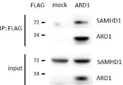
Around 72 kDa, two strong bands were observed; one of them was identified to be HSP70 protein as previously reported (Seo et al., 2016). The other anonymous band was subjected to mass spectrometry for identification, which was revealed to be endogenous SAMHD1 protein (Figure 12). To confirm this result, FLAG mock or ARD1 expressing vectors were transfected to HEK293T cells and the lysates were immunoprecipitated using
FLAG-specific resin. When precipitates were separated by SDS-PAGE and immunoblotted by SAMHD1 specific antibody, the endogenous SAMHD1 proteins were found to be co-immunoprecipitated, in accordance to the previous result (Figure 13). These data demonstrates that SAMHD1 is a novel binding partner of ARD1.
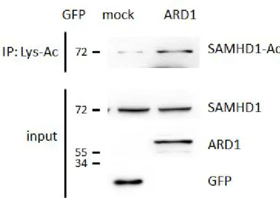
Next, as ARD1 is an acetyltransferase, I investigated whether it could acetylate SAMHD1. Recombinant GST-SAMHD1 proteins were induced and purified from E. coli, then subjected to an in vitroacetylation assay with or without His-ARD1 in the presence of Acetyl-CoA. The protein was found to be acetylated when His-ARD1 was present, demonstrating that ARD1 directly acetylates SAMHD1 in vitro (Figure 14). To test this acetylation in vivo, GFP-mock or ARD1 vectors were overexpressed in HEK293T cells.
The lysed proteins were incubated with Lys-Acetylation specific antibody to selectively immunoprecipitate Lys-Acetylated proteins. When the products were separated and blotted with SAMHD1 antibody, the intensity of endogenous SAMHD1 acetylated forms was observed to be stronger in GFP- ARD1 overexpressing cells than the mock cells, confirming the in vitrodata
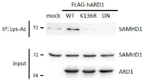
Our lab has previously reported that ARD1 is autoacetylated at K136 residue to enhance its catalytic activity (Seo et al., 2010). To test whether this affects SAMHD1 acetylation, I overexpressed K136R mutant FLAG- ARD1, which cannot be autoacetylated therefore shows less acetyltransferase activity. In addition, dominant negative (DN) mutants of ARD1 which does not exhibit any enzymatic activity at all were also expressed in HEK293T cells. When the Lys-acetylation intensity of endogenous SAMHD1 from these cells were evaluated, they were significantly decreased compared to the ARD1 wildtype expressing cells (Figure 16). This result indicates that the two ARD1 mutants blocked SAMHD1 acetylation; therefore it is suggested that ARD1-autoacetylation is required for SAMHD1 acetylation as an up- stream signal.
Figure 12. LC-MS/MS analysis reveals SAMHD1 as a novel binding partner of ARD1. HEK293T cells were transfected with FLAG/HA-ARD1
Figure 13. Co-immunoprecipitation of endogenous SAMHD1 and FLAG-ARD1. FLAG-ARD1 was immunoprecipitated from HEK293T cells, and co-precipitation of endogenous SAMHD1 was analyzed by western blot.
assay with ±His-ARD1. The acetylation level was determined with anti-Lys- Ac antibody.
Figure 15. Endogenous SAMHD1 protein is acetylated by ARD1 in vivo.
Lys-acetylated proteins were precipitated from GFP-mock or ARD1 expressing HEK293T cells and were blotted with anti-SAMHD1 antibody.
Figure 16. Endogenous SAMHD1 protein is less acetylated by ARD1 mutants in vivo. FLAG-ARD1 wildtype, K136R and DN mutants were
2. K405 residue of SAMHD1 is acetylated by ARD1.
Next, to identify the target site of acetylation, I constructed three types of deletion mutants of SAMHD1 according to their functional motifs; SAM domain (SAM), HD domain (HD) and C-terminal domain (CTD) (Figure 17).
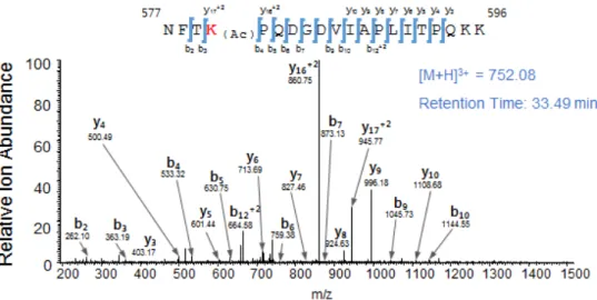
These vectors were transformed in to E.colifor induction and purification of corresponding recombinant proteins. When these proteins were subjected to in vitroacetylation assay in the presence of His-ARD1, only CTD was found to be acetylated, indicating that CTD is the major acetylated domain (Figure 18). To find the specific amino acid residue of acetylation, acetylated and non-acetylated GST-CTD were prepared by incubating with or without His- ARD1. Then, these products were digested into peptides and subjected to micro-liquid chromatography-tandem mass spectrometry (LC–MS/MS).
When the acetylated sites were compared between the two, 2 potential acetylation sites were discovered; K405 and K580 (Figure 19). To verify the
critical site between the two and to establish the causality of this relationship, I mutated K405 and K580 of recombinant SAMHD1 to Arg 405 (K405R) and Arg 580 (K580R) residue respectively, to block their acetylation. When these mutants were subjected to in vitro acetylation, only K405R mutant showed decreased acetylation level, indicating that K405 is the main target site for acetylation and K580 as the minor site (Figure 20). To confirm this in vivo, FLAG-SAMHD1 K405R and K580R were constructed and overexpressed in HEK293T cells. When their acetylation levels were assessed by Lys-Ac antibody, K405R SAMHD1 showed a decreased acetylation level compared to wildtype or K580R expressing cells (Figure 21). Overall, these result demonstrates that ARD1-mediated SAMHD1 acetylation occurs at the K405 residue.
Proteins with key biological functions commonly have homologues from many different tissues and organisms. Therefore multiple sequence alignment of the protein from various species can highlight the conserved residues critical for its common function and key structure (Livingstone and Barton, 1993). Therefore to estimate the importance of K405 residue-specific
were carried out using BLAST (Basic Local Alignment Search Tool), which revealed that K405 residue is highly conserved especially in mammals, from human to Mus Musculus. Also, K405 was conserved in amphibians such as Xenopus, but not in fish such as Danio Rerio. Together, these result indicate that this acetylation site may be critical for its biological functions or protein structure in many species (Figure 22). To identify the structural importance of K405 acetylation, structural analysis was carried out using PyMOL software. The location of K405 was distant from the catalytically critical sites such as allosteric sites or substrate binding regions, but further studies are required to investigate its structural importance (Figure 23). In summary, ARD1-mediated SAMHD1 acetylation occurs at K405 residue, which is highly conserved among various species.
Figure 17. Construction of SAMHD1 deletion mutants. SAMHD1 deletion mutants were constructed according to their functional motifs. SAM;
Figure 18. Recombinant CTD protein of SAMHD1 was mainly acetylated by ARD1 in vitro. Deletion mutants of GST-SAMHD1 were subjected to in vitro acetylation assays with ±His-ARD1 and blotted with
terminal domain.
A. K405
B. K580
Figure 19. Acetylated sites of GST-SAMHD1 CTD. Recombinant GST- CTD were acetylated in vitro in the presence of recombinant ARD1 protein and subjected to LC–MS/MS to identify the acetylated sites. (A) K405 and relative ion abundance (B) K580 and relative ion abundance
Figure 20. K405 is the major site of ARD1-mediated acetylation of SAMHD1 in vitro. GST-SAMHD1 recombinants were subjected to in vitro acetylation assays with His-ARD1. Acetylation levels were determined with an anti-Lys-Ac antibody.
Figure 21. K405 is the major site of ARD1-mediated acetylation of SAMHD1 in vivo. FLAG-SAMHD1 variants were immnunoprecipitated from HEK293T cells, and their acetylation levels in vivo were analyzed using an anti-Lys-Ac antibody.
Figure 22. Sequence alignment of SAMHD1 K405 residue in various species.
Figure 23. Location of K405 residue in SAMHD1 structure.Blue, orange, yellow and green colors represent each SAMHD1 monomers. Red indicates Lys 405 residue.
3. ARD1-mediated SAMHD1 acetylation enhances its dNTPase activity
could regulate this activity directly. By using in vitrodNTPase assay, I tested the ability of recombinant acetylated and non-acetylated SAMHD1 to hydrolyze α-32P labeled thymidine triphosphate (α-[32P]TTP) to deoxythymidine (dT) and α-[32P]PP (Figure 24). In this assay, the released amount of α-[32P]PP is considered as the marker for dNTPase activity; the more α-[32P]PP is released, the stronger the enzymatic activity is. First, the GST-SAMHD1 proteins were subjected to in vitro acetylation assay with or without His-ARD1 to produce acetylated and non-acetylated SAMHD1 proteins, respectively. Then the products were incubated with α-[32P]TTP and separated using thin layer chromatography to compare the amount of released α-[32P]PP. The acetylated SAMHD1 released more α-[32P]PP, indicating its higher enzymatic activity over the non-acetylated protein (Figure 25). To confirm this result, the K405R mutant SAMHD1 were also subjected to acetylation and dNTPase assay. As a result, the increase of activity found in wild type SAMHD1 was diminished in K405R mutant, as it could not be acetylated (Figure 26). Together, these results demonstrates that ARD1-mediated SAMHD1 acetylation directly enhances its dNTPase activity in vitro.
To confirm the phenomena in vivo, FLAG-SAMHD1 wildtype and K405R
mutant proteins were overexpressed and purified from HEK293T cells.
When these proteins were subjected to dNTPase activity, SAMHD1 wildtype showed enhanced enzymatic activity over the K405R mutant, similarly to in vitro results (Figure 27). To test whether this was affected by ARD1, MYC- ARD1 were co-expressed with FLAG-SAMHD1 wildtype or K405R in HEK293T cells. When these SAMHD1 proteins were purified from these cells and their dNTPase activities were assessed, the amount of released α- [32P]PP were increased in FLAG-SAMHD1 wildtype and MYC-ARD1 co- expressing cells compared to MYC-mock expressing cells (Figure 28).
However, SAMHD1 K405R mutant proteins showed no difference whether co-expressed with ARD1 or not. Overall, these data suggest that the ARD1- mediated SAMHD1 acetylation enhances its dNTPase activityin vitroand in vivo.
Figure 24. Schematic representation of in vitrodNTPase assay.
Recombinant GST proteins and GST-SAMHD1 proteins were acetylated in vitro with ±His-ARD1 and blotted with anti-Lys-Ac antibody (lower right).
Products were subjected to in vitro dNTPase assay and imaged using a phosphorimager (Left). The released amount of α-[32P]PP were measured and presented as mean ±S.D. (n=3) (upper right). **P<0.01; t-test
Figure 26. SAMHD1 K405R mutant does not show upregulated
to in vitro dNTPase assay and imaged using a phosphorimager (Left). The released amount of α-[32P]PP were measured and presented as mean ±S.D.
(n=3) (upper right). **P<0.01; t-test
Figure 27. SAMHD1 WT shows stronger dNTPase activity over K405R mutant in vivo. FLAG-mock, SAMHD1 wildtype or K405R mutants were
overexpressed and immunoprecipitated from HEK293T (lower right).
Precipitated proteins were subjected to in vitro dNTPase assay (left) and were quantified (upper right). Shown are mean ±S.D. (n=3). **P<0.01; t- test
Figure 28. SAMHD1 WT shows stronger dNTPase activity in the presence of ARD1 in vivo. FLAG- SAMHD1 WT or K405R mutants were
subjected to hydrolysis (left) and were quantified (upper right). Shown are mean ±S.D. (n=3). *P<0.05; **P<0.01; t- test, N.S.; not significant
4. SAMHD1 acetylation is important for cell proliferation in various cancer cells
In proliferating cell populations, the supply of dNTPs needs to be strictly balanced for proper DNA replication and repair. Indeed, SAMHD1 is reported to control intracellular pool of dNTP via its dNTPase activity and promotes cell cycle progression in monocytes and fibroblasts. Since ARD1 is oncogenic and ARD1-mediated SAMHD1 acetylation enhances its enzymatic activity, I hypothesized that SAMHD1 acetylation may also regulate cell proliferation in cancer. However, little has been investigated about the relationship between SAMHD1 expression and tumorigenesis; thus I first aimed to establish the connection between the two. To investigate the SAMHD1 expression level changes between tumor and normal tissues, the hepatocarcinoma tissues (tumor tissues) and surrounding normal tissues
(non-tumor tissues) were collected from 7 patients; their clinicopathological characteristics are listed in table 1. Then SAMHD1 expression levels were analyzed by western blot, which revealed an increase of SAMHD1 levels in the tumor tissues than non-tumor tissues; statistical analysis confirmed the significance of this increase (Figure 29). This result suggests that SAMHD1 may be related to hepatocellular tumorigenesis. Moreover, the level of ARD1 was also elevated in tumor tissues as previously reported (Shim et al., 2012), implying the possibility that ARD1-mediated SAMHD1 acetylation level may have been upregulated in hepatocarcinoma, and that combined action of the two enzymes may be positively linked to cell proliferation in tumor cells
Next, to investigate whether SAMHD1 shows tumorigenic activity in cancer cell linesin vitro, SAMHD1 was silenced in various cancer cell lines by transfecting SAMHD1-specific siRNA (Figure 30). When siCtrl and siSAMHD1 transfected HeLa cells were subjected to MTS assay to assess their growth rate, SAMHD1-downregulated HeLa showed relatively slow growth compared to the control cells (Figure 31). In accordance, control and SAMHD1-silenced MCF7 cells were subjected to anchorage-dependent
SAMHD1 expression promotes cancer cell growth.
I next investigated whether SAMHD1 acetylation can affect this phenomena. First, stable cell lines expressing SAMHD1 wildtype and K405R mutant were constructed in HeLa, MCF7, A549 and Hep3B cells, which are derived from cervical cancer, breast adenocarcinoma, lung cancer and hepatocarcinoma, respectively (Figure 33). When the growth rate of stable HeLa cells were analyzed by MTS assay, overexpression of SAMHD1 wildtype resulted in increased growth rate over the mock cells, whereas K405R mutant could not (Figure 34). Moreover, wildtype expressing MCF7 cells exhibited increased number of colonies over mock or K405R expressing cells when subjected to anchorage dependent colony formation assay (Figure 35). This result was also confirmed in A549 and Hep3B cells, as they too showed increased colony forming abilities only in the wildtype expressing cells (Figure 36, Figure 37).
Anchorage-independent colony formation assay, or the soft-agar assay is a common method to measure the ability of transformed cells to grow independently of a solid surface; a hallmark of carcinogenesis. (Borowicz et al., 2014) To investigate the role of SAMHD1 acetylation in malignant transformation, stable HeLa cells were subjected to soft agar assay.
SAMHD1 wildtype expressing cells showed increased growth over mock or K405R expressing cells, indicating that SAMHD1 acetylation may contribute to the malignancy of the cancer cells (Figure 38). Taken altogether, these data demonstrate that SAMHD1 acetylation increases its ability to promote cancer cell proliferation.
Table 1. Clinicopathological characteristics of 7 hepatocarcinoma patients.
Pati ent No.
Age Gen
der Etiology
Tumor size, max (cm)
Procedure
Edmo nson grade
Cirrhosis
1 42 M HBV 1.8 Surgical
resection 3 No
2 74 M NBNC 10.5 Surgical
resection 4 No
3 48 M HBV 3.5 Surgical
resection 2 Yes
4 43 M HBV 3.8 Surgical
4 No
6 70 F HBV 5.3 Surgical
resection 3 Yes
7 56 M HBV 2.2 Surgical
resection 3 No
Abbreviations: M, male; F, female; HBV, hepatitis B virus; NBNC, non-B non-C; HCV, hepatitis C virus
Figure 29. Expression of SAMHD1 in hepatocarcinoma tissues and surrounding normal tissues. Tumor and non-tumor tissues from 7 hepatocarcinoma patients were lysed and extracted. Protein expression levels were analyzed by western blot and presented as mean ±S.E.M. (n=7). T, Tumor tissues; N, Non-tumor surrounding tissues;*P<0.05; t- test
and MCF7 cells were transfected with siCtrl or siSAMHD1 and were lysed after 48~72h. The level of proteins were assessed by western blot.
Figure 31. Relative cell growth of SAMHD1-downregulated HeLa cells.
Cell growth of siCtrl or siSAMHD1 treated HeLa cells were monitored over time by MTS assay. Shown are mean ±S.E.M. (n=5). *P<0.05;
****P<0.0001; t test; N.S.; not significant
Figure 32. SAMHD1-downregulated MCF7 cells show decreased anchorage-dependent colony formation. siCtrl or siSAMHD1 treated MCF7 cells were subjected to anchorage-dependent colony formation assays.
Number of colonies were presented as mean ±S.D. (n=3) with representative well pictures. **P<0.01; t test
Figure 33. Establishment of FLAG-SAMHD1 WT or K405R overexpressing stable cancer cell lines. SAMHD1 wildtype or K405R expressing stable cell lines were constructed in HeLa, A549, Hep3B, and MCF7 cells and their protein levels were assessed by western blot.
Figure 34. Relative cell growth of FLAG-SAMHD1 WT or K405R overexpressing HeLa cells. Cell growth of stable HeLa cells expressing SAMHD1 wildtype, K405R or empty vector were monitored over time by MTS assay. Shown are mean ±S.E.M. (n=5). *P<0.05; ***P<0.001;
****P<0.0001; t- test; N.S.; not significant
Figure 35. Anchorage-dependent colony formation of FLAG-SAMHD1 expressing stable MCF7 cells. Stable MCF7 cells expressing SAMHD1 wildtype, K405R or empty vector were subjected to anchorage-dependent colony formation assays. Number of colonies were presented as mean ±S.D.
(n=3) with representative well pictures. *P<0.05;t- test
Figure 36. Anchorage-dependent colony formation of FLAG-SAMHD1 expressing stable A549 cells. Stable A549 cells expressing SAMHD1 wildtype, K405R or empty vector were subjected to anchorage-dependent colony formation assays. Number of colonies were presented as mean ±S.D.
(n=3) with representative well pictures. *P<0.05;t- test
Figure 37. Anchorage-dependent colony formation of FLAG-SAMHD1 expressing stable Hep3B cells. Stable Hep3B cells expressing SAMHD1 wildtype, K405R or empty vector were subjected to anchorage-dependent colony formation assays. Number of colonies were presented as mean ±S.D.
(n=3) with representative well pictures. *P<0.05;t- test
Figure 38. Anchorage-independent colony formation of FLAG- SAMHD1 expressing stable HeLa cells. Stable HeLa cells expressing SAMHD1 wildtype, K405R or empty vector were subjected to anchorage- independent colony formation assays. Number of colonies were presented as mean ±S.D. (n=3) with representative well pictures. **P<0.01;t- test
5. SAMHD1 acetylation promotes G1/S transition in cancer cells