저작자표시-비영리-변경금지 2.0 대한민국 이용자는 아래의 조건을 따르는 경우에 한하여 자유롭게
l 이 저작물을 복제, 배포, 전송, 전시, 공연 및 방송할 수 있습니다. 다음과 같은 조건을 따라야 합니다:
l 귀하는, 이 저작물의 재이용이나 배포의 경우, 이 저작물에 적용된 이용허락조건 을 명확하게 나타내어야 합니다.
l 저작권자로부터 별도의 허가를 받으면 이러한 조건들은 적용되지 않습니다.
저작권법에 따른 이용자의 권리는 위의 내용에 의하여 영향을 받지 않습니다. 이것은 이용허락규약(Legal Code)을 이해하기 쉽게 요약한 것입니다.
Disclaimer
저작자표시. 귀하는 원저작자를 표시하여야 합니다.
비영리. 귀하는 이 저작물을 영리 목적으로 이용할 수 없습니다.
변경금지. 귀하는 이 저작물을 개작, 변형 또는 가공할 수 없습니다.
약학박사학위논문
ARD1-mediated Aurora kinase A acetylation promotes cell proliferation
and cell migration
ARD1 에 의한 Aurora kinase A 아세틸화가 암세포 증식과 전이에 미치는 영향에 대한 연구
2017 년 8 월
서울대학교 대학원 약학과 의약생명과학전공
Vo Thuy Lu Tam
i
ABSTRACT
ARD1-mediated Aurora kinase A acetylation promotes cell proliferation and cell migration
Vo Thuy Lu Tam Division of Pharmaceutical Bioscience College of Pharmacy The Graduate School Seoul National University
Aurora kinase A (AuA) is a prerequisite for centrosome maturation, separation, and mitotic spindle assembly, thus, it is essential for cell cycle regulation. Overexpression of AuA is implicated in poor prognosis of many types of cancer. However, the regulatory mechanisms underlying the functions of AuA are still not fully
ii
understood. Here, we report that AuA colocalizes with arrest defective protein 1 (ARD1) acetyltransferase during cell division and cell migration. Additionally, AuA is acetylated by ARD1 at lysine residues at positions 75 and 125. The double mutations at K75/K125 abolished the kinase activity of AuA. Moreover, the double mutant AuA exhibited diminished ability to promote cell proliferation and cell migration. Mechanistic studies revealed that AuA acetylation at K75/K125 promoted cell proliferation via activation of cyclin E/CDK2 and cyclin B1. In addition, AuA acetylation stimulated cell migration by activating the p38/AKT/MMP-2 pathway. These findings indicate that ARD1-mediated acetylation of AuA enhances cell proliferation and migration, and probably contributes to cancer development.
Keywords: Arrest Defective 1 (ARD1)/ Aurora kinase A (AuA)/
Lysine Acetylation/ Cell proliferation/ Cell migration Student Number: 2014-30555
iii
TABLE OF CONTENTS
ABSTRACT
………...iTABLE OF CONTENTS
………….……..………iiiLIST OF FIGURES
...………. viiLIST OF ABBREVIATIONS
……….…...xINTRODUCTION
……….1I. Protein acetylation………...……….1
II. ARD1………...5
III. Aurora kinases ……..…………...………....9
1. Aurora kinase family……...……….……9
2. Structure of Aurora kinases ………...10
3. Subcellular localizations and functions of Aurora kinases……12
4. Activation and degradation of Aurora kinase A………15
5. Mitoticfunction of Aurora kinase A………18
6. Non-mitotic function of Aurora kinase A……….……….22
iv
PURPOSE OF THIS STUDY
………25MATERIALS AND METHODS
……….271 . Antibodies……….27
2 . Plasmid construction……….……….28
3 . Cell culture……….29
4 . Cell synchronization……….………29
5 . Immunoflourescence……….………..30
6 . Immunoblotting and immunoprecipitation…….…………..31
7 . In vitro acetylation assay ………...32
8 . ATP-binding assay ………..…..……33
9 . ATPase activity assay……….…….33
10. Protein sequence alignment………...34
11. Cell proliferation assay………...34
12. Clonogenic assay………..……….…….35
13. Cell cycle analysis………..………35
14. Wound healing assay……….……….36
15. Transwell invasion assay……….36
16. Statistical analysis ……….37
RESULTS
………...……381 . ARD1 has centrosome-like localization during cell cycle progression ………...….38
2 . ARD1 colocalizes with Aurora kinase A during cell cycle progression...41
v
3 . ARD1 colocalizes with Aurora kinase A during cell migration
………...43 4 . Aurora kinase A is acetylated by ARD1……….45 5 Acetylation of Aurora kinase A is dependent on ARD1 catalytic activity……….49 6 Aurora kinase A is acetylated at at lysine residues at position
at 75 and 125………....…..52 7 Double mutation at lysine 75 and 125 abolished acetylation of
AuA…56
8 Acetylation of Aurora kinase A increases the phosphorylation of Aurora kinase A………..60 9 Acetylation of Aurora kinase A increases the kinase activity of Aurora kinase A………...……62 1 0 Acetylation of Aurora kinase A promotes cell proliferation...68 1 1 Acetylation of Aurora kinase A increases G2/M cells population………...73 1 2 Acetylation of Aurora kinase A promotes cell proliferation by increasing expression level of cell cycle check point proteins………..……….76 1 3 Acetylation of Aurora kinase A increases during cell migration………....79 1 4 Acetylation of Aurora kinase A promotes cell migration………....…81
vi
1 5 Acetylation of Aurora kinase A promotes cell migration via
p38/AKT/MMP2 pathway………..…84
DISCUSSION
………...87REFERENCES
………....94ABSTRACT IN KOREAN ( 국문초록 )
………114vii
LIST OF FIGURES
Figure 1. Comparison between N-terminal acetylation and Lysine acetylation………...4 Figure 2. Various substrates of ARD1...8 Figure 3. Structure of Aurora kinase…………..………...……...……11 Figure 4. Subcellular localizations of Aurora kinase A and B…………..…..14 Figure 5. Crystal structure of C-terminal domain of Aurora kinase A………17 Figure 6. Activities of Aurora kinase A in cancer………...…………21 Figure 7. Non-mitotic functions of Aurora kinase A...……....24 Figure 8. ARD1 expression changes during cell cycle progression …….…39 Figure 9. ARD1 obtains centrosome-like colocalization………40 Figure 10. Aurora kinase A colocalalizes with ARD1 during cell division
………42 Figure 11. Aurora A colocalizes with ARD1 in migrating cell ....…………..44 Figure 12. Aurora kinase A binds to ARD1……….………...……..…..46 Figure 13. AuA is acetylated by ARD1..……….…47 Figure 14. AuA acetylation occurs in a time-dependent manner …..……….50 Figure 15. AuA acetylation is dependent on ARD1 acetyltransferase activity………51
viii
Figure 16. ARD1 acetylates the N-terminus of AuA in vitro.……….54 Figure 17. Lysines at positions 75 and 125 of AuA are acetylated by ARD...55 Figure 18. Double mutation at K75/125 showed negligible acetylation level of AuA………57 Figure 19. Consevation of lysine residues 75 and 125………..59 Figure 20. Acetylation of Aurora kinase A increases phosphorylation of Aurora kinase A………61 Figure 21. Acetylation of Aurora kinase A increases ATP-binding affinity of AuA………63 Figure 22. Acetylation of AuA increases ATPase activity……….65 Figure 23. ARD1 increases ATPase activity of AuA……….67 Figure 24. Acetylation of AuA increases cell proliferation …….…………69 Figure 25. Acetylation of AuA promotes colony formation …..……….71 Figure 26. Acetylation of AuA increases G2/M population ………74 Figure 27 Acetylation of Aurora kinase A promotes cell proliferation via cyclin E/CDK2 and cyclin B1…………..…...78 Figure 28. Acetylation of AuA is upregulated in migrating cell…….……....80 Figure 29. Acetylation of Aurora kinase A promotes cell migration …..…...82 Figure 30. Aceylation of Aurora kinase A promotes cell migration via
p38/AKT/MMP2………..…...85
ix
Figure 31. Schematics for regulation of cell proliferation and migration by ARD1-mediated AuA acetylation ………...……….93
x
LIST OF ABBREVIATIONS
ARD1; Arrest Defective 1
APC; anaphase promoting complex ATP; Adenosine Triphosphate AuA; Aurora Kinase A
DAD box; D-box activating domain GNAT; Gcn5-related N-acetyltransferase hARD1; Human Arrest Defective 1 HATs; Histone Acetyltransferases KATs; Lysine Acetyltransferases KDACs; Lysine Deacetylases Hsp70; 70kDa Heat Shock Protein Hsp90; 90kDa Heat Shock Protein mARD1; Mouse Arrest Defective 1 NATs; Nα-terminal Acetyltransferases NLS; Nuclear Localization Signal TACC; Transforming acidic coiled-coil MMP-2; Matrix Metallopeptidase 2
xi
1
INTRODUCTION
I. Protein acetylation
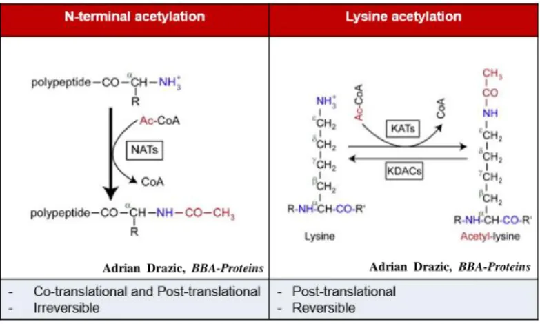
Alongside the existence of phosphorylation, SUMOylation, methylation, acetylation is one of the most abundant and important post-translational modifications in eukaryote cells, in which acetyl group from the dornor is transferred to the target proteins (Drazic, Myklebust, Ree, & Arnesen, 2016). The acetylation of proteins could occur either at their Nα-termini of the nascent polypeptide chains termed as N-terminal acetylation or at specific lysine residues on the polypeptide chains termed as lysine acetylation.
N-terminal acetylation is mainly co-translational modification, catalyzed by a group of highly conserved proteins, the N- acetylatransferases (NAT) (Silva & Martinho, 2015). However, recent studies reveal that N-terminal acetylation also occurs post- translationally (Aksnes, Van Damme, et al., 2015; Helsens et al., 2011;
Van Damme et al., 2011). N-terminal acetylation is an ubiquitous modification in eukaryote as its frequency is 50-70% in yeast and 80-
2
90% in human (Thomas Arnesen et al., 2009), but this is a rare event in prokaryote as only 31 proteins are modified in this manner in Escherichia coli so far (Schmidt et al., 2016). Six different NATs have been identified in mammals including NatA-NatF, whereas five NATs including NatA-NatE have been discovered in yeast. Among the Nats family, NatA, NatB, NatC are the main Nat complexes that are responsible for approximately 80% of N-terminal acetylation in human cells (Aksnes, Hole, & Arnesen, 2015).
The other major type of protein acetylation is lysine acetylation, which is catalyzed by lysine acetyltransferases (KATs). As histones were the first proteins identified being modified in this manner, the lysine acetylatransferases were originally named histone acetyltransferases (HATs) (Allfrey & Mirsky, 1964). Soon after, not only histones but many more nuclear and cytosolic proteins have been elucidated being lysine acetylated (S. C. Kim et al., 2006). The lysine acetylation prevents positive charges from forming on the amino group, and as a result, has a significant impact on the electrostatic properties of the protein, thereby regulates diverse biological events in cytoplasm, nucleus as well as mitochondria such as DNA-binding affinity of transcription factors, protein-protein interactions, protein stability
3
(Glozak, Sengupta, Zhang, & Seto, 2005; Kouzarides, 2000). To date, approximately 22 KATs have been identified in the human genome, grouped in three major families: the GNAT family, the MYST family and the p300/CBP (CREB-binding protein) family (Drazic et al., 2016).
Unlike N-terminal acetylation, which is considered irreversible, lysine acetylation is reversed by lysine deacetylases (KDACs) (Glozak et al., 2005; Taunton, Hassig, & Schreiber, 1996; Yang & Seto, 2008) (Figure 1). There are two main deacetylase mechanism: Zn2 +-dependent HDACs (HDAC1–11) and NAD+-dependent sirtuins (SIRT1–7) (Glozak et al., 2005). The balance between acetylation and deacetylation is highly controlled to ensure proper gene expression and signaling pathway regulation. Both hypoacetylation or hyperacetylation leads to developmental disorders. Decrease of expression of genes essential for synaptic maintenance and neurotransmission, leading to aging and neurodegeneration is caused by hypoacetylation of neuroplasticity genes; whereas hyperacetylation is implicated in many disorders and diseases atherosclerosis, aneurysm, myocardial infarction and tumor angiogenesis or even death because of the growth retardation (Drazic et al., 2016).
4
Figure 1. Comparison between N-terminal acetylation and Lysine acetylation. Left, N-terminal acetylation occurs at the Nα-termini of the polypeptide and is regulated by N-terminal acetyltransferases (NATs).
Right, Lysine acetylation occurs at specific lysine residue of the
polypeptide chain and is regulated by lysine acetyltransferases (KATs).
Adrian Drazic, BBA-Proteins
and Proteomics, 2016
Adrian Drazic, BBA-Proteins
and Proteomics, 2016
5
II. ARD1 (Arrest defective protein 1)
ARD1 was first identified in Saccharomyces cerevisiae (Whiteway & Szostak, 1985) and evolutionarily consevered from yeast to human. Various isoforms of ARD1 from alternative splicing of mRNA have been characterized. In mouse, ARD1 has three alternative splicing isoforms mARD1198, mARD1225 and mARD1235. Whereas, there are two human ARD1 isoforms including hARD1131 and hARD1235 (S.-H. Kim et al., 2006). These variants share a conservered N-acetyltransferase domain, a putative NLS (Nuclear Localization Signal) (KRSHRR), and acetyl-CoA binding domain [(Q/R)XXGX(G/A)], but contain diferent sequences and lengths in C- terminal region (Thomas Arnesen et al., 2005; S.-H. Kim et al., 2006).
ARD1 is known as a catalytic subunit, in combination with Naa15 to form functional NatA complex (Polevoda & Sherman, 2003;
Starheim, Gevaert, & Arnesen). Crystal structure studies reveals that ARD1-Naa15 complex is a very stable complex (Thomas Arnesen et al., 2005). As a subunit of NatA, ARD1 shows N-terminal acetyltransferase activitity, adopting a Gcn5-related N-acetyltransferase (GNAT) fold containing a central acetyl CoA–binding region and
6
flanking N- and C-terminal segments (Liszczak et al., 2013). Besides the functions in complex with Naa15, ARD1 exists independently and has ability to acetylate acidic side chains like aspartate and glutamate in γ- and β-actin (Van Damme et al., 2011). Additionally, tuberous sclerosis 2 (TSC2) is acetylated by ARD1 at the first methionine, whereby stabilizing TSC2 (Kuo et al., 2010).
In addition to N-terminal acetylatransferase activity, ARD1 also has lysine transferase activity (Polevoda & Sherman, 2003). Indeed, variety of ARD1 substrates which are acetylated as specific lysine residues have been documented (Figure 2). Human ARD1235 has lysine acetylatransferase activity on androgen receptor, promoting dissociation from HSP90 complex (DePaolo et al., 2016); acetylates CDC25A, increasing CDC25A phosphatase activity (Lozada et al., 2016). ARD1 mediates lysine acetylation of β-catenin, inducing the binding of TCF4 to cyclin D1 promoter (Lim, Park, & Chun, 2006); as well as induces Runx2 acetylation, facilitating bone formation (Yoon & Park, 2015).
Acetylation of HSP70 at lysine 77 regultes the balance of protein degradation and refolding (Seo et al., 2016). Furthermore, recent study reported that ARD1 could self-acetylate at lysine 136 and this autoacetylation is crucial for its acetylatranserase activity (Seo et al.,
7
2010). This study also reported that arginine residue 82 and tyrosine residue 122 are necessary for ARD1 catalytic activity.
Different subcellular localization of ARD1 displays different cellular functions owing to different regularoty mechanism. mARD1235 was found mainly in nucleus. In contrast, mARD1225 isoform was mainly localized in cytoplasm (Chun et al., 2007). In human, hARD1235 distributes both in cytoplasm and nucleus and possibly shuttles between these two organelles (Park et al., 2014; Seo, Park, &
Lee, 2015) but the distribution of hARD1235 in cytoplasm is dominant over the distribution in nucleus, whereas hARD131 was reported mainly nuclear (Seo et al., 2015).
8
Figure 2. Various substrates of hARD1235. ARD1 could acetylate itself and has acetyltransferase activity on variety of substrates such as CDC25A, Runx2, HSP70, Androgen receptor, β-catenin
.
9
Ⅲ. Aurora kinases
1. Aurora kinase family
Aurora kinases is a family of three serine/threonine kinases that control a number of mitotic events (Carmena & Earnshaw, 2003). The first Aurora was first described in Drosophila. The name “Aurora” was given for the kinase since the mutation of the gene caused the failure of centrosome segregation, causing the monopolar spindle (Glover, Leibowitz, McLean, & Parry, 1995). Only one Aurora kinase exists in fungi, named Ipl1 in S. cerevisiae (Chan & Botstein, 1993; Francisco, Wang, & Chan, 1994) and Ark1 in S. pombe (Nigg, 2001). There are two types of Aurora kinases in Caenorhabditis elegans (J.M.
Schumacher, Ashcroft, Donovan, & Golden, 1998; Jill M. Schumacher, Golden, & Donovan, 1998), Drosophila (Glover et al., 1995), and Xenopus (Roghi et al., 1998). In mammals, there are three members in Aurora kinase family: Aurora kinase A, B, C (Nigg, 2001).
10 2. Structrures of Aurora kinases
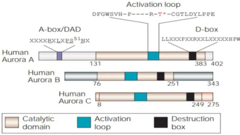
Aurora kinases comprise two domains: N-terminal regularoty domain and C-terminal catalytic domain. The Aurora kinases share 70%
homology in the catalytic domain, whereas the Aurora kinases differ in length and sequence of N-terminal domain (Carmena & Earnshaw, 2003). The catalytic domain of Aurora kinases contains ATP binding sequence and activation loop that account for kinase activity of Aurora kinases. Moreover, there is a key threonine named T-loop residue within the catalytic domain of Aurora kinases which should be phosphorylated by autophosphorylation manner for kinase activity activation. In Aurora kinase A, the T-loop residue positions at 288, and this T-loop residue locates at 232 in Aurora kinase B and 195 in Aurora kinase C (Goldenson & Crispino, 2015) (Figure 3). In addition, the C- terminal domain also contains destruction box (D-box) that mediates Aurora kinases degradation (Goldenson & Crispino, 2015). Besides the D-box, Aurora kinase A contains KEN motif and D-box activating domain (DAD box) in N-terminal domain. These structrures are absence in Aurora kinase B (Carmena & Earnshaw, 2003).
11
Mar Carmena & William C. Earnshaw, Nature Reviews Molecular Cell Biology, 2003
Figure 3. Structure of Aurora kinases. Aurora kinases share 70% homology in catalytic domain which contains activation loop and destruction box (D-box). However, aurora kinases are different in the N-terminal domain. Aurora kinase A contains A- box activating domain (A-box/DAD) at N-terminal domain which is absent in Aurora kinase B and C.
12
3. Subcellular locacalizations and functions of Aurora kinases
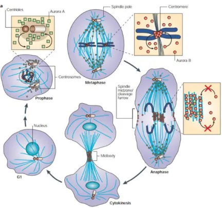
Although Aurora kinases have high similarity in structure and sequence, these kinase proteins have distinct subcellular localizations and functions. Aurora kinase A localizes to the centrosomes from the end of the S phase until mitotic exit. Aurora kinase A also localizes to the mitotic spindle from prophase to telophase (Stenoien, Sen, Mancini,
& Brinkley, 2003; Zhou et al., 1998). Due to its localization, Aurora kinase A has functions in centrosome maturation, separation (Marumoto et al., 2003) and mitotic spindle assembly (Tsai et al., 2003). Aurora kinase B localizes to centromeres from prophase to metaphase, translocates to the midbody when the anaphase begins and remains in the midbody until cytokinesis is completed (Adams et al., 2000; Earnshaw & Bernat, 1991). Aurora kinase B plays important roles in chromosmome condensation regulation (Adams et al., 2000;
Terada, 2001), as well as spindle checkpoints (Lan et al., 2004) and proper cytokinesis (Terada, 2001). Unlike Aurora kinase A and B, which are globally expressed in both somatic and meiotic cells, Aurora kinase C is limited to meiotic germ cells (2000). Aurora kinase C shows
13
centrosomal localization from anaphase to telophase (Tseng, Chen, Hsu, & Tang, 1998). Since its predominant expression in testis, Aurora kinase C is required for spermatogenesis (Yan et al., 2005).
14
Mar Carmena & William C. Earnshaw, Nature Reviews Molecular Cell Biology, 2003
Figure 4. Subcellar localizations of Aurora kinase A and B.
Aurora kinase A (square, green) localizes to centrosomes throughout the cell cycle progression, whereas Aurora kinase B (round, red) localizes to centromeres from prophase to metaphase and relocates to midbody in anaphase till the mitosis exits
15
4. Activation and degradation of Aurora kinase A
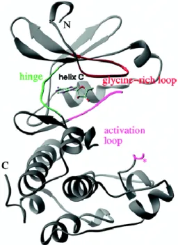
Catalytic domain of Aurora kinase A comprises an N-terminal lobe (residues 123-210) including a β-sheet and two α-helices and a C- terminal α-helical domain (residues 217-387). These two domain are linked together by a hinge region (residues 210-216) (Bayliss, Sardon, Vernos, & Conti, 2003) (). ATP-binding region is a cleft located at the interface of the α-helical and β-strand domains, bound by the glycine- rich loop (residues 140-149) and the activation loop (Figure 5). The moieties threonine 288 (T288) in the activation loop is phosphorylated during activation of Aurora kinase A (Cheetham et al., 2002). The autophosphorylation of Aurora kinase A at T288 is regulated by the binding-partner TPX2. Binding of TPX2 to Aurora kinase A maintains the active conformation of Aurora kinase A and protects phosphorylated Aurora kinase A from dephosphorylation (Bayliss et al., 2003).
The presence of destruction box (D-box) with motif RxxL regulates the degradation of Aurora kinase A by ubiquitination mediated by APC/Fizzy (anaphase promoting complex) (Arlot- Bonnemains et al., 2001). In addition to D-box, KEN motif (lysine/glutamic acid/asparagine) in the N-terminal domain of Aurora
16
kinase A plays an important role in ubiquitin-mediated degradation of Aurora kinase A (Lindon, Grant, & Min, 2016). Anna Castro et al.
investigated an addition destruction signal within the N-terminal domain of Aurora kinase A, which is named D-box-activating domain (DAD-box). The D-box-dependent proteolysis of Aurora kinase A is conferred by DAD box with motif RxLxPSN (Figure 3) (Castrol et al., 2002).
17
Figure 5. Crystal structure of C-terminal domain of Aurora kinase A (residues 122-403). The hinge region (green) is the linker of N- terminal lobe and C-terminal lobe of catalytic domain. The glycin-rich loop (red) and the activation loop (pink) are important for kinase activity of Aurora kinase A. The key residue for activation of Aurora kinase A T288 is marked by a star.
Cheetham et al., Journal of Biological Cheistry,2002
18
5. Mitotic functions of Aurora kinase A
Since the first finding of Aurora kinase A as a crucial factor for centrosome segregation, many studied have revealed the contributions of Aurora kinase A to centrosome regulation. Centrosomes undergo a maturation process to form the poles for bipolar spindle as the cell enter the mitosis (Palazzo, Vogel, Schnackenberg, Hull, & Wu, 1999).
Aurora kinase A is accumulated centrosomes after the duplication of centrosome during the S phase (Hannak, Kirkham, Hyman, & Oegema, 2001). Polo-like kinase 1 is required for the centrosomal targeting of Aurora kinase (Luca, Lavia, & Guarguaglini, 2006). At the duplicated centrosomes, in particular at the pericentriolar mass, Aurora kinase A regulates the recruitment of γ-tubulin, centrosomin, LATS2, TACC, and NDEL1 to the pericentriolar mass for the centrosome maturation. After the maturation process, centrosomes segregate and move toward two opposite poles of the nucleus. The separation of centrosomes requires Aurora kinase A. The depletion of Aurora kinase A causes the monopolar spindle due to centrosome segregation defects (Glover et al., 1995; Liu & Ruderman, 2006; Roghi et al., 1998). In such cells, the monopole contains two centrosomes that fail to separate (Giet, Uzbekov, Kireev, & Prigent, 1999). In Xenopus, it is reported that Eg5,
19
which is critical for proper centrosome separation, is phosphorylated by Aurora kinase A (Giet et al., 1999). In addition, the inhibition of Aurora kinase A or Eg5 causes defects in centrosome separation, suggesting that Aurora kinase may regulate separation via Eg5.
The importance of of Aurora kinase A in controlling mitotic spindle assembly has been intensively studied. Aurora kinase A interacts with TACC (transforming acidic coiled-coil) proteins and forms complexes with these proteins at the centrosomes. Then, interaction of TACC proteins and ch-TOG/XMAP215 at the centrosome stabilize the microtubules and control the plus-end dynamics of microtubules (Kinoshita et al., 2005; LeRoy et al., 2007).
Concomitant with the centrosome maturation, Aurora kinase A also participates in mitotic entry regulation. Aurora kinase A activates CDK1/cyclin B complex to allow nuclear entry. This activation is regulated by phosphorylation of Bora at T210 (Seki, Coppinger, Jang, Yates, & Fang, 2008). Aurora kinase A takes part in G2/M transition control by phosphorylating BRCA1 (breast cancer associated gene 1), which is a requirement for M phase entry (Brodie & Henderson, 2012).
The AuA-encoding gene is located on chromosome 20q13.2- q13.33, a region frequently amplified in many cancers such as rectal,
20
ovarian, prostate, colon, and breast cancers (Bar-Shira et al., 2002; J. R.
Bischoff et al., 1998; Zhou et al., 1998). The overexpression of Aurora kinase A causes the impairment of centrosome segregation, thus results in multipolar spindles, aneuploidy, increased resistance to apoptosis and deficient cell cycle checkpoint functions (Figure 6). Consequently, overexpression of Aurora kinase A promotes tumorigenesis. It is also demonstrated that Aurora kinase A-mediated p53 phosphorylation induces MDM2-dependent degradation of p53, leading to cancer progression.
21
A.S.Nikonova, Cell. Mol. Life Sci.,2013
Figure 6. Activities of Aurora kinase A in cancer.
Overexpression of Aurora kinase A contributes to cancer progression by either causing failure of cytokinesis, impaired cell cycle checkpoints or loss of p53
22
6. Non-mitotic functions of Aurora kinase A
Eventhough localization of Aurora kinase A to the centrosome and the functions of Aurora kinase A in the mitosis have widely studied, the expression of Aurora kinase A in other phases of cell cycle outside the M phase and the presence of Aurora kinase A in the cytosol (Rannou et al., 2008) and the Golgi (Cervigni, Barretta, Persico, Corda,
& Colanzi, 2011) have been described. These clues suggest the non- conventional functions of Aurora kinase A alongside the mitotic functions. Mori et al. proposed that aPKC interacts with Aurora kinase A at post-mitotic neurons and phosphorylate Aurora kinase A at T287 augmenting activation of Aurora kinase A, whereby phosphorylation of NDEL1 by activated Aurora kinase A induces microtubule dynamics modulation, resulting in neurite elongation (Mori et al., 2009).
Furthermore, Aurora kinase A has also been reported to contribute to ciliary disassembly in 2007. Pugacheva and coworkers observed that autophosphorylation of Aurora kinase A at T288 and the expression level of Aurora kinase activator NEDD9 are upregulated along with the increased percentage of ciliated cells. It is shown that Aurora kinase A interacts with HDAC6 at the basal body of cilia, leading to Aurora
23
kinase A – induced HDAC6 phosphorylation, promoting ciliary disassembly (Pugacheva, Jablonski, Hartman, Henske, & Golemis, 2007). Recent studies have been demonstrated the role of Aurora kinase A in cell motility and metastasis. Overexpression of Aurora kinase A has been found in breast cancer metastasis. The overexpression of Aurorkinase induces the dephosphorylation of cofilin, facilitating actin organization and polymerization, resulting in cell migration (L. H.
Wang et al., 2010). The knockdown of Aurora kinase A significantly inhibits cell invasiness and secretion of MMP2, whereas the overexpression of Aurora kinase A causes the elevation of esophageal squamous cell migration and invasion via MMP2 secretion (Wang et al., 2012).
24
A.S.Nikonova, Cell. Mol. Life Sci.,2013
Figure 7. Non-mitotic functions of Aurora kinase A. Several non- mitotic functions of Aurora kinase A have been elucidated such as ciliary disassembly, neurite elongation and cell migration.
25
PURPOSE OF THIS STUDY
Defective control of cell proliferation is the main characteristic of cancer. In normal cells, cell division is rigorously controlled by complex signaling circuits. However, cancer cells can sustain proliferative signaling themselves and can grow unstoppably. Another hallmark of cancer is cell migration. Cell migration is a highly integrated process, and controlled by dynamic regulatory mechanisms.
Hence, understanding the mechanisms of controlling cell proliferation and cell motility could open new horizons in cancer therapy.
Aurora kinase A (AuA) is a member of the serine/threonine kinase family, which has been implicated in controlling the cell cycle and cell division. The overexpression of AuA has been found in many types of metastatic cancers and its correlation with poor prognosis. The non-mitotic functions of AuA, in addition to its role in mitotic regulation, have been elucidated. Recent findings have revealed the
26
contribution of AuA in cell migration and metastasis enhancement.
However, the mechanisms regulating AuA activity in cell movement and cell proliferation are not fully understood.
This study suggested a previously undefined post-translational modification of AuA by ARD1 acetyltransferase. In this study, AuA was found as ARD1 substrate and acetylated at lysines at positions 75 and 125, consequently this contributes to AuA catalytic capacity, then leading to promotion of cell proliferation and stimulation of cell migration.
27
MATERIALS AND METHODS
1. Antibodies
Anti-AuA antibody (ab13824) and Anti-phospho Serine/Threonine (ab17464) antibody were purchased from Abcam.
Anti-acetylated lysine (Lys-Ac) antibody (#9941), anti-AKT antibody (#4691S), anti-phospho AKT (S473) antibody (#9271), anti-p38 MAPK antibody (#9212), anti-phospho p38 MAPK antibody (#4511), anti- p44/42 MAPK (Erk1/2) antibody (#9102) were from Cell Signaling Technology. Anti-cyclin B1 antibody (GNS1, sc-245), anti-cyclin E antibody (HE12, sc-247), anti-CDK2 antibody (9E10, sc-40), anti- Vinculin antibody (H300, sc-5573), anti-GFP antibody (B2, sc-9996), anti-MMP2 antibody (H-76, sc-10736), anti-GAPDH antibody (G-9, sc- 365062), anti p-ERK antibody (E-4, sc-7383), anti-CDK2 antibody (D- 12, sc-6248) were purchased from Santa Cruz. Anti-RFP tag antibody
28
(MA5-15257), anti-phosphor-FAK (S397) antibody (44-624G), and anti-FAK antibody (610087) were obtained from ThermoFisher Scientific, Invitrogen and BD Biolegend respectively.
2. Plasmid construction
Oligonucleotide primers were designed to amplified ARD1 gene (GenBank: NM_003491.3) and AuA gene (GenBank: NM_198433.2).
Each primer covers additional sequences of restriction enzyme. ARD1 PCR product was digested and cloned into pET28b, pEGFP-C3 and pGEX-4T3. AuA PCR products were digested and cloned into pTagRFP-C and pET28b. Deletion mutants of His-AuA were constructed from pET28b-AuA plasmid. cDNAs of AuA corresponding to 1–140 aa, and 126–403 aa were amplified by PCR and inserted into pET28b.
29 3. Cell culture
Human embryonic kidney HEK293T cell line, human breast cancer MCF7 cell line and Non-small lung cancer cell H1299 cell line obtained from ATCC were grown in DMEM medium containing 10%
FBS (fetal bovine serum) with penicillin and streptomycin in a 5% CO2
humidified atmosphere at 37oC. Cells were transfected using Polyethylenimine (PEI) reagent following manufacturer’s
recommendation. For MCF7 stably overexpressing GFP-ARD1, RFP- AuA, MCF7 cells were transfected with pEFGP-C3-ARD1 and pTagRFP-C-AuA using Lipofectamine 2000 (Lifetechnologies) then selected by using G418 (geneticin).
4. Cell synchronization
MCF7 cells were synchronized by treated with cytosine arabinoside in 16h for G1/S phase. After cells were released into fresh medium in 4h, cells were then arrested in early mitosis by using
30
nocodazole at concentration 100ng/ml for 6h. For metaphase, cells were then treated with MG132 for 30 min. And cells were in anaphase and telophase after being released from MG132 30 min and 60 min respectively.
5. Immunoflourescence
Cells were grown on circular glass coverslips plated in 24-well plate. Cells were fixed with formaldehyde in 10 min, permeabilized by incubating in PBS containing 0.25% Triton-X in 10 min and washed 3 times by PBS, then blocked for 1h in 1% BSA. Primary antibody recognizing AuA (1:500) was diluted in blocking buffer and incubated overnight at 4oC. Alexa Flour®546 goat anti-mouse antibody was used at recommended concentration and incubated in 1h in the dark at room temperature. This was followed by counterstaining with Hoechst and mounting with Gel/mount (Biomeda Corp.). Cells were observed under Axiovert M200 microscope (Zeiss).
31
6. Immunoblotting and immunoprecipitation
Total cell lysates were isolated using cell lysis buffer (Cell signaling), which contained 20mM Tris-HCl pH7.5, 150mM NaCl, 1mM Na2EDTA, 1mM EGTA, 1% Triton, 2.5mM sodium pyrophosphate, 1mM beta-glycerophosphate, 1mM Na3VO4, 1μg/mi leupetin and the protease inhibitor cocktail set (Calbiochem). Lysates were separated by sodium dodecyl sulfate gel electrophoresis, and transferred to nitrocellulose membranes for Western blot analysis.
Detection was performed using ECL (enhanced electrochemiluminescence) plus (Amersham Bioscience). For immunoprecipitation, lysates were immunoprecipitated with appropriate primary antibody. The immunoprecipitated complexes were subjected to electrophoresis and immunoblotting.
32 7. In vitro acetylation assay
BL21 cells transformed with plasmids pET28a-ARD1, pGEX-4T3 or pET28a-AuA were grown to and OD600 of 0.6–0.8. Overall, 1 mM IPTG was added to induce His-ARD1, GST-ARD1 or His-AuA, then cells were grown for overnight at 25⁰C. For His-tagged protein
purification, cells were collected and proteins were extracted with a lysis buffer (pH 7) containing 50 mM NaH2PO4, 300 mM NaCl, 10 mM Imidazole and 1% Triton X-100. His-tagged proteins were purified with Ni-NTA and followed by eluted with an elution buffer containing 50 mM NaH2PO4, 300 mM NaCl, and 250 mM Imidazole.
For GST-tagged protein purification, cells were collected and proteins were extracted with lysis buffer containing 250 mM Tris-Cl pH 8.5, 500 mM NaCl, 5 mM EDTA and 1% Triton X-100. Cell lysates were then incubated with Glutathione-Agarose for 4h, following by washing three times with PBS to obtain purified GST-tagged protein. Acetylation
33
assay was performed as described. Freshly prepared recombinant ARD1 and AuA were incubated in the reaction mixture [50 mmol/L Tris-HCl (pH 8.0), 0.1mmol/LEDTA, 1mmol/LDTT, 10%glycerol, and 10 mmol/L acetyl-CoA] at 37⁰C.
8. ATP-binding assay
Total cell lysates were isolated using cell lysis buffer (Cell signaling). 1mg cell lysates were then subjected to ATP-agarose resins (Innova Biosciences) and incubated at 4⁰C for 4h. The ATP-bound
protein complexes were washed three times with the same buffer.
Proteins bound to ATP were eluted by sample buffer and subjected to electrophoresis and immunoblotting.
9. ATPase activity assay
Recombinant proteins were applied to in vitro acetylation assay and ATPase activity were measured using High Throughput Colorimetric ATPase Assay Kit (Innova Biosciences) according to the
34
manufacturer’s instructions. Briefly, recombinant proteins were incubated for 30 min at 30⁰C in a reaction buffer consisting of 50 mM
Tris (pH 7.5), 2.5 mM MgCl2 and 0.5 mM ATP. After that PiColorLock Gold reagent and Accelerator were added to the mixture. Then Stabilizer was added and stabilized for 30 min. The absorbance at 595nm was measured. Each experiment was repeated 3 times.
10. Protein sequence alignment
Protein sequence alignment was conducted using Clustal Omega.
11. Cell proliferation assay
Cells were seeded in 96-well plate at a density of 103 cells per well and cultured for 48h. The proliferation rates were measured using a Non-Radioactive Cell Proliferation Assay Kit (Promega) according to the manufacturer’s instructions. Briefly, 20ul of freshly mixed tetrazolium/phenazine methosulfate was added, and the cells were incubated for 1 hour to allow color development. The absorbance at
35
490nm was measured to indicate the number of viable cells. Each experiment was repeated 3 times.
12. Clonogenic assay
Cells were seeded in 6-well plate at a density of 103 cells per well and cultured for 7 days to form colonies. Colonies then were fixed with 1% formaldehyde and stained with crystal violet (0.5%) and counted.
Each experiment was repeated 3 times.
13. Cell cycle analysis
2x10⁶ cells were harvested and washed twice with PBS, fixed overnight with 70% ethanol. After two washes with cold PBS, cells were incubated with RNAse for 30 min, following up with 30 min incubation with propidium iodine. Cell cycle was analyzed by BD Calibur flowcytometry. Each experiment was repeated 3 times.
36 14. Wound healing assay
Wounds were created in confluent MCF7 cells by 200μl tip and washed twice by PBS to remove non-adherent cells. To inhibit the influence of cell proliferation, cells were treated with 10μM cytosine
arabinoside. After 24h incubation, the wound closure rates were measure under a phase-contrast microscope. Each experiment was repeated 3 times.
15. Transwell invasion assay
Invasion assays were conducted using transwell invasion chambers coated with Matrigel matrix (Corning). Cells were seeded in the upper chambers (104 cells/ well) in FBS-free DMEM. DMEM containing 10% FBS was added to the lower chambers. After 24h of incubation, invaded cells on the lower surface were stained with crystal violet stain and counted under a light microscope. Each experiment was repeated 3 times.
37 16. Statistical analysis
Data analysis and statistical significance were tested using Graphpad Prism 6.0. P values were calculated by applying two-tailed Student’s t test. Statistical significance was determined at P<0.05.
38
RESULTS
1. ARD1 has centrosome-like localization during cell cycle progression
Recent studies showed that ARD1 is essential for cell survival, as knockdown of ARD1 in human cells triggers apoptosis and G1 arrest (T. Arnesen et al., 2006; Lim et al., 2006). To check whether the expression level of ARD1 alters in cell cycle progression, cells were synchronized at differents phases of cell cycle and western blotted.
Interestingly, the expression of ARD1 increased as the cell transitted to mitotic phases and eventually degraded at the end of the mitosis (Figure 8). Then, to figure out the subcellular localization of ARD1 in cell cycle, ARD1 overexpressing cells were synchronized at different stages of cell divion. Surprisingly, ARD1 was observed to have distinct pattern during cell cycle progression. ARD1 seemed to have the centrosome- like localization during cell cycle stages (Figure 9). These results suggest that ARD1 may involve in cell cycle regulation.
39
Figure 8. ARD1 expression changes during cell cycle progression.
Cells were treated with arabinoside C at concentration 5mg/ml in 16h for G1/S synchronization. For synchronizing cells at G2/M phase, cells were treated with Nocodazole at 400ng/ml in 16h following by nocodazloe release at indicated time. Cell lysates were then western blotted for ARD1. Cyclin B was used as mitotic marker and cyclin E was used as G1/S phase marker.
40
Figure 9. ARD1 obtains centrosome-like colocalization.
Overexpressing GFP-ARD1 cells were synchronized, fixed and then mounted. Cells were then visualized by confocal microscope. DNA was counter-stained by Hoechst. Scale bar, 5μm. Subcellular localization of ARD1 is indicated by arrows.
41
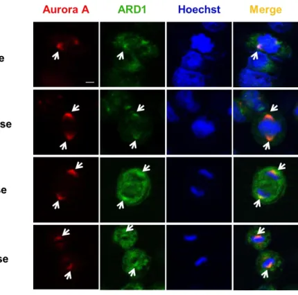
2. ARD1 colocalizes with Aurora kinase A during cell cycle progression
The observation of distinct localization of ARD1 prompted me to examine whether ARD1 localize at centrosomes. AuA is considered as a centrosome marker owing to its localization to the centrosome and its functions in centrosome maturation and separation (Adams et al., 2000;
James R. Bischoff & Plowman, 1999), ARD1 overexpressing cells were synchronized at different stages of cell cycle and stained with Aurora kinase A. Interestingly, ARD1 and Aurora A were observed to colocalize at the centrosome during cell cycle progression, particularly during cell division (Figure 10).
42
Figure 10. Aurora kinase A colocalalizes with ARD1 during cell division. Aurora A colocalizes with ARD1 at centrosomes during cell division in prophase, metaphase, anaphase and telophase. GFP- ARD1 overexpressing cells were synchronized and stained with anti -AuA antibody (red). DNA was counter-stained by Hoechst. Cells were then viewed under a confocal microscope. Scale bar, 5 µm.
Colocalization of ARD1 and AuA is indicated by arrows.
43
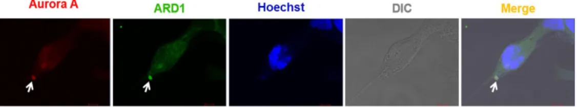
3. ARD1 colocalizes with Aurora kinase A during cell migration The centrosome not only participates in cell cycle progression, but also contributes to cell migration by undergoing reorientation and supporting the forces generated in cells during cell migration (Scliwa &
Höner, 1993). AuA, as a component of the centrosome, plays a part during cell migration. Here, I also observed the colocalization of ARD1 and AuA in migrating cells (Figure 11). This observation suggests that interaction between ARD1 and AuA may play a critical role in cell movement.
44
Figure 11. Aurora A colocalizes with ARD1 in migrating cell.
Monolayer of cells on coverslip was scratched to stimulate migration, then cells were stained with anti-AuA antibody (red). DNA was counter-stained by Hoechst. Scale bar, 5 µm. Colocalization of ARD1 and AuA is indicated by arrows.
45
4. Aurora kinase A is acetylated by ARD1
To determine whether AuA and ARD1 physically interact in cells, I immunoprecipitated ARD1 from lysates of cells overexpressing ARD1 and found that ARD1 was bound to AuA (Figure 12). As ARD1 is an acetyltransferase that catalyzes the transfer of acetyl groups from acetyl-coA onto lysine residues in its substrates, I hypothesized that AuA could be modulated by ARD1 by acetylation. To examine this hypothesis, I performed an in vitro acetylation assay in which recombinant His-tagged AuA was mixed with recombinant His-tagged ARD1 in the presence of acetyl-CoA. Expectedly, AuA was acetylated by ARD1 (Figure 13A). Consistent with the in vitro experiment, the overexpression of ARD1 significantly upregulated the level of AuA acetylation in cells (Figure 13B).
46
Figure 12. Aurora kinase A binds to ARD1. Lysates from HEK293T cells overexpressing GFP-ARD1 were immunoprecipitated with anti- GFP antibody and immunoblotted with anti-AuA antibody or anti-GFP antibody.
47
Figure 13. AuA is acetylated by ARD1. A, AuA is acetylated by ARD1 in vitro. His-ARD1, His-AuA recombinants were subjected to an in vitro acetylation assay with or without presence of acetyl group donor acetyl- coenzyme A (CoA) for 1 h, and acetylation levels of
A
B
48
recombinants were assessed by western blotting using an anti-acetylated lysine antibody (Lys-Ac). Ponceau S staining shows the quantification of the input proteins. B, Acetylated AuA level increases in GFP-ARD1 overexpressing cells. Lysates from GFP-ARD1 overexpressing MCF7 cells were immuprecipitated with anti-Lys-Ac antibody and analyzed by immunoblotting with anti-AuA antibody or anti-GFP antibody.
49
5. Acetylation of Aurora kinase A is dependent on ARD1 catalytic activity
Interestingly, AuA acetylation occurred in a time-dependent manner after autoacetylation of ARD1 (Figure 14), suggesting that the autoacetylation of ARD1 is essential for regulating AuA acetylation.
Previously, we reported that ARD1, in addition to acetylating a variety of substrates, undergoes self-acetylation and that arginine 82 (R82) and tyrosine 122 (Y122) are required for its acetyltransferase activity (Seo et al., 2010). Thus, we examined the levels of AuA acetylation in the presence of functional (wild-type) and R82A/Y122F mutant ARD1 proteins. It was seen that the AuA acetylation level decreased dramatically when ARD1 was mutated at R82 and Y122 (Figure 15).
Taken together, these data indicate that AuA interacts with ARD1, and AuA acetylation is regulated by functional ARD1.
50
Figure 14. AuA acetylation occurs in a time-dependent manner.
His-ARD1 recombinants were subjected to an in vitro acetylation assay for series of time, and acetylation levels of recombinants were assessed by western blotting using an anti-Lys-Ac antibody. Quantification of the input proteins were analyzed by Ponceau S staining.
51
Fig 15. AuA acetylation is dependent on ARD1 acetyltransferase activity. MCF7 cells were transfected with wild type (WT) GFP-ARD1 or GFP-ARD1 R82F/Y122A mutant. The extracts from the overexpressing cells were immoprecipitated with anti Lys-Ac antibody and acetylated AuA levels were analyzed by immunoblotting with anti- AuA antibody.
52
6. Aurora kinase A is acetylated at at lysine residues at position at 75 and 125
AuA comprises 403 amino acids and has two domains, an N- terminal domain spanning residues 1 to 131, and a C-terminal domain spanning residues 132 to 403. The C-terminus includes a catalytic domain that harbors the kinase activity and a destruction box (D-box) that plays a role in ubiquitin-mediated degradation of several mitotic proteins. The N-terminus contains the A-box/D-box activating domain (DAD) that controls AuA degradation (Figure 16). However, the function of the N-terminal domain is yet unclear (Fu, Bian, Jiang, &
Zhang, 2007; Marumoto, Zhang, & Saya, 2005). To identify the target sites on AuA that are acetylated by ARD1, I performed in vitro acetylation assays with recombinant AuA. For this, I constructed two truncated fragments of AuA, an N-terminal domain-containing fragment comprising amino acids 1 to 140 and a C-terminal domain- containing fragment comprising residues 126 to 403 (Figure 16). As shown in Figure 15, the N-terminal domain of AuA was acetylated, but not the C-terminal domain. To further delineate the residues involved in ARD1-mediated AuA acetylation, a series of N-terminal fragments were generated, in which the lysine residues were substituted with
53
arginine to mimic non-acetylated lysine, and in vitro acetylation assays were performed. Lysines at positions 75 and 125 were identified as preferable sites for AuA acetylation (Figure 17).
54
Figure 16. ARD1 acetylates the N-terminus of AuA in vitro. Top, Deletion mutants of His-AuA were subjected to in vitro acetylation assays with His-ARD1. Bottom, Construction of AuA deletion mutants.
AuA-N, AuA N-terminal domain; AuA-C, AuA C-terminal domain.
55
Figure 17. Lysines at positions 75 and 125 of AuA are acetylated by ARD1. Mutagenesis was performed to substitute lysine residues in N- terminus by arginine. Site-mutated AuA recombinants were then applied to in vitro assay.
56
7. Double mutation at lysine 75 and 125 abolished acetylation of AuA
To test whether lysine 75 and 125 play important role in Aurora kinase A, the double mutation at K75/K125 was esbtablished and Auorora kinase A double mutant recombinant proteins were subjected to in vitro acetylation assay. Indeed, AuA acetylation was almost negligible when the K75R/K125R double mutant AuA was subjected to in vitro acetylation (Figure 18A). Similarly, cells overexpressing AuA
double mutant K75R/K125R displayed a dramatically decreased level of acetylated AuA (Figure 18B), suggesting that these sites are critical for the acetylation of AuA by ARD1. These two sites are conserved across species (Figure 19A), indicating that these sites may be essential for regulating AuA activity. However, the sites are not conserved in other members of the Aurora kinase family (Figure 19B), and are probably responsible for the distinct functions of AuA compared to those of other Aurora kinases in regulating cellular processes.
57
Figure 18. Double mutation at K75/125 showed negligible acetylation level of AuA. A, Double mutant AuA K75R/K125R exhibits the negligible acetylation. His-AuA wild type (WT) and K75R/K125R double mutant (DM) were subjected to in vitro acetylation assays with His-ARD1. Acetylation levels were assessed by western blot. B, Double mutant AuA K75R/K125R displays
A
B
58
significantly decreased level of acetylated AuA in cells. The lysates from stable cells overexpressing RFP-AuA WT or RFP-AuA K75R/K125R mutant were collected for immunoprecipitation using anti-Lys-Ac antibody. The immunoprecipitates were examined for acetylated AuA by blotting with anti-AuA antibody.
59
Figure 19. Consevation of lysine residues 75 and 125. A, The lysine residues at positions 75 and 125 are conserved across species. Protein sequence alignment of AuA in various species. B, The lysine residues at positions 75 and 125 are not conserved among Aurora kinase family proteins. Protein sequence alignment of AuA in Aurora kinase family proteins
A B
60
8. Acetylation of Aurora kinase A increases the phosphorylation of Aurora kinase A
The functions of AuA in diverse cellular processes are related to its kinase activity. Several studies have proposed that phosphorylation affects AuA kinase activity. To investigate whether AuA acetylation influences its catalytic activity, I examined the phosphorylation level of AuA. Impressively, AuA phosphorylation was elevated in stable cells overexpressing wild-type (WT) AuA, but not in cells expressing the K75R/K125R mutant AuA (Figure 20). This result demonstrated that acetylation of AuA at K75/125 elevated phosphorylation of AuA.
61
Figure 20. Acetylation of Aurora kinase A increases phosphorylation of Aurora kinase A. A, the collected extracts from MCF7 cells overexpressing RFP-AuA WT or DM were immunoprecipitated with anti-RFP antibody and were then immnublotted with anti-phospho- Serine/Threonine antibody (p- Ser/Thr) for examining phosphorylated AuA. B, phosphorylated AuA levels were analyzed using ImageJ program. **, p<0.005. Error bar indicates S.E.M.
62
9. Acetylation of Aurora kinase A increases the kinase activity of Aurora kinase A
AuA catalytic activity is strictly governed by an ATP cycle comprising ATP binding, ATP hydrolysis, and release of Pi and ADP (Xu, Wang, Xiao, Li, & Wang, 2011; Zhao et al., 2008). This ATP cycle is regulated by the ATP-binding site of AuA, which is located at the interface of the catalytic core (Zhao et al., 2008). Thus, the ability of AuA to bind ATP is critical for AuA activation. To elucidate the impact of AuA acetylation on its kinase activity, I next probed the ability of WT and K75R/K125R mutant AuA to bind ATP. I found that K75R/K125R mutant AuA exhibited significantly reduced ATP-binding capacity than the WT protein (Figure 21). Furthermore, the ATPase activity of WT AuA was significantly enhanced in the presence ARD1, which acetylates AuA; however, the mutant AuA, which was unable to be acetylated, did not show significant change in ATPase activity (Figure 22), indicating that ATP hydrolysis by acetylated AuA is more efficient than that by the non-acetylated mutant. In addition, recombinant his-tagged AuA proteins were subjected to in vitro acetylation assay with the presence of ARD1 WT or ARD1 R82/Y122 or without the presence of ARD1. Then the AuA proteins from the in
63
vitro acetylation assay were applied to ATPase activity assay. The
results showed that ATPase activity of AuA was significantly enhanced by acetylation by ARD1 WT, whereas the ARD1 R82/Y122 did not show the increase of AuA ATPase activity (Figure 23). Taken together, these data suggest that ARD1-mediated AuA acetylation at K75/K125 positively regulates AuA kinase activity.
64
Figure 21. Acetylation of Aurora kinase A increases ATP-binding affinity of Aurora A. Top, Lysates of RFP-AuA WT overexpressing cells and RFP-AuA K75R/125R overexpressing cells were incubated
65
with ATP-agarose for 4h, and ability of AuA binding to ATP was analyzed by western blot using anti-RFP tag antibody. Bottom, ATP- binding capacity was analyzed from western blot density using Image J.
**, p<0.005. Error bar indicates S.E.M.
66
Figure 22. Acetylation of AuA increases ATPase activity. His-AuA WT and His-AuA DM recombinants were acetylated, and subjected to reaction with ATP. ATPase activity was analyzed by measuring absorbance at 595nm. *, p<0.05; n.s, no significant. Error bar indicates S.E.M
67
Figure 23. ARD1 increases the ATPase activity of AuA.
Recombinant AuA proteins were subjected to in vitro acetylation in the presence of ARD1 WT or ARD1 R82/Y122. ARD1 R82/Y122 which lost acetyltransferase activity does not affect the ATPase activity of AuA.
68
10. Acetylation of Aurora kinase A promotes cell proliferation AuA overexpression is correlated with tumorigenesis and cancer progression. Because acetylation of AuA enhanced AuA kinase activity, I sought to elucidate the role of AuA acetylation in cell proliferation.
The MCF7 cells and H1299 cells were seeded in 96-well plates, incubated the cells for 48 h, treated them with MTT, and measured absorbance as an indicator of cell density. After 48 h, cells overexpressing WT AuA showed significant proliferation compared with cells overexpressing K75R/K125R mutant AuA (Figure 24). In confirmation with these data, clonogenic assays displayed the outgrowth of cells expressing acetylated AuA compared with cells expressing nonacetylated mutant AuA in both MCF7 and H1299 cell lines (Figure 25). These results suggested that acetylation of AuA promotes cell proliferation.
69
Figure 24. Acetylation of AuA increases cell proliferation. The
70
overexpressing RFP-AuA WT and RFP-AuA K75R/K125R MCF7 cells (A) and H1299 cells (B) were seeded in 96-well plates and cultured for 48h. Cells were then treated with MTT and measured absorbance at 490nm. ***, p<0.0005; *, p<0.05. Error bar indicates S.E.M.
71
A
B
72
Figure 25. Acetylation of AuA promotes colony formation. The overexpressing RFP-AuA WT and RFP-AuA K75/125 MCF7 cells (A) and H1299 cells (B) were seeded in 6-well plates and cultured for 7 days. Cells were then fixed with formaldehyde and stained with crystal violet to visualize colony formation for quantification. Representative image is shown. **, p<0.005, *, p<0.05. Error bar indicates S.E.M.
73
11. Acetylation of Aurora kinase A increases G2/M cells population
AuA is a key regulator of centrosome maturation and separation, in preparation for cell division. Moreover, various mitotic proteins are regulated by AuA-induced phosphorylation to ensure chromosome segregation and cell cycle progression. Overexpression or downregulation of AuA causes inappropriate cell cycle progression, leading to promotion or inhibition of cell growth. Then, the distribution of cell population in cell cycle were analyzed. The flowcytometry results showed that population of G0/G1 phase of AuA WT overexpressing cells decreased, whereas the number of cells in G2/M phase significantly increased, accompanying with the slight increase in S phase (Figure 26), indicating that acetylated AuA overexpressing cells had higher proliferative proportion than that of non-acetylated AuA mutant overexpressing cells.
74
Figure 26. Acetylation of AuA increases G2/M population. The overexpressing RFP-AuA WT cells shows the decreased population i n G0/G1 phase and increased number of cells in S phase and G2/M phase. RFP-AuA WT and RFP-AuA K75R/K125R overexpressingM
75
CF7 cells were stained with propidium iodide and analyzed by BD Calibur flowcytometry. ***, p<0.0005; **, p<0.005; *, p<0.05. Error b ar indicates S.E.M (n=3).