저작자표시-비영리-변경금지 2.0 대한민국 이용자는 아래의 조건을 따르는 경우에 한하여 자유롭게
l 이 저작물을 복제, 배포, 전송, 전시, 공연 및 방송할 수 있습니다. 다음과 같은 조건을 따라야 합니다:
l 귀하는, 이 저작물의 재이용이나 배포의 경우, 이 저작물에 적용된 이용허락조건 을 명확하게 나타내어야 합니다.
l 저작권자로부터 별도의 허가를 받으면 이러한 조건들은 적용되지 않습니다.
저작권법에 따른 이용자의 권리는 위의 내용에 의하여 영향을 받지 않습니다. 이것은 이용허락규약(Legal Code)을 이해하기 쉽게 요약한 것입니다.
Disclaimer
저작자표시. 귀하는 원저작자를 표시하여야 합니다.
비영리. 귀하는 이 저작물을 영리 목적으로 이용할 수 없습니다.
변경금지. 귀하는 이 저작물을 개작, 변형 또는 가공할 수 없습니다.
이학석사학위논문
A synergistic interaction between human epidermal growth factor receptor 2/neu and c-Jun N-terminal
kinase promotes gastric cancer cell migration and invasion
Human epidermal growth factor receptor 2/neu와 c-Jun N-terminal kinase의 상호작용이 위암세포의
이동과 침윤에 미치는 영향
2016년 2월
서울대학교 대학원
의학과 (협) 종양생물학 전공
최이슬
A synergistic interaction between human epidermal growth factor receptor 2/neu and c-Jun N-terminal
kinase promotes gastric cancer cell migration and invasion
Human epidermal growth factor receptor 2/neu와 c-Jun N-terminal kinase의 상호작용이 위암세포의
이동과 침윤에 미치는 영향
지도교수 이 병 란
이 논문을 의학박사 학위논문으로 제출함.
2015년 10월
서울대학교 대학원
의학과 종양생물학 전공
최 이 슬
최이슬의 의학박사 학위논문을 인준함.
2016년 1월
위 원 장 김 우 호 (인)
부위원장 이 병 란 (인)
위 원 전 양 숙 (인)
i
Abstract
A synergistic interaction between human epidermal growth factor receptor 2/neu and c-Jun N-terminal
kinase promotes gastric cancer cell migration and invasion
Yiseul Choi Department of Cancer Biology College of Medicine Seoul National University
Purpose: Human epidermal growth factor receptor2 (HER2) is a crucial regulator of tumor progression, but the underlying molecular mechanisms remain unclear.
Recent studies reported the association between HER2 and c-Jun N-terminal kinase (JNK) in breast cancer; however, little is studied in gastric cancer (GC).
The present study investigated the relationship between HER2 and JNK in relation to the metastatic potential in GC cells.
Methods: HER2-overexpressing human GC cell lines SNU-216 and NCI-N87 were used. JNK activation was suppressed by treatment with SP600125 and
ii
HER2 expression was silenced by RNA interference. Western blot and semi- quantitative reverse transcription-PCR were used to detect the expressions of pHER2, HER2, pJNK, JNK and epithelial mesenchymal transition (EMT) markers.
Cell growth was determined by crystal violet assay. Cell migration and invasion were assessed by a Transwell assay.
Results: In both GC cell lines, pharmacological inhibition of JNK using
SP600125 reduced cancer cell growth, migration, invasion and actin cytoskeleton organization. It also upregulated E-cadherin and downregulated Snail and
Vimentin. HER2 silencing by lentivirus-mediated HER2 shRNA transfection blocked JNK activation. On the other hand, JNK inhibition reduced HER2
expression at the protein and mRNA levels in GC cells. Moreover, JNK inhibition in HER2-silenced GC cells induced further decrease in GC cell growth, migration and invasion compared to HER2 silencing alone.
Conclusions: The interaction between HER2 and JNK synergistically contributes to the GC cell growth and metastatic potential in HER2-overexpressing GC cells.
Thus, JNK may be an attractive target for the treatment of GC patients with a HER2-overexpressing GC.
Keywords: gastric cancer; HER2; JNK; metastatic potential; EMT
Student Number: 2014-21154
iii
List of figures
Figure 1. Effect of pharmacological inhibition of JNK in GC cell lines SNU- 216 and NCI-N87 cells. ··· 17
Figure 2. Effect of pharmacological inhibition of JNK on cell growth, migration and invasion in GC cell lines. ··· 18
Figure 3. Effect of JNK inhibition on EMT marker expressions and actin cytoskeleton organization. ··· 20
Figure 4. The relationship between HER2 and JNK in GC cell lines. ··· 21
Figure 5. Immunofluorescence staining showing the effect of SP600125 on HER2 expression. ··· 22
Figure 6. Synergistic effects of HER2 and JNK on metastatic potential of GC cells. ···24
iv
List of Abbreviations
DMSO Dimethylsulfoxide
EMT Epithelial-mesenchymal transition ERK Extracellular signal-regulated kinase FBS Fetal bovine serum
GC Gastric cancer
HER2 Human epidermal growth factor receptor 2/neu JNK c-Jun N-terminal kinase
MAPK mitogen-activated protein kinase mRNA Messenger RNA
PBS phosphoate-buffered saline SAPK Stress-activated protein kinase SDS Sodium dodecyl sulfate
shRNA Short hairpin RNA
v
Contents
Abstract ··· i
List of figures ··· iii
List of abbreviation ··· iv
Introduction ··· 1
Materials and methods ··· 4
Results ··· 11
Discussion ··· 26
References ··· 38
국문초록 ··· 46
1 Introduction
Gastric cancer (GC) is the fourth most common malignancy and the second leading cause of cancer death worldwide. Cases of GC in Eastern Asia account for half of the world total, and the highest estimated mortality rates are reported in those countries (Oh et al. 2015). Although metastasis is the main cause of the poor outcome of GC patients, the underlying molecular mechanisms responsible for GC metastasis still remain unclear.
Human epidermal growth factor receptor 2 (HER2/neu/ERBB2) is a member of the epidermal growth factor receptor family of receptor tyrosine kinases (Matsui et al. 2005). HER2 is overexpressed in 6-35% of GC
cases (Lin et al. 2000) and in 15-59% of advanced GC cases (Bang. 2012).
Since previous studies demonstrated that HER2 overexpression contributes to tumor formation and progression of GC (Ko et al. 2015;
Janjigian et al. 2015), targeting HER2 combined with chemotherapy has
2
been the first-line treatment for HER2-overexpressing advanced gastric cancer (Bang et al. 2010). However, chronic exposure to anti-HER2 therapy eventually develop acquired resistance (Kim et al. 2014), indicating the importance of elucidating the molecular mechanism underlying this resistance in HER2-overexpressing GC.
The c-Jun N-terminal kinases (JNK)/stress-activated protein kinase
(SAPK) belong to the mitogen-activated protein kinase (MAPK) family
(Xiao et al. 2012). JNK is activated by phosphorylation at Thr183 and
Tyr185 by the upstream MAP2K protein kinases, and can translocate to
the nucleus followed by regulation of transcription factors (Kyriakis and
Avruch. 2001). Since JNK is involved in a diverse range of tumor-related
functions, including proliferation, survival, tumorigenesis and metastasis
(Bubici and Papa. 2014), the JNK signaling mechanisms in cancer cells is
the subject of the intense ongoing research. Aberrant expression and
activation of JNK have been found in a variety of different cancers,
3
including prostate cancer (Vivanco et al. 2007), hepatocarcinoma (Hui et al.
2008), breast cancer (Han and Crowe. 2010) and esophageal squamous cell carcinoma (Qin et al. 2014). However, the role of JNK in tumor development and progression has been inconsistent depending on the cancer cell type and cell context (Khatalani et al. 2007; Ono et al. 2008;
Wang et al. 2010; Wei et al. 2013; Chen et al. 2015; Sahu et al. 2015).
A previous study (Han and Creowe. 2010) revealed an association
between HER2 and JNK in HER2-overexpressing breast cancer cells with
respect to cancer cell proliferation and survival; however, there has been
no such report on this association for any other cancer. In the present
study, we extended previous studies to confirm the role of JNK in the
metastatic potential in HER2-overexpressing GC cells (SNU-216 and NCI-
N87) and investigated the association between HER2 and JNK in relation
to cancer cell migration and invasion.
4
Materials and methods
Cell culture
Human GC cell lines SNU-216 and NCI-N87 were purchased from the Korean
Cell Line Bank (Seoul, Korea). Cells were cultured in RPMI1640 (Life
Technologies, Grand Island, NY, USA) supplemented with 10% fetal bovine
serum (FBS), 2 mg/mL sodium bicarbonate, 100 U/mL penicillin, and 100 μg/mL
streptomycin (Life Technoloiges) at 37 °C in a humidified 95% air and 5 % CO2
atmosphere.
Pharmacological inhibition of JNK
Cells were seeded and allowed to attach for 24 h. To inhibit endogenous JNK
activity, cancer cells were treated with the indicated concentrations of a specific
JNK inhibitor SP600125 (Cell Signaling Technology, Beverly, MA, USA),
dissolved in dimethylsulfoxide (DMSO).
5 Western blot
Cell lysates were prepared in 100-200 μL of 1 x sodium dodecyl sulfate (SDS)
lysis buffer [125 mM Tris-HCl (pH 6.8), 4% SDS, 0.004% bromophenol blue, and
20% glycerol]. Protein contents were measured using BCA Protein Assay
Reagent (Pierce, Rockford, IL, USA). Equal amounts of proteins were separated
on an 8% discontinuous SDS-polyacrylamide gel and electrophoretically
transferred to PVDF membranes (Millipore Corporation, Billerica, MA, USA)
blocked with 5% nonfat dry milk in phosphate-buffered saline (PBS)-Tween 20
(0.1%, v/v) for 1 h. The membranes were then incubated at 4°C overnight. The
primary antibodies used were against phospho-JNKThr183/Tyr185 (1:1000, Cell
Signaling Technology), JNK (1:1000, Cell Signaling Technology), phosphor-
HER2 Tyr1221/1222 (1:1000, Cell Signaling Technology), HER2 (1:1000, Cell
Signaling Technology), E-cadherin (1:1000, BD Biosciences, San Jose, USA),
Snail (1:1000, Santa Cruz Biotechnology), Vimentin (1:1000, Neomarkers) and β-
actin (1:1000, Santa Cruz Biotechnology). Horse-radish peroxidase-conjugated
anti-rabbit IgG (1:4000, Santa Cruz Biotechnology) or anti-mouse IgG (1:4000,
6
Santa Cruz Biotechnology) was used as a secondary antibody. Enhanced
chemiluminescence was used to detect the immunoreactive proteins. Equal
protein loading was confirmed by β-actin.
Assessment of cell growth
SNU-216 (1 x 10⁴ cells/each well) and NCI-N87 cells (5 x 10⁴ cells/each well)
were seeded into 24-well plates and were allowed to grow for 48 h. Cell numbers
were measured indirectly using the method reported by Kim et al. (Kim et al.
1994). Briefly, cells were stained with 0.2% crystal violet aqueous solution in 20%
methanol for 10 min, dissolved in 10% SDS, transferred into 96-well plates, and
the absorbance was measured at 570 nm using an ELISA reader (Bio-Rad,
Hercules, CA, USA).
Cell invasion and migration assay
A 24-well Insert System with an 8 μm pore size polyethylene terephthalate
membrane was purchased from BD Biosciences. Transwell inserts were coated
7
with Matrigel, followed by rehydration with medium for 2 h. Ten percent FBS-
containing medium was placed in the lower chambers to be used as a
chemoattractant. SNU-216 cells (1 x 10⁴ cells/insert) or NCI-N87 cells (5 x 10⁴
cells/insert) in 300 μL volume of 1% FBS-containing medium. After incubation for
48 h at 37°C, non-invasive cells were removed with a cotton swab. Non-invasive
cells were removed from the top of each insert with a cotton swab. Invasive cells
on the bottom surface of the insert were stained with 0.2% crystal violet in 20%
methanol for 30 min and were photographed with an inverted microscope.
Stained cells were lysed with 10% SDS for 30 min, and absorbance was
measured at 570 nm using an ELISA reader (Bio-Rad) as described previously.
Migration assays were performed the same way as the invasion assays, using
transwell compartment except that Matrigel was not included (Zhang et al. 2013).
Immunofluorescence staining
SNU-216 cells (1 x 10⁴ cells/well) were cultured on 4-well chamber slide
(Thermo Scientific, Rockford, IL, USA). After 24 h, cells were fixed with 4%
8
paraformaldehyde for 10 min, and blocked with 5% normal donkey serum
containing 0.5% Triton X-100 for 5 min. Cells were incubated overnight at 4°C
with mixture of the following primary antibodies: rabbit anti-HER2 (1:200; Cell
Signaling Technology) and mouse anti-pJNK (1:200, Santa Cruz Biotechnology).
Alexa fluor-555-conjugated anti-rabbit IgG (1:200, Life Technologies) and -488-
conjugated anti-mouse IgG (1:200, Life Technologies) were used as secondary
antibodies. To examine whether JNK inhibition reorganizes cytoskeleton,
filamentous actin (F-actin) was visualized. Cells were incubated with 165 nM
Alexa Fluor-633-conjugated phalloidin (Invitrogen, Carlsbad, CA, USA) for 10 min,
followed by DAPI staining. Immunofluorescence was observed under a
fluorescence microscope as described previously (Yoon et al. 2013).
Lentivirus-mediated shRNA silencing of HER2
Lentiviral particles containing non-targeting shRNA or HER2 shRNA were
purchased (Sigma, St. Louis, MO, USA). The sequence of HER2 shRNA was 5’-
CCGGTGTCAGTATCCAGGCTTTGTACTCGAGTACAAAGCCTGGATACTGAC
9
ATTTTTG-3’. The control shRNA particles contain four base pair mismatches
within the short hairpin sequence to any known human or mouse gene. Viral
infection was performed by incubating GC cells in the culture medium containing
lentiviral particles for 12 h in the presence of 5 μg/mL Polybrene (Santa Cruz
Biotechnology). Pooled puromycin (2 μg/mL)-resistant cells were used for further
analysis.
Semi-quantitative reverse transcription-polymerase chain reaction (SQ
RT-PCR)
To quantify mRNA levels of HER2, we used a highly sensitive SQ RT-PCR
method, as previously described (Ko et al. 2015). Total RNAs were isolated using
TRIzol (Invitrogen) and 1 μg RNA was reverse-transcribed at 48°C for 30 min.
Complementary DNAs were amplified over 20 PCR cycles (denaturation at 94°C
for 30 s, annealing at 52°C for 30 s, and extension at 70°C for 30 s) in a reaction
mixture containing 5 μCi (α-32P)dCTP (NEN, Boston, MA, USA). The resulting
PCR fragments (5 μl) were electrophoresed on a 2% agarose gel at 100 V in 1 X
10
TAE, and the gels were dried and autoradiographed. Primer sequences were 5’-
GGGAGAGAGTTCTGAGGATT-3’ and 5’-CGTCCGTAGAAAGGTAGTTG-3’ for
HER2 and 5’-ACACCTTCTACAATGAGCTG-3’ and 5’-CATGATGGAGTTGAAG
GTAG-3’ for β-actin.
Statistical analysis
Data were analyzed using GraphPad Prism software for Windows 7 (version
4; GraphPad Software, San Diego, CA, USA) and the significances of the results
was determined by the two-tailed Student’s t-test. P values of < 0.05 were
considered statistically significant for all statistical analyses.
11
Results
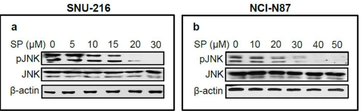
JNK activation was pharmacologically inhibited by treatment with
SP600125
Since HER2 protein expression in GC cell lines varied, GC cell lines SNU-216
and NCI-N87 showing a high level of HER2 expression were selected. In order to
observe the role of JNK in HER2-overexpressing GC cells, cells were treated with
a specific JNK inhibitor SP600125 for 24 h. Western blot confirmed that JNK
activation (manifested by pJNK expression) was inhibited in both GC cell lines in
a dose-dependent manner (Fig. 1).
Pharmacological inhibition of JNK reduces the cell growth, migration and
invasion
Since the effects of JNK on cell growth (Shibata et al. 2008; Ohsawa et al. 2010;
Hong et al. 2015) and metastatic potential in GC have been inconsistent (Fu et al.
2007; Mishra et al. 2010; Li et al. 2012), we first confirmed the effect of JNK on
12
GC cell growth. Treatment of GC cells with SP600125 for 48 h decreased cell
growth by 68% in SNU-216 and by 54% in NCI-N87 compared to control cells
(Fig. 2a, d). These findings indicate that JNK activation induces cell proliferation
in HER2-overexpressing GC cells.
Next, to determine the effects of JNK activation on the cell motility and invasion
in GC cells were determined by transwell migration assay. We found that
SP600125 treatment for 48 h significantly suppressed the cancer cell migration
by 43% (P = 0.0288) in SNU-216 cells and by 38% (P = 0.0033) in NCI-N87 cells
(Fig. 2b, e) compared to control cells. Consistently, cell invasion assay showed
that JNK inhibition decreased cancer cell invasion by 49% (P = 0.005) in SNU-
216 cells and by 43% (P = 0.0132) in NCI-N87 cells (Fig. 2c, f) compared to
control cells.
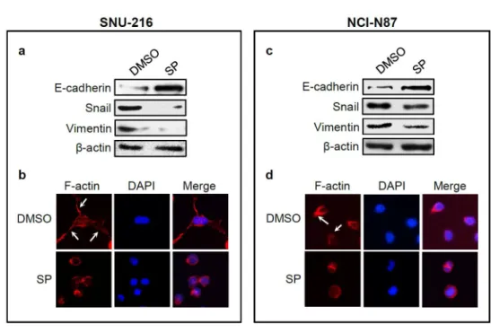
JNK inhibition regulates the expressions of epithelial-mesenchymal
transition (EMT) markers
13
In the initial steps of metastasis of carcinoma cells, epithelial cancer cells change
their phenotype to mesenchymal phenotype and become motile and invasive by
a process called EMT (Guarino et al. 2007). This process includes
downregulation of epithelial markers and upregulation of mesenchymal markers
(Guarino et al. 2007). In the present study, Western blot showed that the
expression of the representative epithelial marker E-cadherin was enhanced,
whereas the expressions of mesenchymal markers Snail and Vimentin were
reduced after SP600125 treatment for 24 h (Fig. 3a, c). These results indicate the
stimulatory role of JNK in the EMT in GC cells. To further confirm these results,
we examined actin organization because actin-dependent membrane protrusions
are regarded as a critical determinant of EMT (Shankar et al. 2010). Staining of
F-actin with FITC-conjugated phalloidin revealed the presence of many filopodia-
like extensions containing actin-rich bundles in control cells with a more
elongated shape, whereas they were absent or less obvious in SP600125-treated
cells with cuboidal morphology (Fig 3b, d).
14
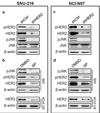
Crosstalk between HER2 and JNK exists in GC cell lines
We previously reported that HER2 downregulation in GC cell lines decreased
cancer cell growth, migration and invasion as well as EMT markers (Ko et al.
2015). We found consistent results in present study (data is not shown). Then,
we investigate the relationship between HER2 and JNK. First, we determined the
effect of HER2 silencing on JNK activation (manifested by pJNK expression) and
expression. Western blot showed that JNK activation and expression were
markedly reduced in HER2 shRNA transfectants compared to control shRNA
transfectants (Fig. 4a, d). On the other hand, Western blot and RT-PCR analysis
showed that the expressions of HER2 protein and mRNA were substantially
decreased by treatment with SP600125 (Fig. 4b, e).
In addition, double immunofluorescence staining for pJNK and HER2 was
performed. We found that, in both cell lines, control cells with pJNK staining in the
nucleus (Fig. 5a, i) showed strong immunofluorescence at the plasma membrane
(Fig. 5b, j). After SP600125 treatment, which drastically reduced the nuclear
15
immunofluorescence for pJNK (Fig. 5e, m), reduced the immunofluorescence for
HER2 at the plasma membrane to a negligible level (Fig. 5f, n). These results
indicate that there is a positive reciprocal regulatory loop between JNK and HER2.
JNK inhibitor exerts synergistic effect when combined with HER2 shRNA
transfection
The above results showed that both HER2 and JNK enhanced metastatic
potential in GC cells and that a positive-crosstalk exists between HER2 and JNK.
Fig. 6a showed that the combination of HER2 silencing and pharmacological
inhibition of JNK markedly reduced the expressions of pHER2 and pJNK. To
investigate whether combination of these two molecules has synergistic effects
on GC cell malignancy, we examined the effect of double inhibition of these
molecules on the cell growth, migration and invasion (Fig. 6b-d). Cell growth
curves (Fig. 6b) showed that HER2 silencing and JNK inhibition reduced cell
number to similar extent (63% reduction in HER2 silencing and 70% reduction in
JNK inhibition, respectively), which was further suppressed by inhibition of both
16
molecules (91% reduction in control). Furthermore, the cell migration capacity
decreased by 49% in cells treated with SP600125, and by 47% in HER2 shRNA
transfected cells compared with control cells. The combination of HER2 down-
regulation and JNK inhibition showed lower migration capacity (61% lower than
that of the control) than those with suppression of either alone (P < 0.005) (Fig.
6c). Similar results were shown in the invasion assay (63% lower invasion ability
in the combined treatment than that of the control) (Fig. 6d). Consistent results
were shown in the experiments using NCI-N87 cells (Fig. 6e-h).
17 Figure 1.
Figure 1. Effect of pharmacological inhibition of JNK in GC cell lines SNU-
216 and NCI-N87 cells. Cells were treated with various concentrations of
SP600125 (SP) for 24 h. Protein expressions of pJNK and total JNK were
determined by Western blot.
18 Figure 2.
19
Figure 2. Effect of pharmacological inhibition of JNK on cell growth,
migration and invasion in GC cell lines. GC cells were cultured in the
presence or absence of SP600125 (SP). a, d Cells were treated with SP600125
(SP) (20 μM for SNU-216 and 30 μM for NCI-N87) for 48 h. Cell growth was
analyzed using crystal violet assay on the indicated times, and absorbance was
measured at 570 nm. b, e The effect of treatment with SP (20 μM for SNU-216
and 30 μM for NCI-N87) on the cell migration was analyzed after plating for 48 h
by Transwell migration assay. c, f The effect of treatment with SP on the cell
invasion was analyzed 48 h after plating by cell invasion assay. Results were
calculated as percentages relative to control cells. Each bar represents the mean
± SD (n = 4 per each group). * P < 0.05 vs control cells.
20 Figure 3.
Figure 3. Effect of JNK inhibition on EMT marker expressions and actin
cytoskeleton organization. SNU-216 and NCI-N87 GC cells were treated with
SP600125 (SP) for 24 h. a, c The effect of SP (20 μM for SNU-216 and 30 μM for
NCI-N87) treatment for 24 h on EMT marker expressions were analyzed by
Western blot for E-cadherin, Snail and Vimentin. b, d Changes in the
organization of the actin cytoskeleton. Cells were stained with Alexa Fluor 633-
conjugated phalloidin to visualize F-actin (red), and the cell nuclei were visualized
by DAPI staining (blue). Photographs were taken with a fluorescence microscope
(x 400 magnification).
21 Figure 4.
Figure 4. The relationship between HER2 and JNK in GC cell lines. SNU-216
cells (a, b) and NCI-N87 cells (c, d) were transfected with HER2 shRNA (a, c) or
treated with SP600125 (SP) (b, d). a, c The effects of HER2 shRNA transfection
on expressions of pHER2, HER2, pJNK and JNK were determined by Western
blot. b, d The effects of SP treatment (20 μM for SNU-216 and 30 μM for NCI-
N87) on the expressions of pJNK, pHER2 and HER2 were determined by
Western blot (WB) and the expression of HER2 were determined by RT-PCR.
22 Figure 5.
23
Figure 5. Immunofluorescence staining showing the effect of SP600125 on
HER2 expression. SNU-216 cells (a-h) and NCI-N87 cells (i-p) were cultured
with vehicle control (DMSO) or SP600125 (SP). The effect of JNK inhibition on
the subcellular localization of HER2 was observed by double
immunofluorescence staining for pJNK (green) and HER2 (red). 4’,6’-diamidino-
2-phenylindole (DAPI) staining was performed for nucleus (blue). (x 400
magnification).
24 Figure 6.
25
Figure 6. Synergistic effects of HER 2 and JNK on metastatic potential of
GC cells. SNU-216 (a-d) and NCI-N87 (e-h) GC cell lines were transfected with
either control shRNA (shCtrl) or HER2 shRNA (shHER2). Then, cells were
treated with vehicle control (DMSO) or SP600125 (SP) (20 μM for SNU-216 and
30 μM for NCI-N87). a, e Transfected cells were treated with SP for 24 h. Protein
expression of pHER2 and pJNK were determined by Western blot. b, f
Transfected cells were treated with SP for 48 h after plating. Cell growth rates
were analyzed using the crystal violet assay on the indicated times, and
absorbance was measured. c, g Transfected cells were treated with SP for 48 h
after plating. Effect of interaction of HER2 and JNK on cell migration capacity was
evaluated by Trans well migration assay. d, h Transfected cells were treated with
SP for 48 h after plating. Effect of interaction of HER2 and JNK on cell invasion
capacity was evaluated by cell invasion assay. Results were calculated as
percentages relative to control cells. Each bar represents the mean ± SD (n = 4
per each group). * P < 0.05 vs control cells.
26
Discussion
Understanding the oncogenic signaling pathways may lead to the development of
therapeutic strategies for cancer treatment. Although the pivotal role of HER2 in
gastric cancer progression has been shown, the link between HER2 and JNK has
not been validated in gastric cancer. This is the first study, to the best of our
knowledge, to show the association between HER2 and JNK and their
significance in the cell growth and metastatic potential in gastric cancer cells.
Two key survival pathways downstream of HER2 included the
phosphatidylinositol 3-kinase (PI3K)/Akt pathway and the MAPK pathway
(Gschwantler-Kaulich et al. 2016). Among these pathways, PI3K/Akt pathway has
gained much attention for the development of an alternative target for anti-HER2
cancer therapy (Chakrabarty et al. 2013). However, the interest has been
diverged to the JNK pathway in experiments using breast cancer cells. JNK,
which belongs to MAPK family, has shown to promote cell survival in HER2-
27
overexpressing breast cancer cells (Han and Crowe. 2010). Moreover, the
current study by Gschwantler-Kaulich et al. (2016) reported that JNK mediated
the anti-HER2 drug resistance in HER2-overexpressing breast cancer cells.
These results suggested that JNK plays a central role in the HER2 signaling in
HER2-overexpressing breast cancer cells.
Metastatic tumor relapses are characterized by rapid proliferating, drug-
resistant cancers (Chakrabarty et al. 2013). With respect to gastric cancer,
implications of JNK in HER2-overexpressing gastric cancer cell growth have
been inconsistent. JNK activation increased the gastric cancer cell survival in
BCG-823 cells (Xia et al. 2006), whereas it decreased the cell survival in SGC-
7901 cells (Xiao et al. (2012). In the present study, we confirmed that JNK, like
HER2, increased the cell growth of SNU-216 and NCI-N87 gastric cancer cells
with HER2 overexpression, which is consistent with the finding of Xiao et al.
shown in breast cancer cells.
28
In previous studies, the role of JNK in HER2-overexpressing gastric cancer
cells still remains elusive. Wang et al. (Wang et al. 2010) reported that JNK
induced gastric cancer cell migration and invasion were increased by JNK in
SGC7901 cells, whereas they were suppressed in SNU-216 cells (Jung et al.
2013). Thus, the role of JNK in GC cell metastasis needs to be further elucidated.
Previously, Ko et al. (Ko et al. 2015) showed that HER2 increased cell
migration, invasion in vitro and metastasis of two HER2-overexpressing gastric
cancer lines (SNU-216 and NCI-N87) in nude mice. However, the association
between HER2 and JNK in these cell lines has not been determined. In the
present study, we investigated implications of JNK activation in HER2-
overexpressing gastric cancer cell lines. We found that the pharmacological
inhibition of the JNK by SP600125 resulted in decreased migration and invasion
ability in both gastric cancer cell lines. Furthermore, analyses of the EMT marker
expressions and actin organization revealed that JNK inhibition suppressed the
expressions of mesenchymal markers (Snail and Vimentin) and increased E-
29
cadherin expression accompanied by appearance of epithelial morphology. Thus,
these results indicate that JNK might contribute to acquisition of malignant
phenotype of GC cells through promotion of EMT. Our data provide evidences for
the role of JNK in the regulation of metastatic potential in GC cells through the
modulation of mesenchymal phenotype.
The success of future trials of combination anti-HER2 therapy will depend on
the selection of molecular biomarker selection. Major survival pathways
downstream of HER2 included phosphatidylinositol 3-kinase (PI3K)/Akt pathway
and MAPK pathway (Gschwantler-Kaulich et al. 2016). Among these pathways,
PI3K/Akt pathway has gained much attention for the development of an
alternative target for anti-HER2 cancer therapy (Chakrabarty et al. 2013).
However, the interest has been diverged to the JNK pathway. JNK, which
belongs to MAPK family, has shown to promote cell survival in HER2-
overexpressing breast cancer cells (Han and Crowe. 2010). Moreover, the
current study by Gschwantler-Kaulich et al. (Gschwantler-Kaulich et al. 2016)
30
showed that JNK was inhibited in lapatinib-sensitive HER2-overexpressing breast
cancer cells, but not in the lapatinib-resistant cancer cells.
In the present study, we investigated the role of JNK in HER2-
overexpressing gastric cancer cell lines. The pharmacological inhibition of the
JNK by SP600125 resulted in decreased migration and invasion ability in HER2-
overexpressing gastric cancer cell lines. Furthermore, analyses of the EMT
marker expressions and actin organization revealed that JNK inhibition
suppressed the expressions of mesenchymal markers (Snail and Vimentin) and
increased E-cadherin expression accompanied by appearance of epithelial
morphology. Thus, these results indicate that JNK might contribute to acquisition
of malignant phenotype of GC cells through promotion of EMT. Our data provide
direct evidence for the role of JNK in the regulation of metastatic potential in GC
cells through the modulation of mesenchymal phenotype.
Since we wondered whether there is a reciprocal regulatory loop between
31
JNK and HER2 in GC, we further analyzed the effect of HER2 silencing on the
JNK activation. We found that HER2 downregulation suppressed JNK activation
(manifested by pJNK expression). In turn, JNK inhibition was accompanied by
decreases in the expression of HER2 and pHER2. These results suggest that
JNK controls and is controlled by HER2, and that there is a positive cross-talk
between HER2 and JNK in GC cells.
Although targeted therapies may offer enhanced efficacy and improved
selectivity, mostly their effects are not durable when they are used alone. For this
reason, combination therapies are often needed to effectively treat many tumors.
In the present study, we found that the combination of HER2 silencing and JNK
inhibition further reduced migration and invasion of gastric cancer cells compared
to inhibition of each molecule. Therefore, HER2 and JNK seems to act in a
synergistic manner in modulating migration and invasion of gastric cancer cells.
In conclusion, the present study found that JNK activation in HER2-
32
overexpressing gastric cancer cell lines increased cell growth, migration and
invasion as well as EMT markers. In addition, the interaction between HER2 and
JNK, which showed positive crosstalk, synergistically contributes to the GC cell
growth and metastatic potential in HER2-overexpressing GC cells. Given the
importance of HER2 overexpression in the gastric tumor development and
progression, the identification of JNK as an important downstream component of
HER2 oncogenic signaling pathway represents an important finding. Further
experiments using anti-HER2 drug-resistant gastric cancer cells and animal
studies might give evidence that this pathway could serve as a useful target for
therapeutic strategies for treatment of HER2-overexpressing gastric
cancer.attractive as therapeutic strategies in gastric cancer (Chen et al. 2010).
HER2 is of particular interest as a drug target in GC therapy because it has been
shown to be amplified and/or overexpressed in a subset of gastric cancers (Chen
et al. 2010). However, due to development of acquired drug resistance,
alternative therapeutic targets are needed to effective treatment of HER2-
overexpressing GC patients. Understanding HER2 signaling pathways may lead
33
to the development of therapeutic strategies for cancer treatment. In present
study, we investigated the relationship between HER2 and JNK in terms of
proliferation and metastatic potential of GC cells. To the best of our knowledge,
this is the first study to show the association between HER2 and JNK in GC.
Major survival pathways downstream of HER2 included phosphatidylinositol
3-kinase (PI3K)/Akt pathway and MAPK pathway (Gschwantler-Kaulich et al.
2016). Among these pathways, PI3K/Akt pathway has gained much attention for
the development of an alternative target for anti-HER2 therapy (Chakrabarty et al.
2013). However, the interest has been diverged to the JNK pathway. JNK, which
belongs to MAPK family, has shown to promote cell survival in HER2-
overexpressing breast cancer cells (Han and Crowe. 2010). Moreover, a recent
study (Gschwantler-Kaulich et al. 2016) showed that JNK was inhibited in
lapatinib-sensitive HER2-overexpressing breast cancer cells, but not in the
lapatinib-resistant cells. These results indicate that lapatinib-induced apoptosis in
HER2-overexpressing breast cancer cells is mediated through JNK inhibition.
34
Metastatic tumor relapses are characterized by rapid proliferating, drug-
resistant cancers (Chakrabarty et al. 2013). With respect to GC growth, the
implication of JNK has been inconsistent (Chen et al. 2002; Xia et al. 2006;
Shibata et al. 2008; Xiao et al. 2012). Previously, in vitro studies (Chen et al.
2002; Xiao et al. 2012) showed that JNK activation decreased the cell survival in
GC cell lines (MGC80-3, SGC-7901). In contrast, JNK activation increased the
survival in GC cell lines BCG-823, MKN-45 and AGS (Xia et al. 2006). Moreover,
Shibata et al. (Shibata et al. 2008) observed that loss of JNK decreased the
tumorigenesis of gastric tumor in mice. Thus, the role of JNK in GC still remains
elusive. In the present study, we confirmed that JNK, like HER2, increased the
GC cell growth, which is consistent with the finding of Han and Crowe (Han and
Crowe. 2010).
Initial studies of JNK examined their ability to induce the phosphorylation of
several transcription factors, such as the oncogene c-Jun. However, recent
evidence indicates that JNK have conserved their evolutionary role in cell
35
movement, cytoskeleton rearrangement and migration. The ability of tumor cells
to migrate from the primary tumor to surrounding tissues is a prerequisite for
metastasis. JNK promotes increased motility of multiple cell types including tumor
cells (Ono et al. 2008). Although findings reported here support a role for JNK in
GC metastasis, the mechanism by which this pathway is activated in GC have
not been elucidated. To address the underlying molecular mechanism involved in
the JNK pathway implicated in cancer cell motility, we used migration and
invasion assay in the HER2-overexpressing GC cells. The inhibition of the JNK
by treatment with SP600125 resulted in a decreased migration and invasion
ability in GC cells. Furthermore, analyses of the EMT marker expressions and
actin organization revealed that SP600125 treatment suppressed the
expressions of mesenchymal markers (Snail and vimentin) and increased E-
cadherin expression, with induction of epithelial morphology. In addition, these
phenotypic changes were attenuated by inhibition of JNK activation. Thus, these
results indicate that JNK might contribute to acquisition of malignant phenotype of
GC cells through promotion of EMT. Our data provide direct evidence for the role
36
of JNK in the regulation of metastatic potential in GC cells through the modulation
of mesenchymal phenotype.
Previous ours (Ko et al. 2015) showed that HER2 increased GC cell
migration and invasion. However, the role of JNK in HER2-mediated cell motility
is less defined. Since we wondered whether there is a reciprocal regulatory loop
between JNK and HER2 in GC, we further analyzed the effect of HER2 silencing
on the JNK activation. We found that HER2 downregulation suppressed JNK
activation (manifested by pJNK expression). In turn, JNK inhibition was
accompanied by decreases in the expression of HER2 and pHER2. These results
suggest that there is a positive cross-talk between HER2 and JNK in GC cells.
Although targeted therapies may offer enhanced efficacy and improved
selectivity, mostly their effects are not durable when they are used alone. For this
reason, combination therapies are often needed to effectively treat many tumors
(Yoon et al. 2010). In the present study, we found that the combination of HER2
37
silencing and JNK inhibition further reduced migration and invasion of gastric
cancer cells compared to inhibition of each molecule. Therefore, HER2 and JNK
seems to act in a synergistic manner in modulating migration and invasion of
gastric cancer cells.
In conclusion, the present study identified HER2/JNK as a signaling pathway
responsible further migration and invasion in GC for the first time. Given the
importance of EMT in the tumor development in the progression the identification
of JNK as a key regulator of HER2-induced EMT represents an important finding.
Further studies might give evidence that this pathway could serve as a useful
target for therapeutic strategies for regard to EMT reversion.
38
Reference
Bang YJ (2012) Advances in the management of HER2-positive advanced gastric and gastroesophageal junction cancer. J Clin Gastroenterol 46:637–648
Bubici C, Papa S (2014) JNK signalling in cancer: in need of new, smarter therapeutic targets. Br J Pharmacol 171:24-37
Chen J, Miller EM, Gallo KA (2010) MLK3 is critical for breast cancer cell migration and promotes a malignant phenotype in mammary epithelial cells. Oncogene 29:4399-411
Chen XY, Zhou J, Luo LP, Han B, Li F, Chen JY, Zhu YF, Chen W, Yu XP (2015) Black Rice Anthocyanins Suppress Metastasis of Breast Cancer Cells by Targeting RAS/RAF/MAPK Pathway. Biomed Res Int 2015:414250
Fu P, Thompson JA, Bach LA (2007) Promotion of cancer cell migration:
an insulin-like growth factor (IGF)-independent action of IGF-binding
39
protein-6. Biol Chem 282:22298-306
Guarino M, Rubino B, Ballabio G (2007) The role of epithelial- mesenchymal transition in cancer pathology. Pathology 39:305–318 Gschwantler-Kaulich D, Grunt TW, Muhr D, Wagner R, Kölbl H, Singer CF
(2016) HER Specific TKIs Exert Their Antineoplastic Effects on Breast Cancer Cell Lines through the Involvement of STAT5 and JNK. PLoS One 11:e0146311
Han JS and Crowe DL (2010) Jun amino-terminal kinase 1 activation promotes cell survival in ErbB2-positive breast cancer. Anticancer Res 30:3407–3412
Hong NR, Park HS, Ahn TS, Jung MH, Kim BJ (2015) Association of a Methanol Extract of Rheum undulatum L. Mediated Cell Death in AGS Cells with an Intrinsic Apoptotic Pathway. J Pharmacopuncture 18:26-32 Hui L, Zatloukal K, Scheuch H, Stepniak E, Wagner EF (2008) Proliferation
of human HCC cells and chemically induced mouse liver cancers
requires JNK1-dependent p21 downregulation. J Clin Invest 118:3943-
40
3953
Janjigian YY (2015) Lapatinib in Gastric Cancer: What Is the LOGiCal Next Step? J Clin Oncol pii:CO642892
Jung MK, Houh YK, Ha S, Yang Y, Kim D, Kim TS, Yoon SR, Bang SI, Cho BJ, Lee WJ, Park H, Cho D (2013) Recombinant Erdr1 suppresses the migration and invasion ability of human gastric cancer cells, SNU-216, through the JNK pathway. Immunol Lett 150:145–151
Kim HP, Han SW, Song SH, Jeong EG, Lee MY, Hwang D (2014) Testican- 1-mediated epithelial-mesenchymal transition signaling confers acquired resistance to lapatinib in HER2-positive gastric cancer. Oncogene 33:3334-41
Kim WH, Schnaper HW, Nomizu M, Yamada Y, Kleinman HK (1994) Apoptosis in human fibrosarcoma cells is induced by a multimeric synthetic Tyr-Ile-Gly-Ser-Arg (YIGSR)-containing polypeptide from laminin. Cancer Res 54:5005–5010
Ko YS, Cho SJ, Park J, Kim Y, Choi YJ, Pyo JS, Jang BG, Park JW, Kim
41
WH, Lee BL (2015) Loss of FOXO1 promotes gastric tumour growth and metastasis through upregulation of human epidermal growth factor receptor 2/neu expression. Br J Cancer 113:1186-1196
Kyriakis JM and Avruch J (2001) Mammalian mitogen-activated protein kinase signal transduction pathways activated by stress and inflammation. Physiol Rev 81:807-869
Li LH, Luo Q, Zheng MH, Pan C, Wu GY, Lu YZ, Feng B, Chen XH, Liu BY (2012) P21-activated protein kinase 1 is overexpressed in gastric cancer and induces cancer metastasis. Oncol Rep 27:1435-1442
Lin W, Kao HW, Robinson D, Kung HJ, Wu CW, Chen HE (2000) Tyrosin kinases and gastric cancer. Oncogene 19:5680-5689
Matsui Y, Inomata M, Tojigamori M, Sonoda K, Shiraishi N, Kitano S (2005) Suppression of tumor growth in human gastric cancer with HER2 overexpression by an anti-HER2 antibody in a murine model. Int J Oncol 27:681-685
Mishra P, Senthivinayagam S, Rangasamy V, Sondarva G, Rana B (2010)
42
Mixed lineage kinase-3/JNK1 axis promotes migration of human gastric cancer cells following gastrin stimulation. Mol Endocrinol 24:598–607 Oh DY, Doi T, Shirao K, Lee KW, Park SR, Chen Y, Yang L, Valota O, Bang
YJ (2015) Phase I Study of Axitinib in Combination with Cisplatin and Capecitabine in Patients with Previously Untreated Advanced Gastric Cancer. Cancer Res Treat 47:687-696
Ono R, Matsuoka J, Yamatsuji T, Naomoto Y, Tanaka N, Matsui H, Matsushita M (2008) M-RIP, a novel target of JNK signaling and a requirement for human cancer cell invasion. Int J Mol Med 22:199-203 Ohsawa R, Miyazaki H, Niisato N, Shiozaki A, Iwasaki Y, Otsuji E,
Marunaka Y (2010) Intracellular chloride regulates cell proliferation through the activation of stress-activated protein kinases in MKN28 human gastric cancer cells. J Cell Physiol 223:764-770
Qin X, Zheng S, Liu T, Liu Q, Liang M, Li X, Sheyhidin I, Lu X (2014) Roles
of phosphorylated JNK in esophageal squamous cell carcinomas of
Kazakh ethnic. Mol Carcinog 53:526–536
43
Sahu SK, Garding A, Tiwari N, Thakurela S, Toedling J, Gebhard S, Ortega F, Schmarowski N, Berninger B, Nitsch R, Schmidt M, Tiwari VK (2015) JNK-dependent gene regulatory circuitry governs mesenchymal fate. EMBO J 34:2162-2181
Shankar J, Messenberg A, Chan J, Underhill TM, Foster LJ, Nabi IR (2010) Pseudopodial actin dynamics control epithelial-mesenchymal transition in metastatic cancer cells. Cancer Res 70:3780–3790
Shibata W, Maeda S, Hikiba Y, Yanai A, Sakamoto K, Nakagawa H, Ogura K, Karin M, Omata M (2008) c-Jun NH2-terminal kinase 1 is a critical regulator for the development of gastric cancer in mice. Cancer Res 2008 68:5031-5039
Vivanco I, Palaskas N, Tran C, Finn SP, Getz G, Kennedy NJ, Jiao J, Rose J, Xie W, Loda M, Golub T, Mellinghoff IK, Davis RJ, Wu H, Sawyers CL (2007) Identification of the JNK signaling pathway as a functional target of the tumor suppressor PTEN. Cancer Cell 11:555–569
Wang J, Kuiatse I, Lee AV, Pan J, Giuliano A, Cui X (2010) Sustained c-
44
Jun-NH2-kinase activity promotes epithelial-mesenchymal transition, invasion, and survival of breast cancer cells by regulating extracellular signal-regulated kinase activation. Mol Cancer Res 8:266-277
Wei W, Li H, Li N, Sun H, Li Q, Shen X (2013) WNT5A/JNK signaling regulates pancreatic cancer cells migration by Phosphorylating Paxillin.
Pancreatology 13:384-392
Yoon J, Cho SJ, Ko YS, Park J, Shin DH, Hwang IC, Han SY, Nam SY, Kim MA, Chang MS, Lee HS, Kim WH, Lee BL (2013) A synergistic interaction between transcription factors nuclear factor-κB and signal transducers and activators of transcription 3 promotes gastric cancer cell migration and invasion. BMC Gastroenterol 13:29–39
Xia HH, He H, De Wang J, Gu Q, Lin MC, Zou B, Yu LF, Sun YW, Chan AO, Kung HF, Wong BC (2006) Induction of apoptosis and cell cycle arrest by a specific c-Jun NH2-terminal kinase (JNK) inhibitor, SP- 600125, in gastrointestinal cancers. Cancer Lett 241:268-274
Xiao F, Liu B, Zhu QX (2012) c-Jun N-terminal kinase is required for
45
thermotherapy-induced apoptosis in human gastric cancer cells. World J Gastroenterol 18:7348-7356
Zhang H, Liu L, Wang Y, Zhao G, Xie R, Liu C, Xiao X, Wu K, Nie Y, Zhang
H, Fan D (2013) KLF8 involves in TGF-beta-induced EMT and promotes
invasion and migration in gastric cancer cells. J Cancer Res Clin Oncol
139:1033-42
46
국 문 초 록
목적: 위암에서 표피성장인자2 (epidermal growth factor receptor2, EGFR2,
HER2)의 과발현은 위암에서 악성도 증가를 초래하므로 HER2는 위암 환자에서 임상적 효과를 나타내는 유일한 치료표적이다. 그러나 획득성 내성으로 인하여 치료 효율의 향상은 제한되어 있다. c-Jun N-terminal kinase
(JNK)는 위암에서 과발현되어 있으나 아직까지 확실한 기능은 알려져 있지
않다. HER2와 JNK의 관련성이 유방암에서 보고되어 있으므로 본 연구의 목적은 HER2를 과발현하는 위암세포주들인 SNU-216과 NCI-N87 세포들을 사용하여 JNK가 위암세포의 성장, 이동 및 침윤에 미치는 영향을 확인하고,
HER2와의 관련성을 관찰함으로써 위암환자의 치료제 개발에 유용한 분자적
기전을 규명하고자 하였다.
방법: SNU-216과 NCI-N87 위암세포주를 JNK 저해제인 SP600125로
처리하여 JNK의 활성을 억제시켰고, HER2 shRNA 이입으로 HER2의 발현을 감소시켰다. Western blot를 시행하여 HER2, phospho-HER2 (pHER2), JNK,
phospho-JNK (pJNK), E-cadherin, Snail 및 Vimentin의 단백 발현 변화를 관찰하였다. 세포수의 관찰을 위하여 Crystal violet assay를 시행하였고, 세포의 이동과 침윤 관찰을 위하여 Transwell migration assay와 invasion assay를 시행하였다.
47
결과: 위암세포들에서 HER2와 JNK는 위암세포의 성장과 전이능을
증가시켰으며 상호작용을 통하여 그 효과를 상승시키는 관련성을 나타내었다.
또한, 이들은 E-cadherin의 발현을 감소시키고 Snail과 Vimentin의 발현은 증가시켰으며, 형태학적으로 간엽세포와 유사한 변화를 유발시켰다.
결론: 위암세포에서 JNK는 HER2와 유사하게 세포성장 및 전이능의 증가를 초래하였으며, HER2와의 상호작용을 통하여 이러한 기능이 상승효과를 나타내었다.
주요어: 위암; JNK; HER2; 전이능; EMT