저작자표시-비영리-변경금지 2.0 대한민국 이용자는 아래의 조건을 따르는 경우에 한하여 자유롭게
l 이 저작물을 복제, 배포, 전송, 전시, 공연 및 방송할 수 있습니다. 다음과 같은 조건을 따라야 합니다:
l 귀하는, 이 저작물의 재이용이나 배포의 경우, 이 저작물에 적용된 이용허락조건 을 명확하게 나타내어야 합니다.
l 저작권자로부터 별도의 허가를 받으면 이러한 조건들은 적용되지 않습니다.
저작권법에 따른 이용자의 권리는 위의 내용에 의하여 영향을 받지 않습니다. 이것은 이용허락규약(Legal Code)을 이해하기 쉽게 요약한 것입니다.
Disclaimer
저작자표시. 귀하는 원저작자를 표시하여야 합니다.
비영리. 귀하는 이 저작물을 영리 목적으로 이용할 수 없습니다.
변경금지. 귀하는 이 저작물을 개작, 변형 또는 가공할 수 없습니다.
의학박사 학위논문
Integrin-FAK 신호전달체계를 조절하여 폐 섬유화를 감소시키는 IL-32γ 의
기전 연구
IL-32γ Attenuates Airway Fibrosis by Modulating the Integrin-FAK Signaling Pathway in Fibroblasts
울 산 대 학 교 대 학 원 의 학 과
박 소 영
[UCI]I804:48009-200000174697 [UCI]I804:48009-200000174697 [UCI]I804:48009-200000174697
Integrin-FAK 신호전달체계를 조절하여 폐 섬유화를 감소시키는 IL-32γ 의
기전 연구
지 도 교 수 조 유 숙
이 논문을 의학박사 학위 논문으로 제출함
2018년 12월
울 산 대 학 교 대 학 원 의 학 과
박 소 영
박소영의 의학박사학위 논문을 인준함
심사위원 김 태 범 인 심사위원 조 유 숙 인 심사위원 유 광 하 인 심사위원 김 상 헌 인 심사위원 송 우 정 인
울 산 대 학 교 대 학 원
2018년 12월
ABSTRACT Background
Fibrosis in severe asthma often leads to irreversible organ dysfunction. However, the mechanism that regulates fibrosis remains poorly understood. Interleukin (IL)-32 plays a role in several chronic inflammatory diseases, including severe asthma. This study investigated whether IL-32 is involved in fibrosis progression in the lungs.
Methods
Murine models of chronic airway inflammation induced by ovalbumin and Aspergillus melleus protease and bleomycin-induced pulmonary fibrosis were employed. The degree of tissue fibrosis after treatment with recombinant IL-32γ (rIL-32γ) was evaluated. Expression of fibronectin and α-smooth muscle actin (α-SMA) was examined and the transforming growth factor (TGF)-β-related signaling pathways was evaluated in activated human lung fibroblasts (MRC-5 cells) treated with rIL-32γ.
Results
rIL-32γ significantly attenuated collagen deposition and α-SMA production in both mouse models. rIL-32γ inhibited the production of fibronectin and α-SMA in MRC-5 cells stimulated with TGF-β. Additionally, rIL-32γ suppressed activation of the integrin-FAK-paxillin signaling axis but had no effect on the Smad and non-Smad signaling pathways. rIL-32γ localized outside of MRC-5 cells and inhibited the interaction between integrins and the extracellular matrix without directly binding to intracellular FAK and paxillin.
Conclusion
These results demonstrate that IL-32γ has anti-fibrotic effects and is a novel target for preventing fibrosis.
Key words: Airway Inflammation; Asthma; Interleukin-32γ; Pulmonary Fibrosis; Subepithelial Fibrosis.
TABLE OF CONTENTS
ABSTRACT ··· i
LIST OF FIGURES ··· iv
INTRODUCTION ··· 1
MATERIALS AND METHODS ··· 3
RESULTS··· 9
DISCUSSION··· 24
CONCLUSIONS ··· 28
REFERENCES··· 29
ABSTRACT IN KOREAN··· 33
LIST OF FIGURES
Figure 1 ··· 10
Figure 2 ··· 12
Figure 3 ··· 14
Figure 4 ··· 16
Figure 5 ··· 18
Figure 6 ··· 20
Figure 7 ··· 22
INTRODUCTION
Fibrosis, characterized by the accumulation of fibroblasts and excess extracellular matrix, is a common feature of various pathological states in many organs, resulting in dysfunction. Interstitial lung diseases and chronic inflammatory airway diseases of the lungs, such as severe asthma and chronic obstructive pulmonary disease (COPD), lead to sub- bronchial fibrosis and pulmonary fibrosis, both of which result in irreversible structural changes that affect patient survival 1-3). Because lung fibrosis is mainly a consequence of chronic inflammation, therapeutic strategies have focused on preventing inflammation by administering immunosuppressive agents or anti-inflammatory drugs, including corticosteroids 4,5). However, recent studies have suggested that inflammation alone is not sufficient for inducing fibrosis development. Many studies showed that immunosuppressive therapies do not prevent lung fibrosis 6). To date, targeting fibrosis itself has been unsuccessful.
Interleukin (IL)-32, initially described as NK4 generated by activated T cells or NK cells 7), is produced by various cells, including epithelial cells, endothelial cells, and macrophages.
IL-32 induces the production of several pro-inflammatory mediators, such as tumor necrosis factor (TNF)-α, IL-1β, and IL-6, by activating the nuclear factor-κB and p38 mitogen- activated protein kinase signaling pathways 8,9). IL-32 is also involved in several chronic inflammatory diseases, such as rheumatoid arthritis and COPD 10-12). In addition to its role in inflammation, recent studies suggested that IL-32 is involved in liver fibrosis in patients with chronic hepatitis by affecting cytokine induction 13). Although the precise effects of IL-32 on tissue fibrosis are largely unknown, IL-32 contains an RGD motif, which is known to bind several integrins 14). Moreover, a 3-dimensional reconstruction model of IL-32 revealed that its structure was highly similar to that of the focal adhesion targeting (FAT) region of focal adhesion kinase (FAK). FAK-related non-kinase, a peptide with a structure similar to the FAT
region, inhibits FAK signal transduction 15). It is known that integrin-FAK signaling axis is critical for the development of tissue fibrosis 16,17). Therefore, it was predicted that IL-32 interrupts the signaling pathway by binding to these molecules, thereby inhibiting FAK activation and alleviating fibrosis.
This study hypothesized that IL-32γ modulates fibrosis in chronic airway and lung diseases by disrupting the integrin-FAK signaling pathway. Here, murine models were used for chronic airway inflammation and bleomycin-induced pulmonary fibrosis to examine the role of IL-32γ in fibrosis of the airways and lungs, respectively. In addition, the role of IL-32γ in mechanisms underlying fibroblast function was evaluated.
MATERIALS AND METHODS
1. Generation of murine models of airway inflammation and pulmonary fibrosis
To generate the bleomycin-induced pulmonary fibrosis model, mice were administered intratracheal injection of bleomycin (1 U/kg body weight) on day 2. To evaluate the effect of IL-32γ treatment, mice were administered 500 ng of human recombinant IL-32γ (rIL-32γ) via intranasal injection on days 1, 2, 14, and 28. In this model, rIL-32γ was injected intranasally;
1 h later, bleomycin was injected intratracheally. Mice were sacrificed at 30 days. To generate the chronic asthma model, wild-type (WT) mice were sensitized by intranasal administration of 22 µg of ovalbumin (OVA) and 8 µg of protease (Aspergillus melleus protease; Sigma, St. Louis, MO, USA) twice per week for 8 weeks, as previously described [23]. Mice were sacrificed at 58 days. To evaluate the effect of IL-32γ treatment, mice were treated with 500 ng human recombinant IL-32γ (rIL-32γ) 2 h before each immunization.
Bronchoalveolar lavage fluid (BALF) and lung tissues were obtained from mice 24 h after the last immunization for further analysis. IL-32γ transgenic (TG) mice on a C57BL/6 background were generated as previously described 18). In brief, the ORF of IL-32γ cDNA was transferred into pCAGGS. The entire sequence was linearized with SalI and microinjected into mouse zygotes. Transgenic mice showed no physical abnormalities and were screened by RT-PCR. Wild-type (WT) C57BL/6 and Balb/c mice were purchased from OrientBio (Gapyong, Gyeonggi-do, Korea) and were used as a control. All mice were bred and maintained in a specific pathogen-free animal facility. The Institutional Animal Care and Use Committee approved all experimental procedures (Animal Utilization Protocol 2014-14- 013).
2. Histopathologic examination and quantification of tissue fibrosis
Lungs collected from mice (N=5) in each treatment group were perfused with 5 mL of PBS through the right ventricle. The lung was inflated by intratracheal infusion of 0.3% low- melting agar at 25 cm H2O. The inflated lung was fixed with 10% neutral buffered formalin.
Fixed lungs were embedded in paraffin and sectioned at 4 μm. Sections were deparaffinized, rehydrated, subjected to antigen retrieval by boiling in 10 mM sodium citrate buffer, pH 6.0, for 15 min, and blocked in 3% H2O2 (DaKo Peroxidase Blocking Solution) for 10 min at room temperature in a humidity chamber. To examine collagen deposition, lung sections were stained with Masson’s trichrome staining. The slides were washed 3 times with PBS, blocked with 0.25% casein in PBS (DaKo Protein Block Serum-Free) for 15 min at room temperature in a humid chamber, and then incubated with antibodies against α-SMA (Cell Signaling Inc., Danvers, MA) overnight in the dark at 4 °C. After washing 3 times with PBS, the slides were incubated with FITC-conjugated goat anti-rabbit IgG, and cy3-donkeyAnti- goat IgG for 1 h at room temperature in the dark, and detected using a DaKo EnVision HRP/DAB system (Dako, Carpinteria, CA, USA). The lung sections used for staining were mounted with mounting media fortified with DAPI (Vector Laboratories Inc, Burlingame, CA).
To quantify tissue fibrosis, hydroxyproline levels in the tissue were measured. To measure hydroxyproline in tissue, a hydroxyproline colorimetric assay kit (BioVision) per the manufacturer’s protocol was used. Additionally, quantification graphs were drawn from intensity measurement data using the Image J program (NIH, Bethesda, MD, USA).
3. Cell culture and study design
The human lung fibroblast cell line MRC-5 was purchased from the American Type Culture Collection (Manassas, VA, USA). Mouse embryonic fibroblasts (MEFs) obtained from IL-32γ TG mice were also used. MRC-5 cells were seeded at 2 × 10⁵ cells/well and
stimulated with recombinant proteins. These cells were cultured in MEM (Welgene, Seoul, Korea) with 10% FBS (Gibco, Carlsbad, CA) and 1% penicillin-streptomycin (Welgene).
Cells were seeded at a density of 2 × 10⁵ cells/well in 60-mm culture dishes and were stimulated with 5 ng/mL of TGF-β (R&D Systems, Minneapolis, MN), 10 ng/mL of TNF-α (R&D Systems), 1 μg/mL of LPS (Sigma), 10 μg/mL of Poly I: C (San Diego, CA), 10 ng/mL of IL-1β(R&D Systems), 50–200 µg RGD peptide (tripeptide Arg-Gly-Asp, R&D Systems), and 150 ng/mL of rIL-32 (YBDY, Seoul, Korea). To silence intracellular IL-32γ expression, small interfering siRNA (siRNA) to IL-32γ (antisense sequence: 5’-UCAUCAGAGAGGA CCUUCGUU-3’) was used. Expression of various cellular molecules was measured by Western blotting, reverse transcription-PCR, and semi-quantitative PCR. All in vitro experiments were conducted at least 3 times.
4. Cell adhesion assay
For crystal violet staining, 96-well culture dishes were coated with collagen (Advanced BioMatrix, Inc., San Diego, CA, USA) and seeded with MRC-5 cells. Plates were incubated for 30, 60, or 180 min. Cells were washed with PBS to remove non-adherent cells, and adhered cells were stained with crystal violet. Crystal violet-stained cells were dissolved using 33% acetic acid and OD values were measured at a wavelength of 550 nm. For measuring spindle-shaped cells, cells were observed and counted at 30 min after staining under the microscope (magnification: 100×).
5. Western blotting
Cells were lysed on ice in lysis buffer (Cell signaling Inc, Danvers, MA) containing protease inhibitors for 30 min, and then centrifuged at 18,000 ×g for 20 min at 4 °C. Proteins
were detected with antibodies against fibronectin, smad3, p-smad3, TGF-β receptor1, JNK, p-JNK, p38, p-p38, Erk, p-Erk, paxillin, p-paxillin (all from Cell Signaling Inc.), smurf2 and FAK (Santa Cruz Biotechnology, Dallas, TX), α-SMA (Abcam, Cambridge, UK), β-actin (Bioworld, St. Louis Park, MN), IL-32 (Ybdy, Seoul, Korea), and p-FAK (Invitrogen, Carlsbad, CA). Anti-rabbit, -goat, and -mouse secondary antibodies were purchased from Bethyl Laboratories (Montgomery, AL). Protein bands were detected with ECL solution (Genedepot, Barker, TX). The quantification graphs are drawn through intensity measurements using the Image J program.
6. RT-PCR and semi-quantitative PCR
Total RNA was extracted from cells using Trizol reagent (Invitrogen, Carlsbad, CA) and purified using RNeasy Mini Kits (Qiagen, Hilden, Germany) according to the manufacturers’
instructions. Purified RNA (1 μg) was reverse-transcribed to cDNA using oligo(dT) primers and reverse transcriptase (Roche Applied Science, Mannheim, Germany). Target amplification was performed with following primers: IL-32 sense, GACAGTGGCGGCTTATTATGAG; IL-32 antisense, CCTCGGCACCGTAATCCA T; TNF-α
sense, CGCTCTTCTGCCTGCTGCACTT; TNF-α antisense,
AGGCTTGTCACTCGGGGTTCGA; GAPDH sense, TGCACCACCAACTGCTTA; GAPDH antisense, GGCATGGACTGTGGTCAT; integrin α2sense, GGAACGGGACTTTCGCAT;
integrin α2 antisense, GGTACTTCGGCTTTCTCATCA; integrin αv sense, AATCTTCCAATTGAGGATATCAC; integrin αv antisense, AAAACAGCCAGTAGCAACAAT;
integrin β1sense, CGATGCCATCATGCAAGT; integrin β1 antisense, ACACCAGCAGCCGTGTAAC; integrin β3 sense, CCGTGACGAGATTGAGTCA; integrin β3
antisense, AGGATGGACTTTCCACTAGAA; integrin β5 sense,
GGAGCCAGAGTGTGGAAACA; integrin β5 antisense, GAAACTTTGCAAACTCCCTC;
integrin β8 sense, AATTTGGTAGTGGAAGCCTATC; integrin β8 antisense, GTCACGTTTCTGCATCCTTC.
7. His pull-down assay and immunoprecipitation
His-tagged IL-32γ (100 µg) was incubated with Ni-NTA agarose (Qiagen, Valencia, CA) at 4 °C for 2 h. The beads were then washed 3 times with buffer containing imidazole (Sigma). TGF-β-stimulated MRC-5 cells were lysed and centrifuged; The supernatant of TGF-β-stimulated MRC-5 lysate was incubated with IL-32 pre-bound Ni-NTA Agarose at 4 °C for 2 h. Then, proteins associated with the beads were subjected to immunoblot analysis using anti-integrin β3, anti-paxillin, or anti-His. For immunoprecipitation, flag-tagged IL-32γ- overexpressing MRC-5 cells were lysed on ice in lysis buffer (20 mM Tris, pH 7.5, 150 mM NaCl, 0.25% NP-40, 1.5 mM MgCl2, 1 mM phenylmethylsulfonyl fluoride). Anti-flag bound to protein G sepharose (GE Healthcare) was incubated with lysates at 4 for 3 h. Beads were washed 3 times with lysis buffer. Proteins were electrotransferred to PVDF membranes (GE Healthcare) and subjected to immunoblotting.
8. Live cell imaging of IL-32γ
MRC-5 cells were cultured in a μ-Dish 35 mm, High, IbiTreat (Ibidi GmbH, Martinsried, Germany) and treated with Flamma496-labeled IL-32γ. After 10 min, the cells were washed with medium. Fluorescence images were obtained under a Nikon Ti-E inverted I wamicroscope (Tokyo, Japan) equipped with PFS, iXon Ultra 897 EMCCD camera (Andor Technology, Belfast, UK), and excitation and emission filter wheels.
9. Statistical analysis
All data are reported as the mean ± standard error of mean. Means were compared using the Mann–Whitney test in GraphPad Prism software (version 4.0; GraphPad, Inc., La Jolla, CA, USA). A value of P < 0.05 was considered statistically significant.
RESULTS
1. IL-32γ modulates fibrosis in mouse models of airway inflammation and pulmonary fibrosis
First, histopathological analysis of bleomycin-induced lung fibrosis was conducted to determine the effect of IL-32γ on pulmonary fibrosis. Treatment with rIL-32γ significantly reduced collagen deposition and α-smooth muscle actin (SMA) expression (Fig. 1A and 1B).
Hydroxyproline levels showed a tendency to be lower in the bleomycin-induced fibrosis group treated with rIL-32γ than in the group without rIL-32γ treatment (34.01 ± 7.24 vs. 25.52
± 3.66, P = 0.048; Fig. 1C). Next, to determine the effect of IL-32γ on airway remodeling in chronic asthma, a murine model of chronic airway inflammation with subepithelial fibrosis was treated with rIL-32γ. Treatment with rIL-32γ reduced peribronchial collagen deposition (Fig. 1D). This was accompanied by reduced expression of α-SMA, a marker of activated fibroblasts, around the bronchi of treated mice (Fig. 1E). Figure 1F is a graph showing quantification of hydroxyproline. Hydroxyproline levels were significantly lower in the chronic asthma model treated with rIL-32γ (32.35 ± 1.752 vs. 24.20 ± 1.344, P = 0.010).
Fig. 1. Human IL-32γ prevents fibrosis in chronic asthma and bleomycin-induced pulmonary fibrosis models. (A) Evaluation of collagen deposits in the lungs of bleomycin-induced mice using Masson’s trichrome stain (original magnification: 100×). The quantification graphs of histological analysis in bleomycin-induced fibrosis groups. (B) Immunofluorescence analysis of α-SMA (green) expression in the lungs of bleomycin-induced mice. DAPI staining is blue (original magnification: 100×). (C) Hydroxyproline quantification. In the group with bleomycin-induced fibrosis treated with rIL-32γ (N = 5, B + rIL-32γ), hydroxyproline levels tended to decrease compared to in the non-rIL-32γ-treated bleomycin-induced fibrosis model (N = 6, B) (32.40 ± 3.885 vs. 26.70 ± 1.287, P = 0.166). (D) Evaluation of collagen deposition in the lungs of chronic asthmatic mice using Masson’s trichrome stain (original magnification:
×200). (E) Immunofluorescence analysis of α-SMA (red) expression in the lungs of mice with chronic asthma. DAPI staining is blue (original magnification: ×200). (F) Hydroxyproline quantification graph. Similar results were obtained in each independent experiment, each using five mice per group (32.35 ± 1.752 vs. 24.20 ± 1.344, P = 0.010). *P < 0.05.
2. rIL-32γ attenuates fibroblast activation
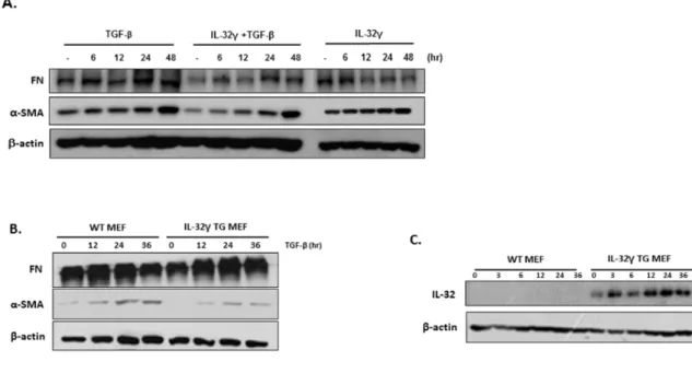
Next, to determine whether IL-32γ affects fibrosis by regulating fibroblast activation, expression of fibronectin and α-SMA was measured in the human fibroblast cell line MRC-5 after treatment with TGF-β in the presence or absence of rIL-32γ. Fibronectin expression in rIL-32γ-treated cells was significantly lower than that in untreated cells, whereas α-SMA expression was slightly lower at early time points (Fig. 2A). However, overexpression of endogenous intracellular IL-32γ did not noticeably affect the production of fibronectin and α- SMA by MEFs from WT or IL-32γ TG mice (Fig. 2B). Endogenous IL-32 expression is shown in Fig. 2C.
Fig. 2. Exogenous, but not endogenous, IL-32γ attenuates fibroblast activation. (A) Fibronectin and α-SMA expression was detected in rIL-32γ (150 ng/mL)-pretreated MRC-5 cells after TGF-β (5 ng/mL) stimulation. (B) Fibronectin and α-SMA expression are shown in IL-32γ-expressing MEFs after TGF-β (5 ng/mL) stimulation. (C) Endogenous IL-32 expression. Data are representative of three independent experiments.
3. Anti-fibrotic effect of rIL-32γ occurs independently of TNF-α
Because IL-32 induces the production of TNF-α and vice versa, this study examined whether IL-32γ exerts anti-fibrotic effects by inducing TNF-α expression. First, it was found that significant expression of IL-32γ mRNA was induced by TNF-α, although no significant change in TNF-α mRNA expression was observed (Fig. 3A and 3B). Similar to IL-32γ, treatment with rTNF-α inhibited the expression of fibronectin and α-SMA in TGF-β-stimulated MRC-5 cells (Fig. 3C). However, IL-32 was not expressed by rTNF-α under IL-32γ- knockdown conditions (Fig. 3D) and an anti-fibrotic effect of TNF-α was not observed in IL- 32γ-knockdown MRC-5 cells (Fig. 3E). Additionally, rIL-32γ inhibited fibronectin and α-SMA expression after TNF-α inhibitor treatment (Fig. 3F).
Fig. 3. Anti-fibrotic effects of rIL-32γ are independent of TNF-α. (A) MRC-5 cells were stimulated with each cytokine including LPS (1 μg/mL), Poly I: C (10 μg/mL), TNF-α (10 ng/mL), IL-32γ (150 ng/mL), TGF-β (5 ng/mL), and IL-1β (10 ng/mL). After 24-h stimulation, mRNA level of IL-32γ and TNF-α (B) were measured by quantitative PCR. (C) Fibronectin and α-SMA expression in MRC-5 cells after 24 h of stimulation with TNF-α (10 ng/mL) and TGF-β (5 ng/mL). (D) MRC-5 cells were transfected with IL-32 siRNA and then stimulated with TNF-α (10 ng/mL) or TGF-β (5 ng/mL). (E) Fibronectin and α-SMA expression in cell lysates was detected. (F) Infliximab-pretreated MRC-5 cells were stimulated with IL-32γ (150 ng/mL) and TGF-β (5 ng/mL), and fibronectin and α-SMA expression in the cell lysate was detected. Results are representative of two independent experiments, each showing similar results.
4. rIL-32γ does not appear to be involved in TGF-β-mediated Smad or non-Smad signaling
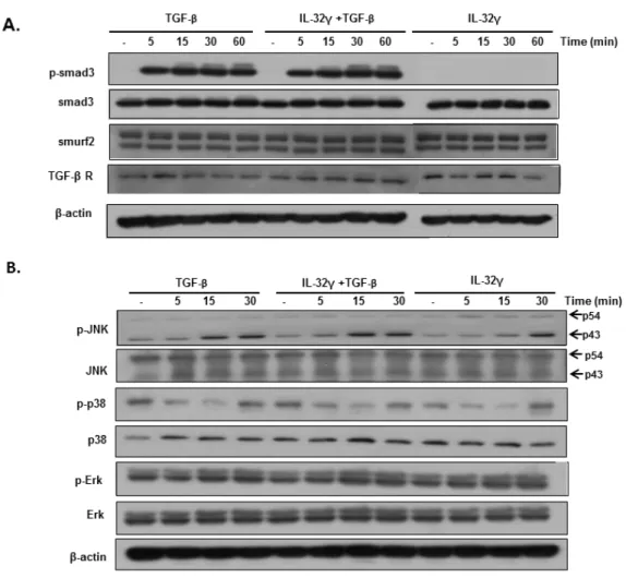
Next, the effect of IL-32γ was examined on activation of the Smad pathway, a well- known TGF-β-mediated signaling pathway. There were no significant differences in the expression of Smad signaling molecules (p-Smad 3, smuf2, and TGF-β receptor 1), regardless of rIL-32γ treatment (Fig. 4A). It was also examined whether the non-Smad pathway plays a role in the anti-fibrotic effects of rIL-32γ. The result showed no significant differences in JNK, Erk, and p38 activation between MRC cells treated with rIL-32γ and untreated cells (Fig. 4B).
Fig. 4. rIL-32γ has no effect on TGF-β-mediated Smad or non-Smad signaling pathways.
MRC-5 wells were stimulated with TGF-β (5 ng/mL) in the presence or absence of rIL-32γ and then harvested at the indicated times. Western blot analysis was performed to examine the expression of proteins in the Smad signaling (A) and non-Smad signaling pathways (B).
Results are representative of three independent experiments.
5. rIL-32γ inhibits integrin-mediated FAK/paxillin activation
Next, integrin-dependent activation of FAK and paxillin was evaluated, a critical pathway in fibroblast activation, after treatment with the RGD tripeptide and integrin blocker. RGD peptide inhibited signaling by both FAK and paxillin in MRC-5 cells stimulated with TGF-β (Fig. 5A). Interestingly, rIL-32γ inhibited FAK and paxillin signaling in a manner similar to that of RGD peptide (Fig. 5B).
To investigate how IL-32γ regulates the integrin-FAK-paxillin signaling pathway, it was performed a protein-protein binding assay to determine whether IL-32 directly binds to integrin β3, paxillin, or FAK. Both integrin β3 and paxillin were detected in the total cell lysate and flow-through lanes, but no bands were detected in the wash and elution fractions (Fig.
5C). This suggests that these proteins do not directly bind to IL-32γ. Additionally, an anti- flag-IL-32γ antibody did not immunoprecipitate with FAK (Fig. 5D).
Fig. 5. rIL-32γ inhibits integrin-mediated activation of FAK/paxillin. Phosphorylation of FAK and paxillin was detected in TGF-β (5 ng/mL)-stimulated MRC-5 cells pretreated with an RGD peptide (A) or rIL-32γ (B). Activated FAK and paxillin were detected after 24 h. Results are representative of three independent experiments. (C) MRC-5 cells were stimulated with TGF-β for 24 h, and His-tagged rIL-32γ was precipitated from cell lysates using Ni-NTA beads. Bound proteins were analyzed by Western blotting with antibodies specific for integrin β3, paxillin, and the His-tag. (D) Flag-tagged IL-32γ-overexpressing MRC-5 cells were stimulated with TGF-β and harvested at 24 h. Flag-tagged IL-32γ was then immunoprecipitated from cell lysates using an anti-flag antibody followed by immunoblotting with an anti-FAK antibody. Similar results were obtained from two independent experiments.
FT, flow-through; W, wash; E, elution.
6. rIL-32γ is localized on the cell surface
To determine the mechanism by which rIL-32γ inhibits activation of the FAK/paxillin pathway, the location of rIL-32γ was examined by live cell imaging for 60 min. rIL-32γ was located outside of MRC-5 cells after 60 min, suggesting that it does not enter cells by endocytosis and is not degraded; therefore, IL-32γ acts extracellularly, at least during the period examined (Fig. 6).
Fig. 6. rIL-32γ localizes extracellularly. Live cell imaging of MRC-5 cells at 10–60 min post- incubation with Flamma496-labeled IL-32γ (magnification, 600×; green color).
7. rIL-32γ modulates the interaction between integrins and the extracellular matrix
To examine the effect of IL-32γ on integrin signaling, the adhesion of MRC-5 cells to collagen-coated plates was examined in the presence/absence of rIL-32γ. MRC-5 cells adhered to collagen within 30 min in the absence of rIL-32γ; however, the process was impeded in the presence of rIL-32γ (Fig. 7A). Moreover, the number of spindle-shaped MRC- 5 cells was much lower in the presence of rIL-32γ, even after 30 min (Fig. 7B). Interestingly, rIL-32γ suppressed integrin/collagen-mediated activation of FAK and paxillin, which is typically induced by cell adhesion to collagen-coated plates in the absence of any other stimulation (Fig. 7C). Finally, the effect of IL-32γ on integrin expression in MRC-5 cells was evaluated according to TGF-β upregulates integrin expression. Semi-quantitative PCR revealed increased expression of integrin β3 and reduced expression of integrin β8 following TGF-β stimulation. This pattern was not altered by IL-32γ treatment (Fig. 7D).
Fig. 7. rIL-32γ modulates the interaction between integrins and the extracellular matrix.
MRC-5 cells were plated on collagen-coated plates in the presence/absence of rIL-32γ. (A) Adherent MRC-5 cells were stained with crystal violet immediately after the adhesion assay (left) and optical density values from the dissolved crystals are shown (right). Similar results were obtained from three independent experiments. *P < 0.05 (B) Adherent cells were observed at 30 min under a microscope (original magnification: 100×). Similar results were obtained from two independent experiments. *P < 0.05, ***P < 0.0001 (C) Phosphorylation of FAK and paxillin was detected after MRC-5 cells attached to collagen-coated plates for 24 h in the presence/absence of rIL-32γ. Similar results were obtained from two independent experiments. (D) Anti-fibrotic effect of rIL-32γ is independent of TNF-α. Anti-fibrotic effect of TNF-α was not observed in IL-32γ-knockdown MRC-5 cells. rIL-32γ suppressed the expression of fibronectin and α-SMA after TNF-α inhibitor treatment.
DISCUSSION
This study demonstrated the anti-fibrotic effect of IL-32γ both in vitro and in vivo. It showed that rIL-32γ regulates fibroblast activation by modulating the integrin-FAK signaling pathway. Thus, rIL-32γ may be useful for inhibiting tissue fibrosis in the clinical setting.
The mechanism of tissue fibrosis is closely related to that of wound repair, which is a normal healing process in injured tissues. However, dysregulated fibrosis can lead to severe organ dysfunction, which is typically irreversible and has a fatal outcome in many disease states. In the lungs, for example, progressive parenchymal fibrosis is a consequence of serious pulmonary fibrotic diseases such as idiopathic pulmonary fibrosis, leading to high mortality. Additionally, bronchial subepithelial fibrosis can cause irreversible fixed airway obstruction, as observed in chronic inflammatory airway diseases such as chronic severe asthma and COPD, which can become critical if untreated.
Although lung fibrogenesis is thought to result from chronic inflammation, numerous studies have suggested that fibrosis is not completely dependent on inflammatory processes and that anti-inflammatory therapeutic strategies are not always effective. Thus, therapeutic trials have shifted their focus from anti-inflammatory targets to anti-fibrotic targets, as many studies demonstrated that such mechanisms underlie the development of fibrosis 19-22). However, therapeutic agents that effectively control fibrosis are lacking; therefore, there is an urgent need to identify novel molecules with potent anti-fibrotic activities.
IL-32, previously considered a pro-inflammatory cytokine, is a multifunctional protein with a potential role in lung diseases 12,23-25). The previous study showed that IL-32γ modulates immune responses by recruiting IL-10-producing monocytic cells in a chronic asthma model [24]. Here, it observed that IL-32γ also exhibits a strong anti-fibrotic effect in a model of sub-bronchial fibrosis. Because chronic inflammation is a major factor driving the progression of fibrosis, its apparent suppressive effect on airway fibrosis may be completely
dependent on the anti-inflammatory effects of IL-32γ. Thus, the modulatory effects of IL-32γ examined in a bleomycin-induced lung injury model, which is considered a prototype of tissue fibrosis but displays a lower accumulation of immune cells in the lungs. This is of interest because IL-32γ is a putative immunomodulatory cytokine. The results of the current study suggest that IL-32γ has a novel function in lung fibrosis, as well as anti-inflammatory effects on chronic airway inflammation.
The human fibroblasts used to further investigate the mechanism underlying the anti- fibrotic effect of IL-32γ, as excessive accumulation of extracellular matrix produced by activated fibroblasts is a major pathological feature in tissue fibrosis, and any possible effects of inflammation in an animal can be excluded. MRC-5 cells were stimulated with TGF-β, which induces fibroblasts to differentiate into fibronectin- and α-SMA-expressing myofibroblasts. It found that IL-32γ effectively inhibited expression of these activation markers upon TGF-β stimulation. Previous studies showed that TNF-α and IL-32γ induce one another. Additionally, TNF-α inhibits the TGF-β-induced Smad signaling pathway 26-29). Thus, the cells were used in which IL-32γ had been silenced and a TNF-α-blocking agent to determine the exact mechanism underlying the suppressive effect of IL-32γ on fibroblast activation. Furthermore, the intracellular pathways linked to the Smad and non-Smad signaling pathways were assessed. The mechanism shows underlying the role of IL-32γ in fibrogenesis was not dependent on TNF-α expression, nor was it associated with activation of TGF-β downstream of the Smad or non-Smad signaling pathways.
Previous studies indicated that TGF-β-induced fibroblast activation depends on the integrin signaling pathway through FAK/paxillin activation 16,30-32). Protein structure modeling suggested that IL-32γ is involved in integrin activation and downstream signaling pathways
14,33). In fact, IL-32γ contains an RGD motif that binds to integrins; indeed, several isoforms of IL-32 bind to integrin αVβ3. In addition, IL-32 has a structure resembling the FAT region of
FAK (similar to an FAK-inhibitory peptide). However, these studies examined only IL-32α and β, although IL-32γ is considered the most active form 34).
rIL-32γ inhibited the phosphorylation of FAK and paxillin in TGF-β-stimulated fibroblasts without directly binding to these molecules. Based on these results, extracellular rIL-32γ regulates TGF-β-mediated fibroblast activation without entering the cell. Indeed, rIL-32γ treatment inhibited integrin-mediated cell adhesion, although rIL-32γ remained outside the cell. These results strongly suggest that IL-32γ is involved in the development of tissue fibrosis, likely by disrupting the binding between integrins expressed in the cellular membrane and the extracellular matrix.
No study has fully identified an IL-32-associated pathway in the context of fibrosis, raising the question of whether IL-32 is released by dead cells or via a specific secretory pathway. Notably, in the early phase of several diseases, IL-32 is produced by activated T cells, monocytes, and NK cells and acts as a pro-inflammatory cytokine that stimulates TNF- α, IL-6, and IL-8 production 8,12,35,36). Because recent studies showed that IL-32 is not secreted 24,37), IL-32γ released from injured epithelial cells in patients with chronic inflammatory diseases, including those with mycobacterium avium complex pulmonary disease and idiopathic inflammatory bowel disease 22), may play a regulatory role in inflammation or tissue remodeling. For instance, our previous study showed that rIL-32γ suppresses chronic airway inflammation, which is closely associated with airway remodeling
24).
There were some limitations to the current study. First, to obtain more convincing and direct evidence to evaluate our hypothesis, mutations or deletions of the RGD motif of IL-32γ should be used. Second, our results do not clearly define the precise function of intracellular and extracellular IL-32γ. Further studies are necessary to resolve these questions.
In summary, IL-32γ has anti-fibrotic effects likely by blocking the integrin-FAK-paxillin
pathway. Therefore, administration of rIL-32γ may play a pivotal role in modulating both inflammation and fibrosis in patients in which inflammation-related fibrosis pathways are activated.
CONCLUSION
The present study suggested that IL-32γ prevents tissue damage by regulating fibroblast activation in the chronic stage. The mechanism underlying this modulatory effect may involve disruption of integrin/FAK signaling cascades, without the need for IL-32γ to directly bind molecules involved in these cascades. Thus, IL-32γ is a new candidate for the treatment of lung fibrosis.
REFERENCES
1. Halwani R, Al-Muhsen S, Al-Jahdali H, Hamid Q. Role of transforming growth factor-beta in airway remodeling in asthma. Am J Respir Cell Mol Biol 2011;44:127-33.
2. Murray LA. Commonalities between the pro-fibrotic mechanisms in COPD and IPF. Pulm Pharmacol Ther 2012;25:276-80.
3. Wilson MS, Wynn TA. Pulmonary fibrosis: pathogenesis, etiology and regulation. Mucosal Immunol 2009;2:103-21.
4. Scotton CJ, Chambers RC. Molecular targets in pulmonary fibrosis: the myofibroblast in focus. Chest 2007;132:1311-21.
5. MichaelKreuter FB, MarliesWijsenbeek,, Toby M. Maher aPS. Pharmacological Treatment of Idiopathic Pulmonary Fibrosis: Current Approaches, Unsolved Issues, and Future Perspectives. BioMed Research International 2015
6. Luzina IG, Todd NW, Iacono AT, Atamas SP. Roles of T lymphocytes in pulmonary fibrosis.
J Leukoc Biol 2008;83:237-44.
7. C A Dahl RPS, H L He and J S Cairns. Identification of a novel gene expressed in activated natural killer cells and T cells. The journal of immunology 1992;148:597-603.
8. Kim SH, Han SY, Azam T, Yoon DY, Dinarello CA. Interleukin-32: a cytokine and inducer of TNFalpha. Immunity 2005;22:131-42.
9. Netea MG, Azam T, Ferwerda G, Girardin SE, Walsh M, Park JS, et al. IL-32 synergizes with nucleotide oligomerization domain (NOD) 1 and NOD2 ligands for IL-1beta and IL-6 production through a caspase 1-dependent mechanism. Proc Natl Acad Sci U S A 2005;102:16309-14.
10. Dinarello CA, Kim SH. IL-32, a novel cytokine with a possible role in disease. Ann Rheum Dis 2006;65 Suppl 3:iii61-4.
11. Joosten LA, Netea MG, Kim SH, Yoon DY, Oppers-Walgreen B, Radstake TR, et al. IL-32, a proinflammatory cytokine in rheumatoid arthritis. Proc Natl Acad Sci U S A 2006;103:3298-
303.
12. Calabrese F, Baraldo S, Bazzan E, Lunardi F, Rea F, Maestrelli P, et al. IL-32, a novel proinflammatory cytokine in chronic obstructive pulmonary disease. Am J Respir Crit Care Med 2008;178:894-901.
13. Terasaki Y, Terasaki M, Urushiyama H, Nagasaka S, Takahashi M, Kunugi S, et al. Role of survivin in acute lung injury: epithelial cells of mice and humans. Lab Invest 2013;93:1147- 63.
14. Heinhuis B, Koenders MI, van den Berg WB, Netea MG, Dinarello CA, Joosten LA.
Interleukin 32 (IL-32) contains a typical alpha-helix bundle structure that resembles focal adhesion targeting region of focal adhesion kinase-1. J Biol Chem 2012;287:5733-43.
15. Nagoshi Y, Yamamoto G, Irie T, Tachikawa T. Expression of FAK-related non-kinase (FRNK) coincides with morphological change in the early stage of cell adhesion. Med Mol Morphol 2006;39:154-60.
16. Thannickal VJ LD, White ES, Cui Z, Larios JM, Chacon R, Horowitz JC, Day RM, Thomas PE. Myofibroblast differentiation by TGF-β1 is dependent on cell adhesion and integrin signaling via focal adhesion kinase. The Journal of Biological Chemistry 2003;278:12384-9.
17. Zhang L, Che C, Lin J, Liu K, Li DQ, Zhao G. TLR-mediated induction of proinflammatory cytokine IL-32 in corneal epithelium. Curr Eye Res 2013;38:630-8.
18. Choi J BS, Hong J, Ryoo S, Jhun H, Hong K, Yoon D, Lee S, Her E, Choi W, Kim J, Azam T, Dinarello CA, Kim S. Paradoxical effects of constitutive human IL-32γ in transgenic mice during experimental colitis Proc Natl Acad Sci U S A. 2010;107:21082-21086.
19. Royce SG, Cheng V, Samuel CS, Tang ML. The regulation of fibrosis in airway remodeling in asthma. Mol Cell Endocrinol 2012;351:167-75.
20. investigates KG. At the frontiers of lung fibrosis therapy. Nat Biotechnol 2013;31:781-3.
21. Beckett PA1 HP. Pharmacotherapy and airway remodelling in asthma. Thorax
2003;58:163-174.
22. Bai X, Ovrutsky AR, Kartalija M, Chmura K, Kamali A, Honda JR, et al. IL-32 expression in the airway epithelial cells of patients with Mycobacterium avium complex lung disease. Int Immunol 2011;23:679-91.
23. Bo-Ram Bang H-SK, Soo-Hyun Kim,Sun-Young Yoon, Ji-Da Choi, Gyong Hwa Hong,Sunjoo Park,Tae-Bum Kim,, Cho H-BMaYS. IL-32γ Suppresses Allergic Airway Inflammation in Mouse Models of Asthma. American Journal of Respiratory Cell and Molecular Biology 2014;50:1021-1030.
24. Meyer N, Christoph J, Makrinioti H, Indermitte P, Rhyner C, Soyka M, et al. Inhibition of angiogenesis by IL-32: possible role in asthma. J Allergy Clin Immunol 2012;129:964-73 e7.
25. Kenichi Yamane HI, 2 Yoshihide Asano, Masatoshi Jinnin, and Kunihiko Tamaki.
Antagnistic effects of TNF-a on TGF-b sifnaling through downregulation og TGF-b receptor type II in human dermal fibroblast The Journal of Immunology 2003;171:3855–3862.
26. Verrecchia F MA. TGF-beta and TNF-alpha : antagonistic cytokines controlling type I collagen gene expression. cell signal 2004;16:873-880.
27. Verrecchia F, Pessah M, Atfi A, Mauviel A. Tumor Necrosis Factor- Inhibits Transforming Growth Factor- /Smad Signaling in Human Dermal Fibroblasts via AP-1 Activation. Journal of Biological Chemistry 2000;275:30226-30231.
28. Franck Verrecchia CT, Erwin F. Wagner§, and Alain Mauviel. A Central Role for the JNK Pathway in Mediating the Antagonistic Activity of Pro-inflammatory Cytokines against Transforming growth factor beta-driven SMAD3/4-specific Gene Expression. THE JOURNAL OF BIOLOGICAL CHEMISTRY 2003;278:1585–1593.
29. Mamuya FA, Duncan MK. aV integrins and TGF-beta-induced EMT: a circle of regulation.
J Cell Mol Med 2012;16:445-55.
30. Munger JS, Sheppard D. Cross talk among TGF-beta signaling pathways, integrins, and the extracellular matrix. Cold Spring Harb Perspect Biol 2011;3:a005017.
31. Greenberg RS, Bernstein AM, Benezra M, Gelman IH, Taliana L, Masur SK. FAK- dependent regulation of myofibroblast differentiation. FASEB J 2006;20:1006-8.
32. Joosten LA, Heinhuis B, Netea MG, Dinarello CA. Novel insights into the biology of interleukin-32. Cell Mol Life Sci 2013;70:3883-92.
33. Choi JD, Bae SY, Hong JW, Azam T, Dinarello CA, Her E, et al. Identification of the most active interleukin-32 isoform. Immunology 2009;126:535-42.
34. Kim S. Interleukin-32 in inflammatory autoimmune diseases. Immune Netw 2014;14:123- 7.
35. Hong J, Bae S, Kang Y, Yoon D, Bai X, Chan ED, et al. Suppressing IL-32 in monocytes impairs the induction of the proinflammatory cytokines TNFalpha and IL-1beta. Cytokine 2010;49:171-6.
36. Keswani A, Chustz RT, Suh L, Carter R, Peters AT, Tan BK, et al. Differential expression of interleukin-32 in chronic rhinosinusitis with and without nasal polyps. Allergy 2012;67:25- 32.
37. Shioya M, Nishida A, Yagi Y, Ogawa A, Tsujikawa T, Kim-Mitsuyama S, et al. Epithelial overexpression of interleukin-32alpha in inflammatory bowel disease. Clin Exp Immunol 2007;149:480-6.
국문초록
연구배경
중증 천식에서 기도의 섬유화는 비가역적으로 현재까지 적절한 치료가 없는 것이 사실이다. 그 이유는 이러한 기도 및 폐의 섬유화의 기전이 명확하게 밝혀지지 않았기 때문이다. IL-32는 중증 천식을 비롯한 다양한 만성 염증성 질환에서 주요한 역할을 하는
것으로 알려져 있다. 이 중 IL-32γ는 구조적으로 FAK (focal adhesion kinase)의 FAT
부분과 매우 유사하고, IL-32γ는 integrin과 결합하는 것으로 잘 알려져 있는 RGD motif를
가지고 있다. 그러므로 이 연구에서 IL-32γ가 기도 및 폐의 섬유화에 어떠한 기전으로
작용하는지 연구하였다.
연구방법
bleomycin 유발 폐 섬유화 모델과, ovalbumin 및 Aspergillus melleus protease 유발 만성
천식 모델을 구축하여 연구를 진행하였고, 재조합 rIL-32γ를 처리하여 섬유화 진행
정도를 비교하였다. rIL-32γ를 처리한 활성화 된 섬유아세포 (MRC-5)에서 fibronectin과
α-smooth muscle actin (α-SMA) 발현을 분석하고 TGF-β 신호전달 체계를 통한 폐
섬유화 정도를 분석하였다.
연구결과
rIL-32γ는 폐 섬유화 및 만성 천식 동물모델에서 조직학적으로 collagen 침착 및 α-SMA
발현을 유의하게 억제하였다. rIL-32γ를 처리한 경우 TGF-β로 자극한 MRC-5세포에서도
fibronectin과 α-SMA 발현이 억제되었다. rIL-32γ는 integrin과 FAK-paxillin 신호 전달
축의 활성화를 억제하였지만, Smad 및 non-Smad 신호전달 경로에는 영향을 미치지
않았다. rIL-32γ는 MRC-5 세포의 바깥에서 작용하였으며, 세포 내부의 FAK와 paxillin에
직접적으로 결합하지 않고 외부에 국한되어 integrin과 ECM 간의 상호작용을 억제하는
것으로 확인되었다.
결론
이러한 결과는 IL-32γ가 세포 밖에서 작용하여 integrin과 ECM 간의 상호작용을
억제함으로서 섬유화 진행을 방해하는 것을 증명하며, 섬유화를 예방할 수 있는 새로운 치료 후보물질로의 개발 가능성을 증명하는 결과이다.
중심단어: 중증 천식, 기도 염증, 폐 섬유화, 인터류킨 32-감마