전이성 대장암 환자의 화학요법 중 신체 구성 및 예후의 변화. 기존 대장암의 체성분에 관한 연구들은 주로 전이가 없는 환자를 대상으로 하거나 단일 시점에서 측정한 체성분만으로 예후를 분석해 왔다. 이에 대한 예후는 잘 알려져 있지 않습니다. 이 중 완화암 치료 시작 전과 치료 종료 후 촬영한 CT 영상을 분석에 포함시켰고, 치료 기간 전반에 걸쳐 서로 다른 시점에서 연속적인 CT 영상을 수집했다.
이러한 치료 기간 동안의 체성분 변화는 치료 시작 당시의 상태와 상관없이 치료 종료 후 기대수명과 유의한 상관관계를 보였다. 질이 감소했거나 내장지방이나 피하지방이 감소한 환자는 치료 종료 후 생존 기간이 더 짧습니다. 결론: 완화적 화학요법을 받는 대장암 환자의 체성분 이상.
소견은 흔하며, 화학요법 중에도 변화가 자주 발생합니다.
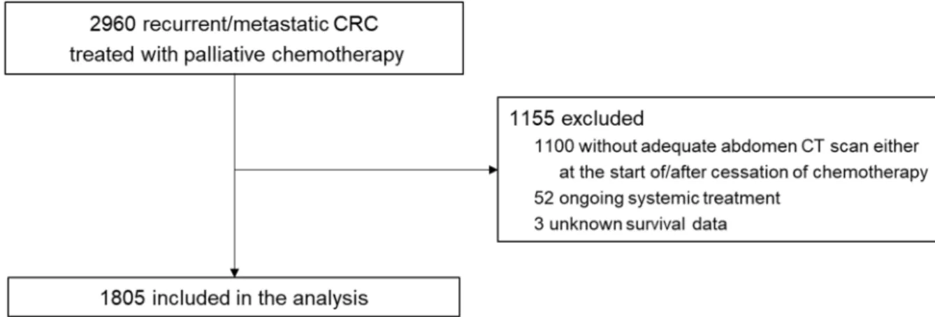
Patients
Here, we selected a homogeneous group of patients with recurrent/metastatic colorectal cancer treated with palliative chemotherapy and assessed the abnormalities in muscle quantity (sarcopenia), quality (myosteatosis) and distribution of fat (visceral and subcutaneous obesity) using serially collected computed tomography (CT) images . By implementing these methods, we aimed for a comprehensive evaluation of body composition abnormalities, their serial changes during systemic chemotherapy, and their prognostic implications in a real patient population with advanced colorectal cancer. This study was approved by the Asan Medical Center Institutional Review Board and conducted in accordance with the ethical standards of the Institutional Research Committee and the Declaration of Helsinki.
Measurement and definition of body composition markers
In this study, four categories of body composition abnormalities were defined as follows: 1) sarcopenia, which represents low muscle mass in terms of muscle quantity, was defined as a T-score <-2.0 calculated from the SMA/BMI index ;142) myosteatosis, which reflects the fatty infiltration of muscles in terms of muscle quality, was defined as a T-score <-2.0 calculated from the LAMA/TAMA index;15 3) visceral obesity, which represents an excessive amount of visceral representing fat was defined as the visceral fat area (VFA). 100 cm2;16 4) subcutaneous obesity, which represents an 'excessive amount of subcutaneous fat', was defined as the height-adjusted subcutaneous fat area index (SFAI) ≥ 50.0 cm2/m2 in men and ≥ 42.0 cm2/m2 in women . 17The status of these four body composition markers was assessed for each CT image collected for analysis. Finally, a BMI ≥ 25 kg/m2 was defined as obese, and a BMI < 18.5 kg/m2 was defined as underweight according to the World Health Organization guidelines for the Asia-Pacific region [18].
Statistical analysis
The T-scores for NAMA/TAMA were calculated based on values measured in the young reference group of Koreans.15. Changes in body composition markers during the treatment course were compared using a linear mixed model. For survival analyses, time-dependent Cox regression was used to estimate the effect of body composition markers measured at multiple time points on OS.
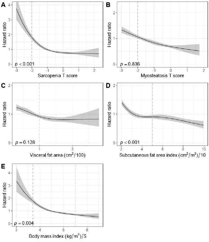
A four-node restricted cubic model was used to assess non-linear associations of body composition markers as covariates with OS. Pearson's R was used to assess correlations between body composition markers, BMI, and laboratory values measured at each time point.
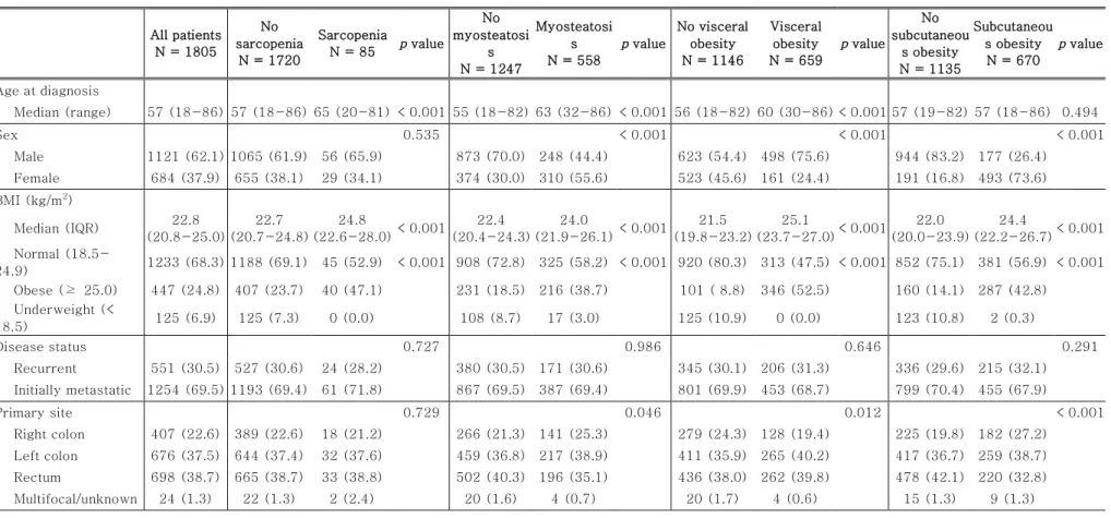
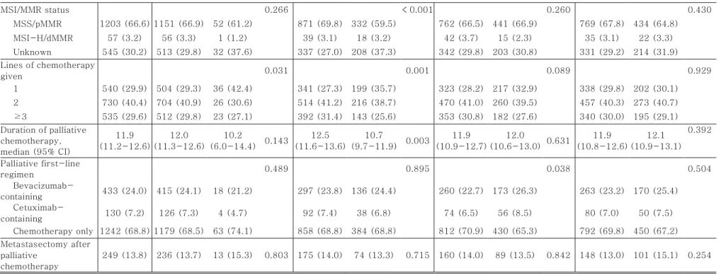
Patients and incidence of abnormal body composition at baseline
Of note, the median BMI of the sarcopenic patients at diagnosis was higher than that of the non-sarcopenic patients (22.7 vs. The median age at diagnosis was higher in patients with sarcopenia, myosteatosis and visceral obesity compared to patients without them (Table) 1) The median T-scores for sarcopenia and myosteatosis were decreased and the median VFA was increased with age.
Abbreviations: BMI, body mass index; CI, confidence interval; dMMR, deficient mismatch repair; IQR, interquartile range;.
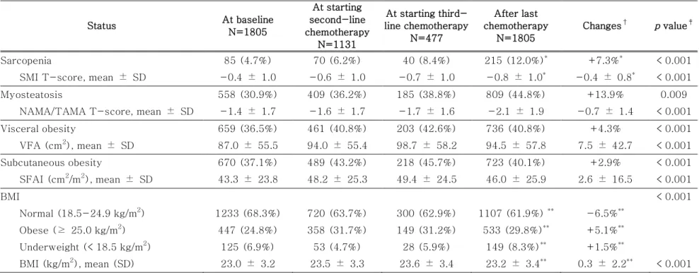
Changes in the body composition during treatment and survival after
Correlations with body composition, body mass index and laboratory testsCorrelations between each body composition marker and correlations Correlations between each body composition marker and correlations of the body composition markers with BMI or laboratory are shown as correlation matrices in Figure. In this study, we evaluated the comprehensive landscape of body composition during palliative chemotherapy and their changes in metastatic colorectal cancer. Although these body composition markers showed prognostic significance, they were only weakly correlated with BMI or other laboratory blood tests, indicating a need for separate assessments of body composition.
To our knowledge, this is the largest study to evaluate the comprehensive, longitudinal landscape of body composition and its prognostic implications in a homogeneous series of metastatic colorectal cancer treated with palliative chemotherapy. These findings suggest that monitoring body composition during treatment may provide prognostic information about patients. 20 Of note, we included all longitudinal body composition data measured at multiple time points and estimated pooled hazards rather than using data obtained at a single time point.
Finally, markers of body composition, particularly myosteatosis and sarcopenia, showed only weak correlations with BMI or laboratory nutritional/inflammatory markers. The prognostic significance of abnormal body composition, together with their low correlation with other measures, highlights the need for special assessments of body composition in patients with metastatic colorectal cancer. The benefit of CT-based body composition analysis, particularly in cancer patients, is that no additional testing is required since most patients undergo regular CT evaluation.
Furthermore, we evaluated various aspects of body composition and comprehensively analyzed their changes during palliative chemotherapy, which have been rarely reported. Furthermore, longitudinal data acquisition with time-dependent survival analyzes with further adjustment for other important clinical characteristics helped to improve methodological limitations in previous studies8 and to understand the overall independent effect of body composition during treatment. We believe that our study can shed light on the landscape of body composition in patients with metastatic colorectal cancer and their clinical implications.
Future studies investigating the effectiveness of therapeutic interventions on body composition, especially myosteatosis, and the mechanistic background of the body composition changes are needed.29-31. In conclusion, abnormalities and changes in body composition were common during palliative chemotherapy in patients with advanced colorectal cancer, especially myosteatosis. Body composition and time course changes in regional distribution of adipose and lean tissue in unselected cancer patients on palliative care - correlations with food intake, metabolism, exercise capacity and hormones.
Body composition markers showed a weak association between themselves, body mass index, and laboratory.
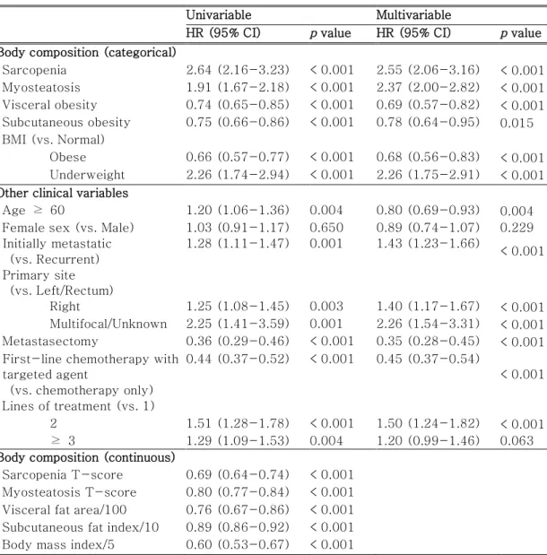
Time-dependent hazards ratios of body composition markers for
Correlations with body composition, body mass index, and laboratory
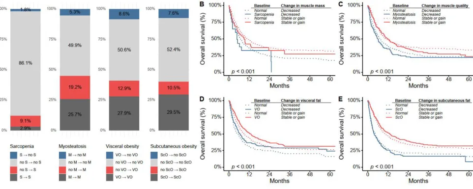
We found that myosteatosis affected the majority of patients and was the most prevalent change during chemotherapy, rather than sarcopenia. Survival analysis using serial data obtained at several time points showed that sarcopenia and myosteatosis were associated with poor survival outcomes, whereas obesity, regardless of fat distribution (subcutaneous or visceral), was associated with a favorable survival outcome. Interestingly, the prevalence of sarcopenia at baseline in our cohort (5%) was similar to that of the healthy Korean population (4–9%).14 Even after recent chemotherapy, most patients remained nonsarcopenic.
In contrast, the prevalence of myosteatosis (31%) was higher than that of the healthy population and 19% of patients developed new myosteatosis during treatment. These data suggest that although reduction in muscle mass is undoubtedly an important change in patients with cancer, qualitative changes in muscle are more common in colorectal cancer and require clinical attention. Patients who lost muscle mass during chemotherapy or had increased fatty infiltration in the muscles showed shorter survival after the last chemotherapy, regardless of the presence of sarcopenia or myosteatosis at baseline.
This contrasts with previous studies that showed poor survival outcomes in obese patients with colorectal cancer.22-24 However, these studies mostly included non-metastatic patients who were treated curatively. Furthermore, the inferior survival outcome observed in patients who lost body fat (both visceral and subcutaneous) during chemotherapy, regardless of baseline obesity status, further supports the protective effect of obesity in these patients. Further investigation is warranted to determine whether measuring body composition can aid treatment decision-making, such as the decision to administer chemotherapy at later lines or to enroll a patient in a clinical trial.
Visceral fat is a risk factor for poor prognosis in colorectal cancer patients receiving adjuvant chemotherapy. Body mass index is prognostic in metastatic colorectal cancer: pooled analysis of patients from first-line clinical trials in the ARCAD database. Methods: Patients with recurrent/metastatic colorectal cancer treated with palliative chemotherapy from 2008 to 2017 were retrospectively identified.
Additionally, 21.5% and 18.1% of patients experienced either resolution or newly developed visceral and subcutaneous obesity, respectively. These changes during treatment were associated with survival after discontinuation of chemotherapy, regardless of baseline status.