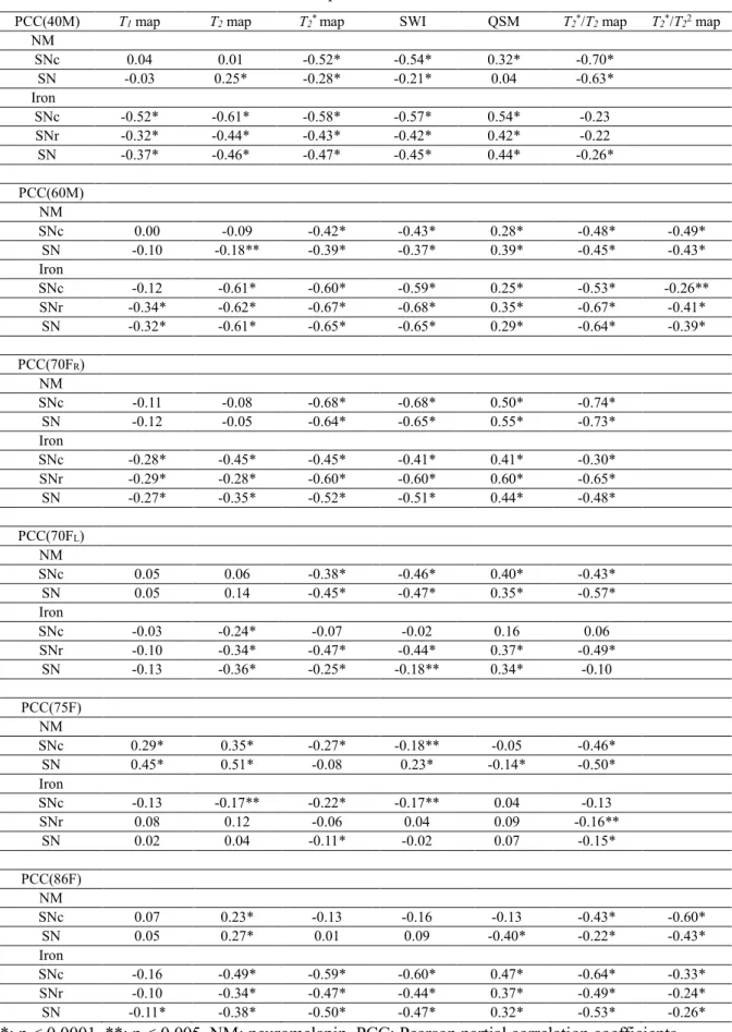
However, it is still impossible to classify the states of the iron and separate the different forms of iron deposited in the brain. Separate segmentations of the areas of iron detected in the T2 map and neuromelanin observed in the T2*/T2 map (or T2*/T22 map) were available in the substantia nigra. The dorsal linear mismatch of T2 and T2* was consistently detected in the brain of healthy controls.
In the second part, iron deposition along the myelinated white matter fiber in the diseased brain was identified. The iron-rich white matter in the frontal subcortical region contributes to the positive susceptibility in patients to leukoencephalopathy with axonal spheroids and pigmented glia in adults. Susceptibility-weighted imaging presented the prominent phase signal showing the tree-like structure in the white matter of the frontal brain, with prominent atrophy.
48 Figure 3.2.4 Postmortem multimodal MRI of three subjects Figure 3.2.5 Postmortem multimodal MRI with direct histological confirmation ………..……50 Figure 3.2.6 Segmentation of neuromelanin according to T2* and T2 boundary from tissue sample obtained from a 75-year-old normal female ………51 Figure 3.2.7 Segmentation of neuromelanin by T2* and T2 cutoff from tissue sample obtained from an 86-year-old normal female ……….52 Figure 3.2.8 Area overlay of MRI T2* and T2 mismatch and binarized distribution of neuromelanin segmented from cryosectioned block faces co-registered from normal controls ……….53 Figure 3.2.9 Segmentation of neuromelanin by T2* and T2 cutoff from tissue sample taken from a 51-year-old male patient with SN depigmentation ………54 Figure 3.2.10 Iron chelation with neuromelanin pigments ……….……55 Figure 3.2.11 In vivo multimodal MRI for two intermediate levels in normal subjects …… …..…57 Figure 3.2.12 Multimodal in vivo MRI for two intermediate levels in patients with Parkinson's disease ..…58 Figure 3.2.13 Schematic flow diagram for postmortem and in vivo MRI analysis ..…59 Figure 3.2. 14 In vivo T2 and T2* ratios for two levels of normal subjects and patients with Parkinson's disease.
Introduction
Purpose
The aim of this study is the development of multi-colour iron magnetic resonance (MR) imaging and the investigation of its in vivo feasibility and implication. Using a new concept of MR imaging, the states of iron molecules were classified non-invasively and the different forms of iron deposition in the brain were separated. The analysis of the spatial distribution and concentration of iron molecules in the brain is also important.
Outline
Abbreviations
Background
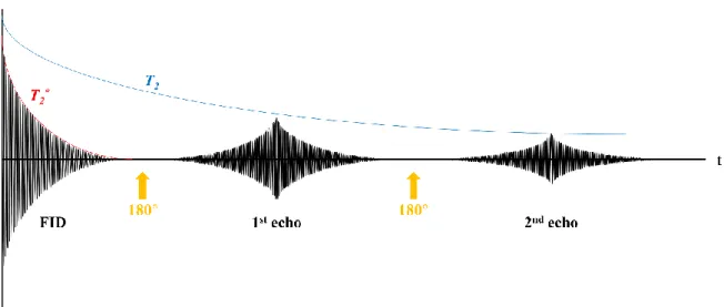
Quantitative susceptibility mapping
The induced magnetic field is determined by the sensitivity of the material in accordance with the applied magnetic field. The paramagnetic susceptibility sources induce internal magnetic fields in the same direction as the applied magnetic field. Therefore, an inverse calculation is required to reconstruct the sensitivity value from the magnetic field map.
Quantitative susceptibility mapping (QSM) is a novel technique that solves an inverse computation to reconstruct the 3D distribution of the tissue magnetic susceptibility value using an MR phase image acquired from gradient-echo based sequences. Since each voxel contains different sources of sensitivity depending on the environment, the bulk sensitivity in each voxel was estimated by the weighted sum of the magnetic susceptibility of the respective element with its fractional volume as the following equation. Even though the T2* or R2* map is the quantitative analysis using multi-magnitude images of gradient echo-based sequences, the calcification and iron deposition in the tissue cannot be distinguished from each other based on the T2 * or R2* values, because both conditions use the shortened T2*. increased R2*) values due to the local difference in magnetic susceptibility.
On the other hand, QSM can provide differentiation of cases such as positive values for iron deposition and negative values for calcification.
Neurodegenerative disease …………………………………..………………………………..16-18
- Progressive supranuclear palsy …
- Perry syndrome
- Adult-onset leukoencephalopathy with axonal spheroids and pigmented glia
Detection of neuromelanin pigments in the substantia nigra
Methods
Results
Discussions and Conclusions
Determination of neuromelanin distribution in the dorsal area of substantia nigra pars compacta
- Methods
- Results
- Discussions and Conclusions
Iron deposition on the myelinated fibers
Methods
The phase shift value in this study was for a left-handed coordinate system, which was positively correlated with iron levels. To confirm SWI contrast, postmortem 7T MRI (Bruker, Karlsruhe, Germany) and a histopathological correlation study were performed in one case (Case 1) after obtaining informed consent for the use of brain tissue before death. A formalin-fixed cortical tissue block of one of the frontal lobes was used for ex vivo experiments.
For SWI, the four times of positive mask produced from phase image (-π to π) was multiplied to the magnitude image. For comparison, postmortem SWI was also obtained from a control brain of an individual without a history of neurological disease who joined the Pusan National University Anatomical Donation Program and signed the informed consent. After MRI scanning, the tissue block was sectioned at 8-μm thickness corresponding to 500-μm-thick ex vivo MR images.
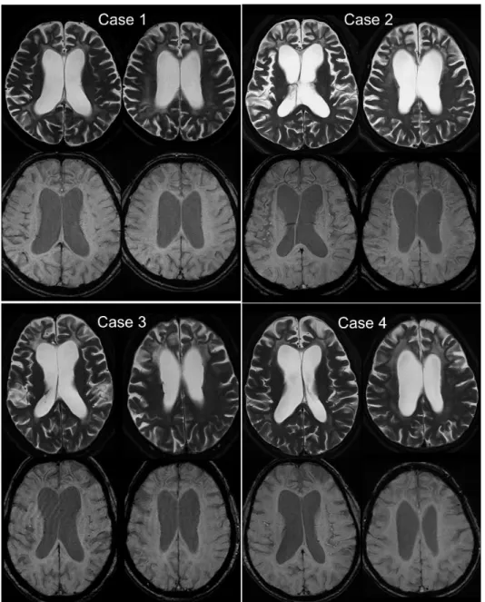
Olympus virtual slide microscopy (Olympus, Tokyo, Japan) was used to scan all stained slides with a pixel size of 0.6836 μm2. In all ALSP cases, T2-weighted images show symmetric bifrontal white matter hyperintensities with sparing of the subcortical U-fibers and frontal-predominant atrophy. Susceptibility-weighted images demonstrate the characteristic 'tree silhouette-like' hypointense configuration of the frontal subcortical white matter.
Results
In all patients, brain MRI showed predominant frontal atrophy and bifrontal, callosal, and deep WM periventricular lesions with sparing of subcortical U fibers (Figure 4.1.1). In case 1, WM changes, cortical atrophy, and enlargement of the lateral ventricles were more prevalent. In case 4, periventricular WM changes were asymmetric, with more severe involvement on the contralateral side of the more severely affected limb.
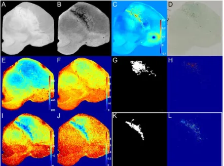
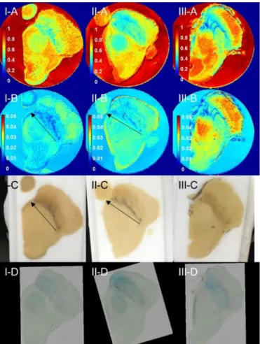
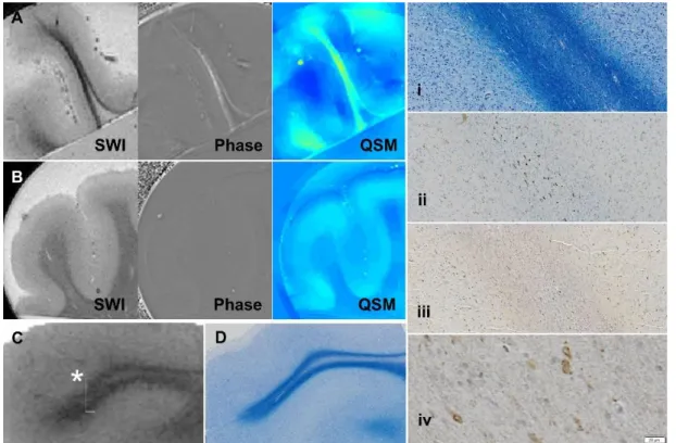
On phase images, areas corresponding to linear hypointensity on SWI were seen as hyperintense (positive phase) suggesting a paramagnetic nature (Figure 4.1.2). Postmortem MRI for ALSP brain (A) manifests marked hypointensity on susceptibility-weighted imaging (SWI) with a positive phase shift on phase imaging and positive susceptibility values on quantitative susceptibility map (QSM) in contrast to control brain (B) ). Signal hypointensity on postmortem SWI (C) correlates with Luxol fast blue-stained white matter density (LFB, D).
The white box (*) on the SWI indicates where the histological images are taken at higher magnification (i~iv). i) LFB staining shows symmetrical central myelin pallor with relatively preserved U fibers. ii) CD68-positivity is most often located in the central demyelinating white matter of the brain. iii, iv). Ferritin immunoreactivity is observed mainly in CD68-positive zones and the junction of white matter and cortical gray matter. Postmortem MRI of ALSP brains revealed paramagnetic properties of the WM layer in contrast to control brains (Figure 4.1.3a and Figure 4.1.3b).
Ferritin IHC staining was observed mainly in CD68-positive zones and at the WM–cortical gray matter junction (Figure 4.1.3g and Figure 4.1.3h). Co-registration between postmortem MRI and histology showed that signal hypointensity on postmortem SWI correlated with the density of LFB-stained WM (Figure 4.1.3c and Figure 4.1.3d). However, positive iron staining with Perl's Prussian blue staining was rarely found in the corresponding WM paramagnetic lesions.
Discussions and Conclusions
Accumulated iron on the myelinated fibers of the oculomotor nerve in the brain of progressive
- Methods
- Results
- Discussions and Conclusions
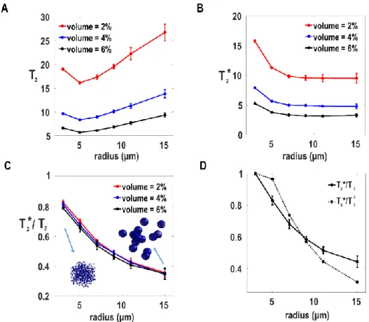
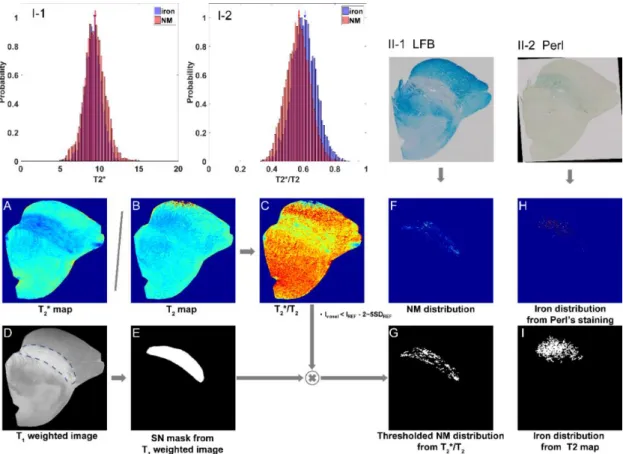
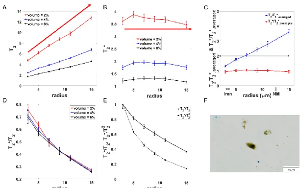
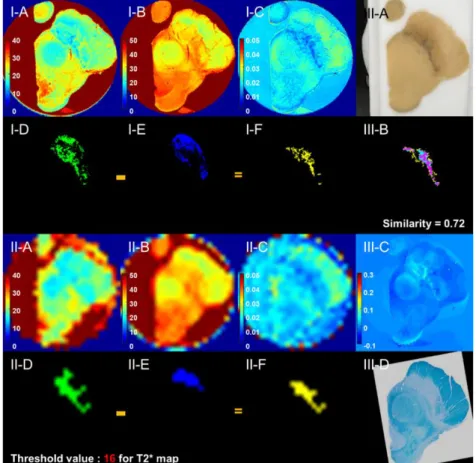
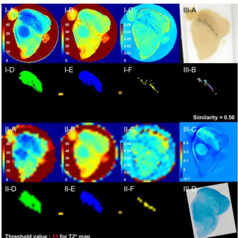
The size-dependent T2 and T2* values from the MC simulations are shown in Figure 3.1.2A and Figure 3.1.2B at different fractional turbulence volumes of 2%, 4% and 6%. The clear relative shift of the T2*/T2 distribution of the neuromelanin-iron complex region with respect to that of the ferric iron region was noted in Figure 3.1.3I-2. The ferric iron region was also obtained by thresholding the T2 map, which is shown in Figure 3.1.3I.
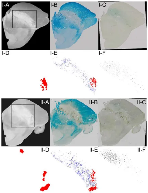
In the brains of two normal subjects, QSM was determined indicating paramagnetic components (positive receptivities) within the SN (Figure 3.2.4I-D and Figure 3.2.4II-D). An iron-positive signature was detected along myelinated fibers in Perls Prussian blue staining (Figure 3.2.5III-D). However, the area of low T2 values was mostly localized in the ventromedial area of normal SNs (Figure 3.2.13I-C and Figure 3.2.13I-c).
Accordingly, the mismatch regions (Figure 3.2.13I-D and Figure 3.2.13I-d) determined by subtracting the T2-segmented mask from the T2*-segmented mask were consistently seen as a linear pattern in the dorsal area of the normal SNs. The measurements along the entire length of the linear T2 and T2* mismatch were both significantly different between normal and G1 and between normal and G2 (Figure 3.2.16A). The spatially overlapped region (red) between segmented myelinated fibers of Luxol fast blue staining (blue) and hypointense regions of SWI (green) is shown in Figure 4.2.3G.
On the other hand, such iron distribution along the myelinated fibers in the PSP brain is not sensitively stained in the Perls' Prussian blue staining (Figure 4.2.3B). Hyperintense areas were also maintained in the white matter region between the SN and RN in patients with PD (Figure 4.2.5C-II and Figure 4.2.5C-V).
Conclusions
Limitations and Future works
Iron, neuromelanin and ferritin content in the substantia nigra of normal subjects at different ages: implications for iron storage and neurodegenerative processes. Visualization of neuromelanin in the Substantia nigra and locus ceruleus at 1.5 T using a 3D gradient echo sequence with magnetization transfer contrast. Neuromelanin-sensitive 3D magnetic resonance imaging with semi-automated substantia nigra pars compacta volume measurement for the diagnosis of Parkinson's disease.
The role of iron and copper molecules in the neuronal vulnerability of locus coeruleus and substantia nigra during aging. The absolute concentration of nigral neuromelanin, tested by a new sensitive method, increases throughout life and is dramatically reduced in Parkinson's disease. Binding of iron to neuromelanin from human substantia nigra and synthetic melanin: an electron paramagnetic resonance spectroscopy study.
Neuromelanin-associated redox-active iron is increased in the substantia nigra of patients with Parkinson's disease. Specific visualization of neuromelanin-iron complex and ferrous iron in human postmortem substantia nigra using MR relaxometry at 7T. Reactive microglia are HLA-DR positive in the substantia nigra of Parkinson's and Alzheimer's disease brains.
New pattern of iron deposition in the nigral fascicula in patients with Parkinson's disease: a pilot study. In vivo assessment of brainstem depigmentation in Parkinson's disease: potential as a severity marker for multicenter studies. Changes in the levels of iron, ferritin and other trace metals in Parkinson's disease and other neurodegenerative diseases affecting the basal ganglia.
Prevalence of apraxia of eyelid opening in the general population and in patients with extrapyramidal disorders. Progressive supranuclear palsy: high-field MR microscopy of the human substantia nigra and globus pallidus.