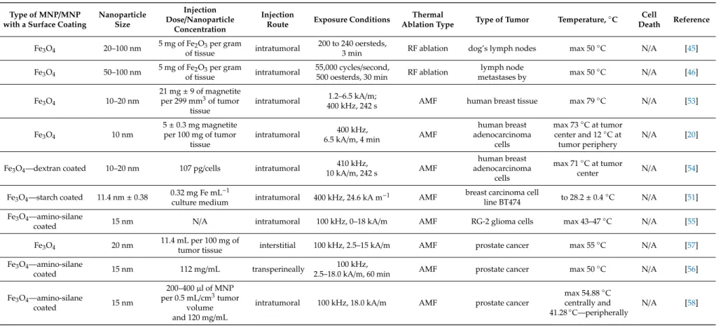
This parameter is proportional to the temperature rise, which is defined as the rate of absorption of electromagnetic energy per unit mass of biological material, and is defined as follows, as in equation (1), where λ – the thermal conductivity of. Among various MNPs, the iron oxide nanoparticles are widely used in thermal ablation therapy due to its high value of magnetic moment density, which is 220 emu/g. The structure of MNPs is one of the key aspects that enable the comprehensive customization of magnetic thermal ablation [39].Nanomaterials 2019, 9, x FOR PEER REVIEW 6 of 35. a) The TEM image of iron oxide magnetic nanoparticles (MNPs) synthesized by the thermal decomposition method; (b) The SEM image of the magnetic iron oxide nanoparticles synthesized using a solvothermal method.
Relaxation losses can be defined by two Figure 5. (a) TEM image of magnetic iron oxide nanoparticles (MNPs) synthesized by the thermal decomposition method; (b) SEM image of magnetic iron oxide nanoparticles synthesized by the solvothermal method. a) TEM image of magnetic iron oxide nanoparticles (MNPs) synthesized by the thermal decomposition method; (b) SEM image of magnetic iron oxide nanoparticles synthesized by the solvothermal method. Relaxation losses can be defined by two mechanisms: the Neel relaxation mechanism and the Brownian motion of particles. The first groundbreaking research on the use of magnetic materials to enhance thermal therapy was conducted by Gilchrist et al.
Gold Nanoparticles (AuNP)
The choice of anti-EGFR antibody conjugation to label cells with AuNPs is justified by the overexpression of EGFR in most tumor cells [77]. In a study by El-Sayed et al., laser photothermal therapy of HaCaT epithelial carcinoma cells (a human keratinocyte cell line) was performed using anti-EGFR antibodies conjugated to AuNPs with an average particle size of 40 nm at 530°. -nm maximum absorption. This study showed that HaCaT cells, after incubation with AuNPs conjugated with anti-EGFR antibodies, could induce complete tumor destruction at a laser intensity higher than 57 W/cm2 compared to control groups of two oral squamous cell carcinoma cell lines (HSC 313 ( Hematopoietic stem cells) and HOC 3 (Cellosaurus cell line).
The study showed not only the importance of the surface functional groups of the nanomaterial, but also the value of the laser power density [72]. Another study with 20-nm-sized anti-EGFR-functionalized AuNPs was performed by the group of Glazer et al. A further clinical in vivo study of thermal therapy of pancreatic cancer was performed by the same group in which two human pancreatic carcinoma cell lines were exposed to the RF field, Panc-1 and Capan-1, using PAM4 hemi-antibody conjugated and C225 antibody conjugated with 20-nm size AuNPs.
The authors reported the absence of the intrinsic cytotoxicity of AuNP, and the range of malignant cell destruction was 99.8 ± 3.1% for Hep3B cell death and 96.5 ± 8.4%. The significance of RF field power for thermal therapy was investigated by Cardinal et al. This group studied the non-invasive RF ablation of liver cancer with HepG2 cancer cells using 13 nm citrate-coated AuNPs.
To determine the effect of RF field power on heating, solutions were exposed to the RF field at variable powers from 10 W to 100 W for 3 min. An overview of AuNP application and conditions in thermal therapy of cancer is presented in Table 2 .
CuS Nanoparticles
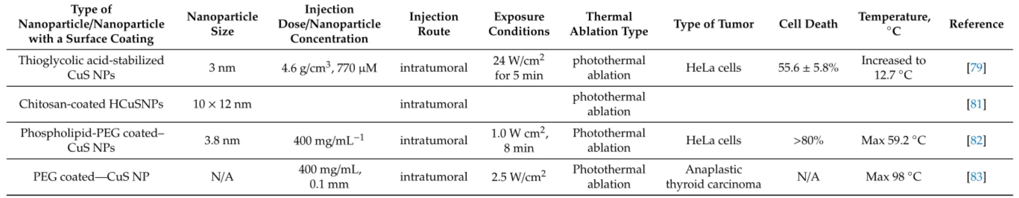
Table 3 summarizes the CuS NP size, exposure conditions, and temperature rise in the thermal therapy of cancer.
Nanorods
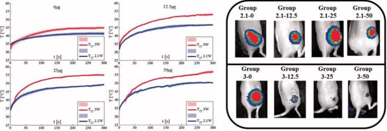
However, the lower temperature change was achieved at the crop depth, comparatively, at 2.5 mm and 5 mm, for which the temperature was approximately 48–58◦C and 43–48◦C, respectively. Finally, the concentration of gold nanorods at the periphery of the tumor area led to a uniform temperature distribution and demonstrated a better heat release to the deeper tumor area, reaching the temperature of 48-52◦C. The photothermal radiation intensity of 1.25 W/cm2 for 300 s at a volume fraction of 0.001% was determined as an optimal parameter to obtain the temperature value higher than 53◦C over the entire tumor area [93].
The results showed that the higher the concentration, the higher the temperature rise during the laser ablation. The temperature increase also influenced the effects of the laser ablation: in the control group, the laser ablation was unsuccessful, while the group with 50 µg nanorods experienced complete resorption of the tumor (Figure 10) [95,96]. However, the lower temperature change was achieved at the depth of the tumor, relatively at 2.5 mm and 5 mm, where the temperature was approximately 48-58 °C and 43-48 °C, respectively.
Finally, the concentration of gold nanorods in the periphery of the tumor area led to a uniform temperature distribution and demonstrated better heat release to the deeper tumor region, reaching the temperature of 48–52 °C. The photothermal irradiation intensity of 1.25 W/cm2 for 300 s at a volume fraction of 0.001% was determined as an optimal parameter to achieve the temperature value higher than 53 °C over the entire tumor region [93]. The increase in temperature also affected the effects of the laser ablation: in the control group, the laser ablation was not successful, while the group with 50 μg of nanorods experienced a full resorption of the tumor (Figure 1).
For example, the temperature at the upper part of the phantoms reached more than 60◦C, while 35◦C was recorded at a distance 6 mm from the surface of a phantom. The study concluded that time and power laser irradiation help to control the temperature in the deep region of a phantom [98].
Carbon Nanotubes (CNTs)
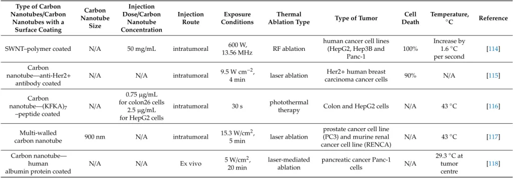
This study showed that Her2+ cells internalized with CNTs were more susceptible to NIR-mediated photothermal destruction compared to cells with CNTs on their surface [115]. Photothermal treatment of colon cancer using SWNTs was investigated by Hashida et al. Surface-functionalized samples at concentrations of 0.75 µg/mL for colon 26 and 2.5 µg/mL for HepG2 cells, respectively, were subjected to NIR laser irradiation.
The research showed a high rate of cell death rate and temperature rise up to 43◦C in 30 s for both cell lines [116]. A human androgen-independent prostate cancer cell line (PC3) and a murine renal cell carcinoma cell line (RENCA) were laser-irradiated at 15.3 W/cm2 and a beam diameter of 5 mm for 1.5 and 5 minutes using MWNT 900 nm. Larger temperature increases were observed for both samples at 5 minutes of ablation and reached 43◦C compared to the control sample without MWNTs, where the temperature increase was observed at 7◦C.
The ablation of the tumor was performed for 30 minutes using a continuous laser generator of 5 W/cm2808 nm. The temperature changes in 20 minutes were recorded as 4.2◦C in the surrounding healthy tissue, 25.6◦C at the edge of the tumor and 29.3◦C in the central area of the tumor, showing that the temperature in the center is higher. The high temperature value that could cause extensive tumor necrosis was reached in 20 minutes [118].
Nanoshells/Nanocomposites 1. Nanoshells
Nanoshells/Nanocomposites
Another study on heat therapy of cancer cells using silica-gold nanoshells was conducted by Stern et al. The experiments studied the treatment of prostate tumors in mice using 110-nm silica core and 10-nm gold-coated nanoshells by intravenous injection and using a NIR laser at 808 nm. Tumors were observed for 21 days and showed 93 percent tumor necrosis and regression in the high-dose group.
The results showed that the approach used can selectively kill medulloblastoma cells that express HER2 without killing cells that do not express HER2. Furthermore, the research showed that the dual requirement of the presence of nanoshells and laser light to induce cell death is particularly promising for use in brain tumors [125]. The use of nanoshells in imaging and therapy has shown promising results for specific cancer cells.
The study showed the destruction of 63% of the cells within 10 minutes after RF ablation under the 350 kHz and 5 kW parameters [129]. The term nanocomposite describes a class of composite materials consisting of two or more materials where one of the phases has at least one dimension less than 100 nanometers. A SEM image morphology of polymer-coated iron oxide superparamagnetic nanocomposite [Reproduced from [130], with permission from Elsevier, Copyright 2008].
Female BALB/c mice bearing 4T1 tumors were intratumorally injected with 40 mL of 50 mg/mL pegylated GO-IONP-Au-PEG nanocomposite and then exposed to 808-nm laser irradiation at a power density of 0.75 W/min for 5 minutes. cm2. . A study showed that the surface temperature of a tumor injected with GO-IONP-Au-PEG rapidly increased to about 55 °C within 5 min of laser irradiation [ 132 ].
![Figure 13. A SEM image morphology of polymer-coated iron oxide superparamagnetic nanocomposite [Reproduced from [130], with permission from Elsevier, Copyright 2008]](https://thumb-ap.123doks.com/thumbv2/azdokorg/10954072.0/25.892.249.650.787.1092/figure-morphology-superparamagnetic-nanocomposite-reproduced-permission-elsevier-copyright.webp)
Conclusions
Magnetic Nanoparticle Heating and Microscale Heat Transfer: Basic Principles, Reality, and Physical Limitations of Hyperthermia for Tumor Therapy. Int. Designing cancer nanomedicine: Effect of particle shape on nanoparticle travel in vivo. Nanomedicine. Magnetic nanoparticles as bimodal tools in magnetically induced labeling and magnetic heating of tumor cells: an in vitro study. Nanotechnology.
Thermotherapy of prostate cancer using magnetic nanoparticles: feasibility, imaging and three-dimensional temperature distribution.Eur. Morbidity and quality of life during thermotherapy using magnetic nanoparticles in locally recurrent prostate cancer: Results of a prospective phase I trial. A/C magnetic hyperthermia of melanoma mediated by iron(0)/iron oxide core/shell magnetic nanoparticles: A mouse study.BMC Cancer2010,10, 19.
Localization and quantification of magnetic nanoparticles by multichannel magnetorelaxometry for in vivo hyperthermia studies in carcinoma models.IFMBE Proc. Selective laser photothermal therapy of epithelial carcinoma using anti-EGFR antibody conjugated gold nanoparticles. Cancer Lett. Combinatorial photothermal and immunocancer therapy using chitosan-coated hollow copper sulfide nanoparticles.ACS Nano.
Phospholipid-PEG-coated copper sulfide nanoparticles for in vivo near-infrared photothermal cancer therapy.Chem. Optimization of surface chemistry on single-walled carbon nanotubes for in vivo photothermal ablation of tumors. Biomaterials. Ex-vivo selective photothermal ablation of human pancreatic cancer with albumin-functionalized multiwalled carbon nanotubes.Int.
Cobalt nanoparticles coated with graphite shells as localized radiofrequency absorbers for cancer therapy. Nanotechnology.

![Figure 7. The Neel relaxation and Brownian mechanism representation of heat generation via MNPs under the atomic magnetic moment [Reproduced from [8], with permission from Elsevier, Copyright 2016]](https://thumb-ap.123doks.com/thumbv2/azdokorg/10954072.0/7.892.177.719.212.550/relaxation-brownian-mechanism-representation-generation-reproduced-permission-copyright.webp)