GENE EXPRESSION ANALYSIS OF SELECTED RIBOSOMAL PROTEIN GENES IN
NASOPHARYNGEAL CELLS
Ng Kiaw Kiaw
Bachelor of Science with Honours (Resource Biotechnology)
2008
Faculty of Resource Science and Technology
Gene Expression Analysis of Selected Ribosomal Protein Genes in Nasopharyngeal Cells
NG KIAW KIAW
A Thesis submitted in partial fulfillment of the requirement for the degree of Bachelor of Science with Honours (Resource Biotechnology)
Faculty of Resource Science and Technology UNIVERSITI MALAYSIA SARAWAK
2008
i
ACKNOWLEDGEMENTS
First of all, I would like to express my highest thanks and deepest appreciation to my supervisor, Dr Edmund Sim Ui Hang for his excellent supervision and guidance throughout the entire duration of my project. I am very grateful that he has imparted me with many invaluable knowledge and experiences in this project. In addition, I would also like to grab this opportunity to express my utmost thanks and gratefulness to Dr. Awang Ahmad Sallehin for his timely advice and helpful guidance in protein study.
Apart from that, I would also like to express my sincere gratitude to the postgraduate students of the Human Molecular Genetics Laboratory, UNIMAS – Ma Xiang Ru, Chia Sze Wooi, Nur Diana Anuar, Chua Suk Ngo, Johnson Chong and Ang Chow Hiang - for their invaluable advice and generous help throughout the whole duration in completing my project.
Besides, I would like to dedicate my special thankfulness to Ang Chun Huap from Molecular Genetic laboratory in assisting me the protein works. Not forgetting, the laboratory assistants - Miss Limjatai Kadin Patrick and Mr Ajis Ajim - for their generous assistance in the laboratory.
Last but not least, I would like to express my special gratitude to my parents for their moral support and encouragement since the commencing of this project. Besides, I would also like to extend my thankfulness and appreciation to my coursemates and juniors for their generous support, care and concern during the course of the project.
ii
TABLE OF CONTENTS
Acknowledgements i
List of Tables v
List of Figures vi
List of Abbreviations vii
Abstract xii
Abstrak xii
Chapter 1 Introduction 1
Chapter 2 Literature Review
2.1 Nasopharyngeal Carcinoma (NPC) 4
2.2 Ribosomal Proteins (RPs) 9
2.3 Implication of Ribosomal Proteins Defects in Human Diseases 12 2.4 Regulatory Mechanism of Gene Expression of Ribosomal Proteins 16 Chapter 3 Materials and Methods
3.1 Cell Cultures 18
3.2 mRNA Expression Analysis
3.2.1 Total RNA Extraction from Cell Lines 18
3.2.2 Agarose Gel Electrophoresis (AGE) 20
iii
3.2.3 Quantitation of RNA samples 20
3.2.4 Reverse Transcription (RT) 20
3.2.5 Polymerase Chain Reaction (PCR) 22
3.2.6 Agarose Gel Electrophoresis (AGE) 24
3.2.7 Expression Analysis 25
3.3 Protein Expression Analysis
3.3.1 Preparation of Lysis Buffer 25
3.3.2 Preparation of Cell Lysates from Cell Lines 26
3.3.3 Denaturation of Proteins 26
3.3.4 Sodium Dodecyl Sulphate-Polyacrylamide Gel Electrophoresis
(SDS-PAGE) 26
3.3.5 Western Blotting (WB) 27
Chapter 4 Results
4.1 Cell Cultures 30
4.2 Total RNA Extraction from Cell Lines 31
4.3 Polymerase Chain Reaction (PCR) 32
4.4 mRNA Expression Analysis 35
Chapter 5 Discussions 41
Chapter 6 Conclusions and Recommendations 44
iv
Chapter 7 References 46
Appendices
Appendix A Second Report of National Cancer Registry 52 Appendix B PCR Optimization Attempts for RPS26 gene 54 Appendix C PCR Optimization Attempts for RPS27 gene 56
v
LIST OF TABLES
Table 1 Contents of a 20 µl RT mixtures 21
Table 2 Contents of a 25 µl PCR mixtures using GoTaq®DNA
polymerase 23
Table 3 Contents of a 25 µl PCR mixtures using DNA
Taq polymerase 23
Table 4 Full length cDNA primer sets used to amplify
GAPDH, RPS26 and RPS27 genes 24
Table 5 The band density values obtained from the amplification of GAPDH, RPS26 and RPS27 genes on normal
nasopharyngeal carcinoma, NPC tissues and NPC cell lines 35 Table 6 t-test showing the significance of gene expression for RPS26
and RPS27 between normal nasopharyngeal tissue (sample H81)
and NPC tissues (sample H96) 37
Table 7 t-test showing the significance of gene expression for RPS26
and RPS27 between normal nasopharyngeal tissue (sample H81) and NPC cell lines (HONE1, SUNE1 and TWO1) 38
Table 8 One-way ANOVA showing the significance of gene expression
for RPS26 and RPS27 among three NPC cell lines (HONE1,
SUNE1 and TWO1) 39
Table 9 Parameters used in PCR attempts in optimizing annealing temperature and condition for RPS26 for samples H81, H96,
HONE1, TWO1 and SUNE1. 54
Table 10 Parameters used in PCR attempts in optimizing annealing temperature and condition for RPS27 for samples H81, H96,
HONE1, TWO1 and SUNE1. 56
vi
LIST OF FIGURES
Figure 1 Location of nasopharynx 6
Figure 2 Pathogenesis model of NPC 8
Figure 3 Flow chart of gene expression analysis 29
Figure 4 The growth conditions of cell lines.
a) The 8th passage of HONE1 cell line.
b) The 9th passage of TWO1 cell line.
c) The 5th passage of SUNE1 cell line. 30
Figure 5 Agarose gel electrophoresis of RNA extraction from cell lines 31 Figure 6 Agarose gel electrophoresis of PCR amplification of GAPDH
on normal nasopharyngeal mucosa, NPC tissues and NPC cell
lines 32
Figure 7 Agarose gel electrophoresis of PCR amplification of RPS26 on
normal nasopharyngeal mucosa, NPC tissues and NPC cell
lines 33
Figure 8 Agarose gel electrophoresis of PCR amplification of RPS27 on
normal nasopharyngeal mucosa, NPC tissues and NPC cell
lines 34
vii
LIST OF ABBREVIATIONS
~ approximate
< less than
% percentage
0C degree Celcius
µg microgram
µl microlitre
AGE agarose gel electrophoresis AMV Avian Myeloblastosis Virus ANOVA Analysis of Variance
bp basepair
BSA bovine serum albumin
cDNA complementary deoxyribonucleic acid
cm centimeter
CO2 carbon dioxide
Conc. concentration df degree of freedom
dT deoxythymidine
Da Dalton
DEPC diethyl pyrocarbonate DNA deoxyribonucleic acid
viii dATP deoxyadenosine triphosphate dCTP deoxycytidine triphosphate dGTP deoxyguanosine triphosphate dTTP deoxythymidine triphosphate dNTP deoxyribonucleotide triphosphate EBV Epstein-Barr virus
EDTA ethylenediamine tetra-acetic acid EST expressed sequence tag
EtBr ethidium bromide
g gram
F forward
FBS fetal bovine serum
GAPDH glyceraldehydes-3-phosphate dehydrogenase GLD gel loading dye
HCl hydrogen chloride
HEPA high-efficiency particulate air HLA human leukocyte antigen
H2O water
kDa kilo Dalton
L large
LMP 1 latent membrane protein 1
M molar
ix MgCl2 magnesium chloride
Min minutes
mg milligram
MgCl2 magnesium chloride
ml mililitres
mM milimolar
M-MLV Moloney Murine Leukimia Virus MPS-1 metallopanstimulin-1
mRNA messenger ribonucleic acid NaCl sodium chloride
nm nanometer
NaOH sodium hydroxide NP-40 Nonident P-40
NPC nasopharyngeal carcinoma PBS phosphate-buffered saline PCR polymerase chain reaction pmol picomolar
PMSF Phenylmethylsulfonyl flouride PVDF polyvinylidene difluoride
R reverse
RNA ribonucleic acid RNase ribonuclease
x RP ribosomal protein
rRNA ribosomal ribonucleic acid rpm revolution per minutes
RPMI Roswell Park Memorial Institute RT reverse transcription
RT-PCR reverse transcription polymerase chain reaction
S small
SCC squamous cell carcinoma SDS sodium dodecyl sulphate
SDS-PAGE sodium dodecyl sulphate-polyacrylamide gel electrophoresis
Sec seconds
T-flask Tissue culture flask TAE tris acetate EDTA Temp. temperature
Tris tris (hydroxymethyl) methylamine
U unit
µl microliter
µg microgram
UV ultraviolet
v volume
V volts
w weight
xi WB Western blotting
WHO World Health Organization
xii
Gene Expression Analysis of Selected Ribosomal Protein Genes in Nasopharyngeal Cells Ng Kiaw Kiaw
Resource Biotechnology Programme Faculty of Resource Science and Technology
Universiti Malayasia Sarawak
ABSTRACT
Ribosomal proteins (RPs) are the essential constituents of ribosomes in all organisms. The investigations, to date, have reported the implication of ribosomal proteins in tumourigenesis. Recently, a preliminary study revealed the down-regulation of RPS26 and RPS27 genes in nasopharyngeal carcinoma (NPC) relative to normal nasopharyngeal tissues. Hence, it is essential to study the gene expression of both RPS26 and RPS27 in NPC in order to obtain an insight into the actual roles and precise mechanism of these two ribosomal proteins in tumourigenesis of NPC. This project aimed to evaluate the gene expression patterns of both RPS26 and RPS27 in NPC at transcript and protein level using comparative reverse-transcription polymerase chain reaction (RT-PCR) and Western analysis. In this study, total RNA isolated from normal nasopharyngeal mucosa tissues, NPC tissues and NPC cell lines were successfully amplified with RT-PCR. Specific bands of PCR products with estimated size of 572 bp and 300 bp were obtained for RPS26 and RPS27 genes respectively. RPS26 mRNAs was found to be up-regulated in NPC cell lines and tissues relative to normal nasopharyngeal mucosa. However, there is no significant difference for RPS26 and RPS27 mRNA expression in both NPC tissues and NPC cell lines relative to the normal nasopharyngeal mucosa. Moreover, no significant variation was observed for RPS26 and RPS27 mRNA expression among three different cell lines.
Keywords: RPS26, RPS27, RT-PCR, Western analysis, NPC
ABSTRAK
Bagi semua organisma, protein-protein ribosoma (RPs) merupakan komponen penting dalam pembentukan ribosoma. Sehingga kini, beberapa kajian telah melaporkan penglibatan protein-protein ribosoma dalam perkembangan kanser. Kebelakangan ini, satu kajian awal telah mendapati bahawa gen RPS26 dan gen RPS27 mengalami penurunan pengekspresan dalam karsinoma nasofaringeal berbanding dengan tisu nasofaringeal yang normal. Maka, kajian pengekspresan gen bagi RPS26 dan RPS27 adalah sangat penting untuk mendapat satu petunjuk terhadap peranan dan mekanisma sebenar mereka dalam perkembangan NPC. Projek ini bertujuan untuk menilai pengekspresan gen bagi RPS26 and RPS27 dalam NPC pada peringkat transkrip dan protein dengan menggunakan RT-PCR dan analisis Western. Dalam kajian ini, keseluruhan RNA yang dipencilkan daripada tisu nasofaringeal yang normal, tisu NPC dan sel-sel temurun NPC telah bejaya diamplifikasikan melalui RT-PCR. Jalur spesifik bagi RPS26 dan RPS27 gen dengan size anggaran sebanyak 572 bp dan 300 bp masing-masing telah diperolehi. mRNA bagi RPS26 telah menunjukkan pengekspresan keterlaluan dalam tisu NPC dan sel-sel temurun NPC berbanding dengan tisu nasofaringeal yang normal.
Namun, RPS27 menunjukkan pengekpresan yang sama dalam semua tisu nasofaringeal yang normal, sel-sel temurun NPC dan tisu NPC. Selain itu, mRNA bagi kedua-dua RPS26 dan RPS27gen tidak menunjukan sebarang pembezaan dari segi pengekspresan antara sel-sel temurun NPC.
Kata kunci: RPS26, RPS27, RT-PCR, analisis Western, NPC
1
CHAPTER 1
INTRODUCTION
According to Kenmochi et al. (2000), the effects of ribosomal mutation and their roles in human diseases have been poorly explored. It is not yet fully understood how mutation in ribosomal protein (RP) genes occur and how it might lead to specific cellular changes.
However, the emergence of evolutionary and genetic studies leads the investigators to predict the association of ribosomal mutation with human congenital disorders (Kenmochi et al., 1998). Few previous studies have demonstrated the correlation of RP genes deficiency with human diseases. For instance, mutant RPL6 gene was associated with Noonan Syndrome (Kenmochi et al., 2000), RPS4 deficiency was observed in Turner Syndrome (Kenmochi, et al., 1998) and RPS19 gene has been mutated in patients with Diamond-Blackfan anaemia (Draptchinskaia et al., 1999; Willig et al., 1999). Kenmochi et al. (1998) has suggested that any quantitative deficiency in ribosomal protein genes will result in reduced protein synthesis and thereby yield individuals with specific, abnormal phenotype. Furthermore, there are some evidences to show that the expression level of ribosomal protein genes is closely associated with neoplasia. A study conducted by Pogue-Geile et al. (1991) has revealed the up-regulation of RPS3, RPS6, RPS8 and RPS12 mRNA in both colorectal carcinoma and polyps. Moreover, Wang et al. (2000) has found that the RPL7a was up-regulated in colorectal carcinoma. Thus, the findings have revealed that the role of ribosomal protein genes in tumourigenesis can no longer be ignored.
2
Recently, a preliminary study (Sim, E. unpublished data) has reported that two ribosomal protein genes, RPS26 and RPS27 are over-expressed in normal nasopharyngeal mucosa relative to those of nasopharyngeal carcinoma (NPC). The up-regulation of both RPS26 and RPS27 mRNA in normal nasopharyngeal tissues was demonstrated by modified RT-PCR differential display assay (Gene FishingTMTechnique). This preliminary discovery provides an insight into the involvement of RPS26 and RPS27genes in tumourigenesis of NPC. Previous studies reported the involvement of RPS27 in tumourigenesis of prostatic carcinoma (Fernandez-Pol, et al., 1997) as well as in regeneration and oncogenesis of hepatocellular carcinoma (Ganger et al., 2001). Moreover, RPS27 has been found to be differentially expressed in normal breast tissues and tumour tissues (Atsuta et al., 2002).
However, no established information is currently available regarding the implication of RPS26 in cancer progression.
To date, the actual role and precise mechanism of ribosomal proteins in progression of cancer remain largely unclear. Furthermore, there is no established data that correlates the RP mutation to NPC. The identification of RPS26 and RPS27 genes in preliminary finding (unpublished report) only gives the first clues on the implication of these genes in NPC. The complete genetic mechanism of these two genes in NPC development still remains unknown.
In order to obtain a better understanding on biochemical roles and molecular mechanisms of RPS26 and RPS27 in tumourigenesis of NPC, we looked for the differential gene expression of RPS26 and RPS27 genes in NPC cell lines and normal nasopharyngeal tissues. The gene expression patterns of RPS26 and RPS27 genes may serve as a unique characteristic of NPC.
3
Hence, this project aims to evaluate the gene expression patterns of RPS26 and RPS27 in different cell lines at both the transcript and protein level.
In this project, total RNAs were isolated from HONE1, TWO1 and SUNE1 cell lines and amplified with reverse-transcription polymerase chain reaction (RT-PCR). Then, the mRNA expression levels in different cell lines, NPC tissues as well as the normal nasopharyngeal mucosa were analyzed by Alpha Ease FC software (Alpha Innotech, California) and compared to detect for any variations present. For further validation, total proteins were extracted from those three types of cell lines and subjected to sodium dodecyl sulphate-polyacrylamide gel electrophoresis (SDS-PAGE) and Western blotting.
4
CHAPTER 2
LITERATURE REVIEW
2.1 Nasopharyngeal Carcinoma (NPC)
Nasopharyngeal carcinoma (NPC) is a squamous cell carcinoma arising from the epithelium that covers the surface of nasopharynx (Brennan, 2006). Most of the NPC originates from the fossa of Rosenmuller, often in the recess (Tao & Chan, 2007).
NPC is a rare cancer disease in most part of the world. It has remarkably distinct racial and geographic distribution. The highest incidence is observed in Southern China and Southeast Asia. In Southern China, NPC occurs in 30 per 100000 persons annually (Tao &
Chan, 2007). The incidence rate is 100-fold higher than that in Caucasians (Chi, 2007). In USA and Canada, the annual incidence rate is < 1 per 100000 persons annually, accounting for 0.2% of all cancer incidences (Tao & Chan, 2007). In addition, men are twice likely compared to women in developing NPC. In Malaysia, NPC is the second most common cancer among men after lung cancer by constituting 8.8% of total male cancers (Lim &
Halimah, 2004). In 2003, the Malaysian Chinese men had the highest age-standardized incidence rate of 18.1 per 10000 persons annually (Lim & Halimah, 2004).
5
The nasopharynx is a box-like chamber that lies at the back of nose toward the skull base and above the soft palate (roof of the mouth). The nasopharynx consists of several types of tissues; each of them contains different types of cells. The World Health Organization (WHO) has classified NPC into three histopathological types based on the degree of differentiations (Kwok, et al., 2004). WHO Type I is keratinizing squamous cell carcinoma (SCC) which is characterized by well-differentiated cells that produce keratin. SCC also consists of intracellular bridges similar to typical squamous cell carcinoma. WHO Type I is prevalent in Western countries as it accounts for more than 75% of the NPC incidences (Marks et al., 1998). WHO Type II is non-keratinizing carcinoma in which the cells varied from mature to anaplastic in morphology and produced minimal amount of keratin. It only represents 12% of NPC in North America and ~3% in Southern China (Tao & Chan, 2007).
WHO Type III is undifferentiated carcinoma which has typical morphology with a prominent lymphoplasmacytic infiltrate. This type of carcinoma is referred as “lymphoepithelioma”.
More than 97% of the NPC occur in endemic region such as Southern China is WHO Type III (Marks et al., 1998).
6
Figure 1: Location of nasopharynx. Nasopharynx is a box-like chamber located at the back of nose toward the base of skull. Taken from American Cancer Society (2006). Detailed guide:
Nasopharyngeal carcinoma. Retrieved from World Wide Web: http://www.cancer.org.
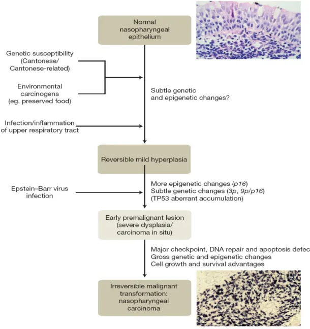
To date, the molecular basis of NPC is still poorly studied. However, it has been suggested that the pathogenesis mechanism of NPC is a complex multistep process. Few epidemiological studies have proposed the involvement of three major aetiological factors in the development of NPC, which includes genetic susceptibility, environmental factors and latent Epstein-Barr Virus (EBV) infection (Tao & Chan, 2007). The ethnic clustering of NPC in Southern China has suggested the strong association between ethnic and genetic influences.
Approximately 10 % of NPC incidences are familial cancers (Tao & Chan, 2007). An NPC susceptibility locus has been identified at the human leukocyte antigen (HLA) region. The researchers postulated that some of the HLA antigens have reduced efficiency in combating EBV infection. Prevalent Chinese A2 subtype (HLA-A*0207), instead of prevalent Caucasian
7
A2 subtype (HLA-A*0201) has been strongly associated with increased NPC risk in a high- resolution genotyping study (Kwok et al., 2004). Moreover, the frequent consumption of salted fish and preserved food as well as the occupational exposure to formaldehyde or wood dust are strongly linked to development of NPC. A study reported that the intake of preserved food contributes to two-fold increment in NPC risk whereas non-preserved food intake leads to 36 % decrement in NPC cases (Tao & Chan, 2007). NPC is very different from other head and neck squamous carcinoma in the fact that it is strongly associated with EBV infection.
Most of the NPC patients have higher EBV antibody titers in their serum. In earliest premalignant lesion, latent and clonal viral genomes as well as viral oncoproteins such as latent membrane protein 1 (LMP 1) are detected. This indicated that EBV infection occurred before the clonal expansion of tumour cells. Hence, it is proposed that EBV plays important roles in transforming normal nasopharyngeal epithelial cells into invasive carcinoma.
8
Figure 2: Possible model of nasopharyngeal carcinoma. The whole pathogenesis of NPC might take approximately 40 years. However, the latent EBV infection has accelerated the transition from carcinoma in situ to an NPC case. Taken from Tao & Chan (2007).
Nasopharyngeal carcinoma: molecular pathogenesis and therapeutic developments. Expert reviews in molecular medicine, 9 (12).
9 2.2 Ribosomal Proteins (RPs)
A complete cytogenetic map of 80 structurally different mammalian ribosomal protein genes and four rRNA molecules have been constructed (Uechi et al., 2001). This map consists of information on chromosomal locations of all genes encoding RPs and rRNAs. Thus, this cytogenetic map acts as a useful tool in studying association of RP genes with human disorders. However, the specific functions of each RP still remain unknown.
In order to provide equimolar supply of ribosomal components, RPs are synthesized stoichiometrically, in conjunction with rRNAs (Li et al., 2002). The rRNAs catalyze the protein synthesis while RPs facilitate rRNA folding, protecting them from nuclease degradation as well as manipulating the multistep process of protein synthesis. Some RPs even have substantial extra-ribosomal functions (Wool, 1996).
Most mammalian ribosomal protein genes, like those in prokaryotes, are organized into a small number of operons, with approximately 11 ribosomal proteins under the regulation of a single promoter (Nomura et al., 1984). Indeed, in a eukaryotic genome, RP genes exist as a cluster of a single expressed gene and some inactive, processed pseudogenes throughout the genomes (Kondoh et al., 1992). According to Kenmochi et al. (1998), both human sex chromosomes and at least 20 autosomes would have one or more RP genes. This is possible as RP genes are widely dispersed throughout the genome. Since RPs are highly conserved among eukaryotes and prokaryotes during evolution, almost all mammalian RPs
10
share 40%-80% similarity with yeast RPs (Kenmochi et al., 1998). The sequence studies, together with conservation of ribosomal proteins among eukaryotes have revealed that the human ribosomes are extremely similar with ribosomes in Drosophila (Kenmochi et al., 1998). Thus, the effects of mutation in human ribosomal protein genes have been investigated thoroughly in Drosophila.
RPS26 and RPS27 are components of small 40S subunit of ribosome. RPS26 is a 13.0 kDa protein which belongs to S26E family (Human Protein References Database, 2007). The protein is encoded by RPS26 gene located on chromosome 12q13 (Human Protein References Database, 2007).
According to Vincent et al. (1993), RPS26 gene which is 600 – 700 bp long is highly and constantly expressed in various human adult tissues. Therefore, it could potentially act as an internal control for all experiment involving RNA quantification and gene expression. In recent years, RPS26 has been widely used as an endogenous control for gene regulation and gene expression study, including expression analysis of Matrilin-2 in human skin (Piecha et al., 2002), differential expression analysis of Gcn512 in mesodermal defects during mouse development and and expression pattern evaluation of homeobox gene in myelomocytic differention acute myeloid leukemia.