These results suggest that STM alone plays a critical role in septum fusion during fruit development. The shoot apical meristem (SAM), a group of undifferentiated, rapidly dividing cells at the tip of the shoot, gives rise to almost all of the aboveground organs (Takano et al., 2010). The peripheral regions of the floral meristem give rise to the primordia of the floral organ, which create the fruit.
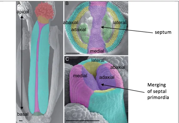
Even at early stages of development, the medial and lateral domains of the gynoecium can be distinguished. In loss-of-function stm mutants, cells in the central zone of the shoot meristem are lost, resulting in termination of the primary shoot meristem at the seedling stage of development ( Endrizzi et al. 1996 ). The phenotype of ath1-5 flowers resembles that of the ath1-3 mutants (Malone, 2018; Leary, 2018; Roth, 2018; Palmer, 2018); therefore, both are thought to represent loss-of-function alleles (Gomez-Mena and Sablowski 2008; Liljegren, unpublished results).
When CUC2 expression was analyzed throughout the Arabidopsis life cycle, it was found at the tips of septal primordia just before septum fusion (Ishida et al. 2000). Another gene, SPATULA (SPT), works synergistically with CUC1 and CUC2 to ensure proper septum and ovary development (Nahar et al., 2012). Within the CMM, cytokinin promotes the proliferation of cells that will become the septum, replum, placenta, ovules, and transmitting tract (Reyes-Olalde et al. 2017).
First, since STM is known to activate cytokinin biosynthesis and thereby promote growth of the central margin meristem (Reyes-Olalde et al. 2017), I hypothesized that stm mutants would exhibit septal fusion defects moderate to heavy.
Planting and Growth Conditions
Water was added to Promix BX soil (Premier Tech Horticulture, Quakertown, PA) to moisten it before loosely compacted soil was added to each pot. Seedlings were then thinned to approx. twelve to fifteen plants per pot, and Marathon 1% granular pesticide (OHP, Inc., Mainland, PA) was added. Plants were watered Monday, Wednesday, and Friday, alternating water with or without Miracle-Gro® water-soluble all-purpose plant food (Miracle-Gro® Lawn Products, Inc., Marysville, OH) diluted to 200 ppm.
The plants were exposed to 16 hours of light and 8 hours of darkness at an average temperature of 23°C with 70%.
Genotyping
For elution, 100 µL of a low-salt buffer (Buffer AE) was added, and the samples were incubated for five minutes at room temperature. Polymerase chain reaction (PCR) is required to amplify the STM and ATH1 gene regions using the genomic DNA obtained in the DNA extraction protocol as the template. To each PCR tube, 18 µL of the master mix and 2 µL of genomic DNA were added to create a 20 µL reactions.
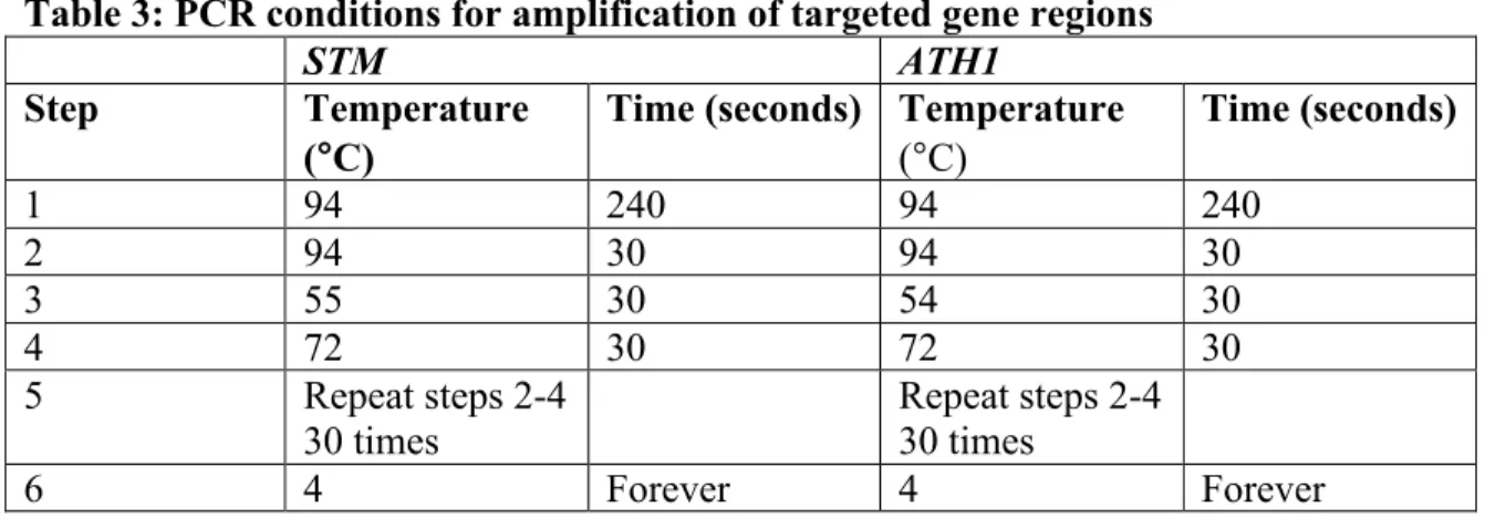
Depending on the genotype, samples were run either on an STM-specific PCR cycle or on an ATH1-specific PCR cycle in an S1000 Thermal Cycler (Bio-Rad, Hercules, CA). The pellet was then resuspended in 20 µl ddH2O prior to use of the desalted DNA in restriction enzyme digests. Homozygous stm plants were distinguished from wild-type and heterozygous stm/+ plants using a BsrI restriction site present only in the wild-type allele of the STM PCR product.
The BsrI enzyme cuts the wild-type PCR product into 106 base pairs (bp) and 29 bp fragments, whereas the uncleaved stm mutant PCR product remains 135 bp. The digestion reaction ratio is 17 µL of sample PCR product, 2 µL of 10X NEBuffer 3.1, and 1 µL of BsrI (New England BioLabs, Ipswich, MA). After gel electrophoresis of digested PCR products (see below), it was possible to diagnose the genotype of the samples.
If the plant was homozygous for the STM mutant allele, only the 135 bp product appeared;. However, if the plant was heterozygous for the STM mutant allele, both the 135 bp product and the 106 bp product appeared. To distinguish between homozygous ath1-5 mutant and wild-type plants, an MluCI restriction site was used because only one of two sites is present in the mutant ath1-5 PCR product compared to the wild-type ATH1 PCR product.
After digestion was complete, the genotype of each DNA sample was determined using gel electrophoresis, which separates DNA fragments by length. 3% agarose gels were used to separate the STM PCR products because there is less difference in size between these products. To determine DNA fragment sizes, a 50 bp scale was used for 3% gels and a 1 kb scale was used for 1% gels.
Fruit Data Collection
The gels were viewed with ultraviolet (UV) light to detect the fluorescence of ethidium bromide, a chemical that intercalates with DNA fragments.
Fruit Measurements
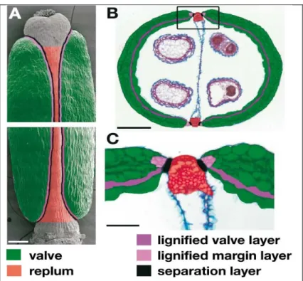
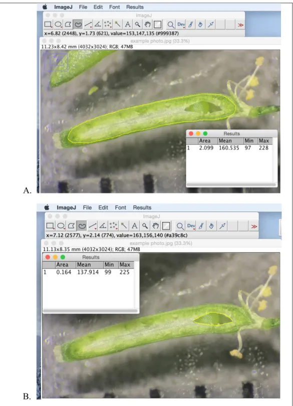
NIH ImageJ software was used to trace and measure (A) the total area of the septum and (B) the area of the septum that was missing for each fruit.
Data Analysis
Each of the 254 fruits was characterized as having a fully developed septum or with fusion defects and then classified by genotype. To assess the extent of septal defects in mutant fruit, each of the 254 fruits in this study was imaged and measurements of 1) the potential septal area and 2) the area of any missing portion of the septum were taken using NIH ImageJ (This was done by taking the missing area of the septal surface (Figure 8), dividing it by the potential septal area (Figure 7) and multiplying by 100 for each fruit.
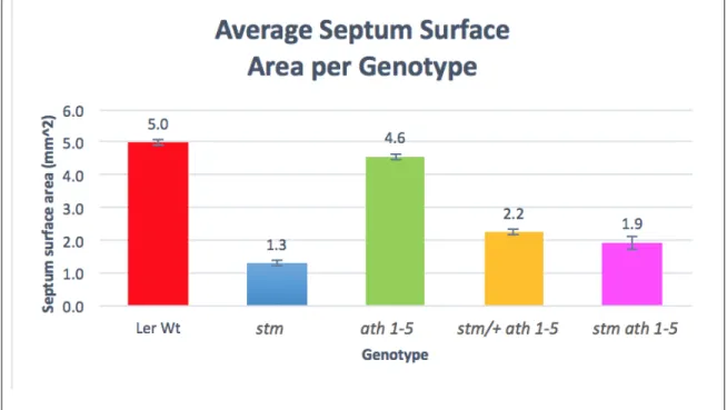
While Figure 9 shows the average percentage of the septum present for the fruit of each genotype, Figure 10 shows the average percentage of the septum present per plant. This analysis reveals the phenotypic diversity among stm, stm/+ ath1-5 and stm ath1-5 plants and, in contrast, the consistency of complete fusion of the septum in wild-type and ath1-5 plants. The aim of this experiment was to analyze the septa of Arabidopsis thaliana wild-type and mutant fruits to gain a better understanding of the roles of STM and ATH1 in promoting development and fusion of the septum.
My first hypothesis was that stm single mutant fruit would exhibit moderate to severe septal defects due to the role of STM in promoting central margin meristem growth via cytokinin synthesis. Therefore, partial loss of STM function is sufficient to interfere with growth and complete septal fusion. My second hypothesis was that stm ath1-5 double mutant fruit would have a heavier phenotype than stm mutant fruit, with less septum present and.
Instead, the stm and stm ath1-5 fruits showed 58 and 60% of septa present, respectively (Figure 9). An intriguing result in my study was that stm/+ ath1-5 fruits showed septal fusion defects, with an average of 86% of the septa present. However, my results do not suggest variable penetrance, as all stm and stm ath1-5 plants produced fruits with septal defects.
A further question prompted by my study is whether STM promotes septal fusion throughout the gynoecium or predominantly at its base or apex. I have noticed that most stm fetuses with partially missing septa seem to be most defective towards the lower quadrant of the fetus. To test the hypothesis that STM primarily promotes septal fusion at the fetal base, Childers (unpublished results).
In the spt mutant fetus, septal fusion defects were found to affect the apical end of the fetus. Further experiments investigating the interactions between STM and SPT may provide further insight into the mechanisms that regulate septal fusion in the Arabidopsis fruit.