I would like to thank all past and present members of the Mayo Lab for establishing and maintaining a friendly, helpful, and supportive environment. In particular, I would like to thank Bassil Dahiyat for his support and motivation during the years we overlapped in the laboratory. Additionally, I would like to thank Scott Ross and Cathy Sarisky for help with the NMR structure determination aspect of my project, as well.
I want to thank Tracy Johnson, Jan Aura and Massimo D'Apuzzo for their support during my last years at Cal tech. I would like to thank my family for their support during the last five years when I was so far from horn. I would like to thank my non-Caltech friends Charis Simms and Heidi Watson for their support and many trips to visit me during my graduate student career.
I would also like to thank my roommate, Michelle Rojas Soto, for her friendship and her generous offer to proofread my dissertation. I cannot help but thank my mother, Alrita Morgan, for her unconditional love and support throughout my life.
Protein function
They reduce the activation energy required to convert reactants into products without changing ~Grxn· Proteins are prepared for very specific catalysts due to the many three-dimensional arrangements of twenty amino acids. Consequently, it binds long, positively charged side chains in the pocket and cleaves polypeptides specifically at Arg and Lys residues. Proteins are adept at recognition because of the variety of tertiary forms or structures and side chains available.
Some examples of recognition by proteins are the recognition of DNA to regulate transcription, carbohydrates to regulate cell-cell interactions, and other proteins and small molecules to stimulate immune system responses. The structure of tangled homeodomain bound to DNA has been solved at 2.2 A resolution and shows that the N-terminal arm of the protein binds in the minor groove and the third helix binds in the major groove, allowing specific binding to DNA with ' enabling a K0 of 7.9. x 10-11 M as shown in Figure 1-12,3. Two examples of protein-protein recognition are present in the major histocompatibility class I immune system proteins.
The T-cell receptor protein probes the top surface of the MHC-peptide complex, initiating a cascade of immune system events if the bound peptide or MHC protein is not recognized as self. Recognition of ion channels by toxins benefits the venomous organism by inhibiting the nervous system of the stung organism.
Protein structure (with an emphasis on helices)
The residues that break the helix without helical dihedral angles, but participate in helical i, i + 4 hydrogen bonding, are labeled as the N-eap and C-cap positions. On the other hand, a priori calculations predicted that the amide hydrogen at the N-terminus and carbonyl oxygen at the C-terminus having unsatisfied hydrogen bond donors and acceptors, a partial positive character at the helix N-terminus and a partial negative charge . Experimental studies have shown that the effect of the helix dipole at the N-terminus is independent of helix length8,9.
At the N-terminus of a helix, the first three amide hydrogens have unsatisfied hydrogen bonds because no hydrogen bond acceptors precede them in the helix. Therefore, experimental and theoretical results suggest that the helical dipole is an electrostatic effect localized at the ends, rather than a sum of the dipole moment of individual peptide bonds. The relationship between the charge of the side chain and the tendency to occupy certain regions of the helix was first observed in a statistical analysis of the seven globular proteins of known structure in 1969 by Ptitsyn14.
In addition to helical residues, propensities have been calculated for the helix N-eap position, the position before N1 which has non-helical dihedral angles but participates in i, i + 4 hydrogen bonding and for the C-cap position which follows the helix11,13,15. Motifs can consist of only helices and turns, such as the helix-turn-helix motif of homeodomains or only of ~-sheets and turns, such as the light chain of MHC class I proteins, ~2m.
Protein design
The arrangement of the motifs of oligomeric proteins in relation to each other is referred to as the quaternary structure. Using rotamer descriptions of the side chains24,25 and a fixed backbone, a fast scoring algorithm based on the Deadlock Elimination Theorem26-28 was used to find the global optimal sequence in the optimal rotamer conformation. Initial design efforts in the Mayo Lab included designing the core residues30 of the GCN4-p1 coiled coil using a van der Waals potential to account for steric interactions31.
A quantitative structure activity relationship analysis of the experimental results was used to determine that a potential dependent on the hydrophobic and polar buried surface would significantly improve the scoring ability of the algorithm. A subsequent design study of the core residues of the G-1 domain of streptococcal protein was used to determine the effect of specific steric constraints and the effect of punishing exposed hydrophobic surface33. Residue positions located at the boundary of core and surface positions are designed by a combination of the core and surface energy scoring functions.
Design of the core residues and half of the border residues of streptococcal protein G ~1 domain led to the sequence of a hyperthermophilic protein variant34. Optimizing protein stability was chosen as the success criterion, because the design of stable proteins is indicative of a good understanding of the physicochemical forces that govern protein structure.
Internal sharp effect measurement of the electric field at the amino terminus of an alpha helix. The side cap of the N-eap residue (Thr) is shown making a hydrogen bond to the i + 2 amide proton. The effect of the helix dipole and N-coverage on the surface design of an a-helical protein.
The effect of the helix macrodipole on protein stability has been studied in both model peptides and proteins. Each of the four mutations (S38D, N144D, Tl09D, N116D) results in a protein which has a stable pH-dependent growth, i.e., mutants. The identity of the residue at the N-eap position also has an effect on helix stability.
Thirty of the 51 enh residues were determined to be solvent exposed as described in the Methods. Therefore, the increases in stability are most likely largely due to the effect of the side chains on the helix dipole. All 1029 possible combinations of the hydrophilic amino acids allowed at the surface were considered for the remaining twenty-nine surface positions in the One calculation.
Measurement of the intrinsic Stark effect of the electric field at the amino terminus of an a-helix. Analysis of the interaction between charge side chains and the α-helix dipole using engineered thermostable mutants of phage T4 lysozyme. Of the forty designed positions in the surface core (scl) plan, there are twenty-nine mutations.
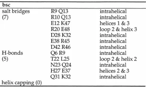
The combinatorial complexity of the computation was made manageable by computing the entire sequence design incrementally. The boundary positions were allowed to be any of the twelve boundary residues listed above. Both forms of bsc are hyperthermophilic, i.e. Tms is greater than 99 °C and could not be measured by CD thermal denaturation techniques.
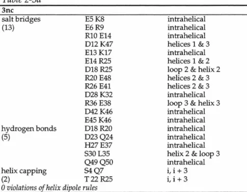
Comparison of the backbone of the average constrained minimized structure12,13 with the wild-type engrailed homeodomain crystal structure reveals excellent agreement for residues 9–48 with an r.m.s. In the model structure, none of the three selected rotamers form hydrogen bonds in these positions. Finally, addition of the boundary residues results in a protein that is a 39-fold mutant of wild type and has a melting temperature of 114 °C.
Crystal structure of the b-glycosidase from the hyperthermophilic archaeon Sulfolobus solfataricus: resilience as a key factor in thermostability.
Circular Dichroism Determination of Class I MHC- Peptide Equilibrium Dissociation Constants
We demonstrate that the thermal stability of the MHC-peptide complex depends directly on peptide binding affinity. In the case of the murine class I MHC molecule H-2Kd, peptide equilibrium dissociation constants were measured by equilibrium dialysis assays (Fahnestock et al., 1994). Approximately 30% of the Kd molecules as purified from transfected cell supernatants were occupied by endogenous peptides (Fahnestock et al., 1992).
Despite the presence of endogenous peptides in a fraction of the MHC proteins, we found a logarithmic. The heavy chain is shown in purple, the light chain is shown in red, and the bonds of the peptide are shown. Design success was assessed by experimentally determining the dissociation constants of the designed peptides.
If the complex is not recognized as self, either because of the peptide or MHC. Peptide design of the MHC-peptide complex must involve both the MHC molecule and the peptide. Together, these energies were used to generate an energy of peptide binding, which could be correlated with the peptide dissociation constant and the melting temperature of the Kd-peptide complex.
Experimental determination of the Tm of CPl complexed to Kd showed that CPl did not bind very tightly; the dissociation constants were calculated to be 1.9 x lQ-3 M (Table 5-1). CP3 was synthesized, complexed to Kd, and found to increase the Tm of the MHC-peptide complex to 65 °C. However, many of the peptide designs have not been successful, and there are several approaches to improve the computational design methodology.
To investigate the importance of disulfide bonds in folding and function, several groups have systematically replaced two half-cystines with unnatural amino acids L-α-aminobutyric acid (Aba) or alanine residues. However, removal of the C12-C28 bond (analogous to C18-C35 in agitoxin) results in an inactive molecule with a disordered structure. Removal of the analogous disulfide bridge in charybdotoxins and leiurotoxins resulted in functional folding mutants.
Similarly, the C14-C33 agitoxin disulfide bridge could be redesigned, resulting in a functional molecule, but with a high incidence of improper pairing of the other two disulfide bonds. It is likely that removal of the C18-C35 disulfide bridge from agitoxin would result in an unfolded, nonfunctional protein with the other disulfide bonds mispaired.