Selain itu, semua ekstrak kasar Calophyllum lanigerum dan senyawa terisolasi diselidiki potensi antioksidannya melalui uji DPPH. Selain itu, ekstrak kasar Calophyllum lanigerum dan senyawa murninya diuji potensi antioksidannya menggunakan metode DPPH. Disertifikasi bahwa MONG LAI WAN (ID No: 15ADB07643) telah menyelesaikan proyek tahun terakhir ini berjudul "STUDI FITOKIMIA DAN ANTIOKSIDAN DARI CALOPHYLLUM LANIGERUM".
INTRODUCTION
General Introduction
- Taxonomy
- Morphology
- Ethnomedical Uses and Pharmacological Studies
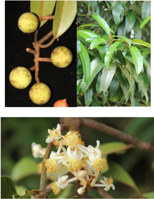
Calophyllum lanigerum can be found in the tropical rainforest of Asia such as Singapore, India and Malaysia (Dweck and Meadows, 2002). In this study, we are going to study the antioxidant activity of Calophyllum lanigerum by means of DPPH test using kaempferol and ascorbic acid as positive controls. To study the antioxidant activity of isolates and crude extracts of Calophyllum lanigerum via DPPH assay.
Phytochemical Studies
- Coumarins
- Xanthones
- Terpenes
- Flavonoids
- Calophyllum brasiliense
- Calophyllum inophyllum
- Calophyllum symingtonianum
10 chemical synthesis yielding a wide variety of compounds with various health benefits (Naithani et al., 2010). They are compounds of the benzopyrone family that consist of a benzene ring linked to a pyrone ring as shown in Figure 2.1, giving the molecular formula C9H6O2 (Rohini, 2014). Based on their chemical structure, coumarins can be classified into four main types, which are simple coumarins, pyranocoumarins, pyrone-substituted coumarins and furanocoumarins.
In 1994, a pyranocoumarin with anti-HIV activity was isolated from the latex of Calophyllum teysmannii (Spino, Dodier and Sotheeswaran, 1998). They are polyphenolic compounds which consist of two benzene rings linked by a carbonyl group and an oxygen bridge as shown in Figure 2.2 and have an essential molecular formula of C13H8O2. Xanthones can be categorized into five main groups which are simple xanthones, prenylated xanthones, glycoside xanthones, bisxanthones and xanthonolignoids.
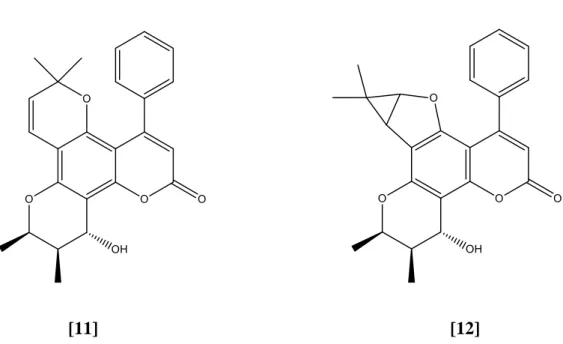
Terpenes are a large class of phytochemicals made up of isoprene with a molecular formula of (C5H8)n, as shown in Figure 2-3. Flavonoids have a general structure consisting of a heterocyclic ring and two phenyl rings, as shown in Figure 2.4. Based on previous studies, Calophyllum species have been shown to be rich in xanthones, coumarins, triterpenoids and flavonoids (Su et al., 2008).
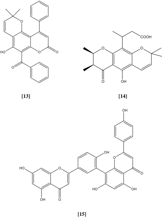
This may be due to the high number of hydroxyl groups present in compound 15 (Aminudin et al., 2015).
Summary of Literature Review on the Genus Calophyllum
Materials
- Plant Materials
Chemical Reagents
- Sequential Solvent Extraction, Isolation and Purification of Chemical Constituents from Calophyllum lanigerum
Solvents/Materials Molecular formula Source Country Dichloromethane CH2Cl2 QReC (Malaysia) Ethyl acetate CH3COOC2H5 Fisher Scientific, United Kingdom. Dichloromethane was used to soak the stem bark powder in a sealed container at room temperature for two days. The dichloromethane crude extracts were then filtered and the aqueous residue in the filtrate was removed by treatment with anhydrous sodium sulfate.
The stem bark material was then extracted with methanol twice following the procedures above to give dried methanol crude extracts. The two crude extracts of Calophyllum lanigerum were separately subjected to gravity column chromatography and gradient elution was used to separate the chemical constituents into a series of fractions. 27 potential fractions showing more than one spot on TLC were combined and subjected to a second column chromatography for further purification.
If a single spot is shown on the TLC plate, the compound was then subjected to spectroscopic analyzes for structural identification.

Chromatography
- Column Chromatography
- Gel Permeation Chromatography
- Thin Layer Chromatography
A thin layer of anhydrous sodium sulfate was then applied on top of the sample layer to act as a desiccant and protective layer. The collected fractions were analyzed for their chemical content by thin layer chromatography (TLC). The sample was dissolved in an appropriate amount of methanol before being placed in the gel permeation column.
The sample solution was introduced on top of the packed column to form a thin layer of sample solution. During the elution, the larger size compounds will be eluted first while the smaller size molecules will be eluted later. The smaller sized molecules stayed longer in the column as they were trapped in the pores of the packing.
29 materials and traveled a greater distance along the column compared to the larger molecules. The TLC plate used was an aluminum plate coated with a layer of silica gel 60 F254 Merck on top and cut to a size of 4 cm x 8 cm. First, the sample was diluted and then spotted on the marked baseline of the TLC plate using a microcapillary tube.
The plate was then placed in a developer chamber containing an appropriate amount of solvent mixture.
TLC Detection Methods
- UV Detection
- Iodine Vapor Detection
The solvent moved up to the solvent front of the plate through capillary action. At short wavelength the spots appeared as dark gray spots on a light green background, while at long wavelength the spots appeared as fluorescent color spots on a pale purple background. The brown spot that appeared on the TLC plate was marked quickly as it will disappear after removal from the iodine chamber.
Instruments
- Nuclear Magnetic Resonance (NMR)
- Infrared (IR) Spectroscopy
- Melting Point Apparatus
An appropriate amount of deuterated solvent such as methanol-d4, acetone-d6 or deuterated chloroform was used to dissolve the sample. The sample was first ground with potassium bromide, KBr in a ratio of 1:10 to form a homogeneous mixture followed by compression under high pressure to form a pellet. KBr pellet was analyzed by Perkin Elmer 2000-Fourier Transform Infrared (FTIR) spectrophotometer and IR spectrum was obtained in the range of 4000 to 400 cm-1.
Ultraviolet-visible (UV-Vis) spectroscopy was used to analyze the presence of conjugated system in a compound. An appropriate amount of analytical grade solvent was used to dissolve sample, then transferred into a quartz cuvette for analysis via a dual beam spectrophotometer, Perkin Elmer Lambda 35. One of the techniques used to determine the purity of a compound , its melting point is determined. .
A pure compound should have a narrow melting range compared to compound that is not pure. The purity of a compound can be determined by comparing the experimental melting point with the literature value of the pure compound. Sample was filled into a hematocrit capillary tube and heated until melting using the Stuart SMP 10 melting point apparatus.
Antioxidant Assay
Then, the stock solutions were sonicated for 5 min to ensure that all the sample was completely dissolved and mixed homogeneously. Similarly, the DPPH powder was dissolved in methanol and followed by sonication for 5 min to prepare 2 mg/ml DPPH solution. All the prepared stock solutions and the DPPH solution were stored in a 4℃ cooler in dark condition.
The positive control used for this test was kaempferol and ascorbic acid, while the wells containing DPPH solution and methanol without the test sample were used as blanks. After addition of reagents, the plate was immediately covered and wrapped with aluminum foil to prevent solvent evaporation. The absorbance of the contents in each well was measured with a microplate reader at 520 nm.
Each test sample was run in triplicate and the average absorbance for each concentration was calculated and recorded. The following equation was used to calculate the inhibition percentages of the test compounds. A graph of inhibition rate versus test sample concentration was plotted to determine the IC50 value of the test samples.
The IC50 is defined as the concentration of sample needed to inhibit 50% of DPPH radicals.
Chemical Constituents Isolated from Calophyllum lanigerum
In the 1H NMR spectra (Figures 4.2 and 4.3), a total of eight proton signals were observed, including two highly shielded proton signals at 12.69 and 9.06, which were assigned to the chelated hydroxyl proton, 1-OH and a free hydroxyl proton, 7 - Oh, respectively. 38 A total of 13 carbon signals were shown in the 13C NMR spectrum (Figure 4.4), which were consistent with the number of carbon atoms in the proposed structure. The HMQC spectrum (Figures 4.6 and 4.7) showed the heteronuclear 1J couplings between protons and their respective carbon atoms, whereas in the HMBC spectra (Figures and 4.11) it indicated the long-range heteronuclear 2J or 3J coupling between protons and their neighboring carbons .
A broad band at 3303 cm-1 appeared on the IR spectrum (Figure 4.12) and corresponded to the O-H stretch in the compound. The IR spectrum (Figure 4.14) showed absorption bands at 1715 and 2927 cm-1, indicating the presence of a carbonyl group and sp3 C-H stretching. This was due to the n π* transition corresponding to the excitation of an electron from the lone pair to the π* orbital.
Moreover, the assignment of these proton signals was done by comparing with the literature values reported by Abbas et al. The 13C NMR spectra (Figures 4.17 and 4.18) showed a total of 30 carbon signals consistent with the number of carbons in the proposed structure. Among the carbons, the keto carbon C-3 gave the most deshielded signal at 213.3 and it was found that the remaining 29 carbon signals appeared in the region below 70.0 assigned to the sp3 hybridized carbons.
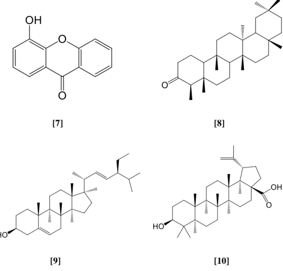
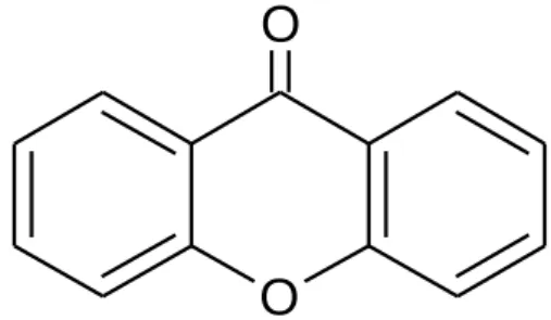
The remaining assignment of carbon signals was completed by comparing with the literature values reported by Abbas et al., in the year 2007. The IC50 value of test samples was determined from the graph of the inhibition rate against concentration of the sample as shown in Figure 4.20. Meanwhile, euxanthone [18] is a phenolic compound, but the hydroxyl group in the molecule is chelated to the keto group, so it is unable to donate hydrogen.
![Table 4.1: Summary of NMR assignment for euxanthone [18]](https://thumb-ap.123doks.com/thumbv2/azpdforg/10232829.0/57.892.349.653.102.326/table-summary-nmr-assignment-euxanthone.webp)
Conclusion
In future studies, it is strongly recommended to use more advanced separation techniques such as high-performance liquid chromatography (HPLC) to isolate small compounds from the stem bark of Calophyllum lanigerum. Bioguided fractionation and isolation of natural inhibitors of advanced glycation end products (AGEs) from Calophyllum flavouramulum. Trends in chemical and pharmacological research on the tropical trees Calophyllum brasiliense and Calophyllum inophyllum, a global context.
Screening of inophyllums against HIV-1 by HPLC-DAD of Calophyllum inophyllum leaf extracts from the islands of French Polynesia. A terpenoid and two steroids from the flowers of Mammea siamensis, Journal of Science and Technology, 27(2), pp.