This thesis titled “THE CONTRIBUTION OF rshA MUTATIONS AND EFFLUX PUMPS IN TIGECYCLINE RESISTANCE IN MYCOBACTEROIDES ABSCESSUS” was prepared by AW KAR MEN and submitted in partial fulfillment of the requirements for the degree of Master of Medical Science at Universiti Tunku Abdul Rahman. It is hereby declared that AW KAR MEN (ID no.: 19UMM07057) has completed this thesis titled "THE CONTRIBUTION OF rshA MUTATIONS AND EFFLUX PUMPS IN TIGECYCLINE RESISTANCE IN MYCOBACTEROIDES ABSCESSUS" under the supervision of Prof. dr.
Background
Interestingly, three efflux genes were also revealed to be upregulated in a tigecycline-resistant rshA mutant (Ng, et al., 2018). This indicated that efflux genes may very likely contribute to tigecycline resistance seen in the mutant.
Problem statement and hypothesis
Therefore, this study was initiated to further elucidate the role of rshA mutations and efflux mechanisms in tigecycline resistance in M.
Objectives
The outcome of the study may contribute to the development of new and effective therapeutic strategies against M.
Nontuberculous and rapid growing Mycobacteria
Mycobacteroides abscessus taxonomy
M. abscessus environmental sources
The persistence of mycobacteria and sharing of habitation with the human population creates the ideal setting for human infection (Degiacomi, et al., 2019; . Ratnatunga, et al., 2020).
The underplayed player, M. abscessus
Vulnerable groups for M. abscessus infections
Clinical manifestations and outbreaks of M. abscessus
In healthy hosts, SSTIs are often associated with post-traumatic injuries and after cosmetic or surgical procedures involving the use of contaminated medical devices or implants (Brown-Elliott and Wallace, 2002; Petrini, 2006; Furuya, et al., 2008; Wongkitisophon, et al. ., 2011; Sfeir, et al., 2018). In South Korea, there was a report of a spontaneous outbreak of skin infections following acupuncture therapy (Song, et al., 2006).
Diagnosis of M. abscessus infections
The Clinical and Laboratory Standards Institute (CLSI) recommended testing for antimicrobial susceptibility by determining minimum inhibitory concentration (MIC) using broth microdilution assay (To, et al., 2020; Weng, et al., 2020). The American Thoracic Society and Infectious Disease Society of America had previously recommended macrolide-based multidrug therapy with one or more parenteral antibiotics based on antibiotic susceptibility test results (Griffith, et al., 2007; Novosad, et al., 2016).
Antimicrobial resistance
As the situation worsened, the World Health Organization identified antibiotic resistance as one of the greatest threats to public health (WHO, 2021). Resistance can also be acquired through genetic mutations or possession of genetic determinants through horizontal gene transfer (Blair, et al., 2015).
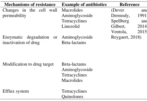
Antimicrobial resistance mechanisms in M. abscessus
This fearsome phenotype that limits antibiotic selection is conferred by the natural or acquired resistance of the mycobacteria (Nessar, et al., 2012). The lipid-rich cell wall works in synergy with other natural mechanisms, making most drugs ineffective (Nessar, et al., 2012).
Tigecycline
Tetracyclines, first discovered in the 1940s, inhibit protein synthesis by interfering with the binding of aminoacyl-tRNA to the ribosomal acceptor (A) site. Unfortunately, the emergence of resistance to tetracyclines has followed the widespread use of these antibiotics in clinical practice as well as in animal feed (Chopra and Roberts, 2001). Increased research efforts were then made to improve the understanding of tetracycline resistance mechanisms in the hope of resolving the use of tetracyclines as therapeutic agents.
Unlike tetracycline, this glycylcycline does not induce the expression of MabTetX, a drug-modifying enzyme responsible for tetracycline resistance in M. As a broad-spectrum antibiotic, tigecycline is used in the treatment of patients with methicillin-resistant Staphylococcus aureus, extended-spectrum beta-lactamase producing bacteria and vancomycin-resistant enterococcal infections (Noskin, 2005).
Tigecycline resistance
Tigecycline works by inhibiting protein synthesis in bacteria as it binds to the bacterial 30S ribosome and prevents the integration of amino acids into the peptide chains (Greer, 2006; Zhanel, et al., 2012). Unfortunately, the induction of whiB7 (a transcriptional activator of natural antibiotic resistance) (Pryjma, et al., 2017) and RshA mutations (Ng, et al., 2018) had also been associated with resistance to tigecycline in M. 2021) showed that the sigH gene was associated with phenotypic resistance to tigecycline in M.
RshA and SigH
Under unstressed conditions, the RshA binds to SigH and represses the SigH-dependent transcription (Song, et al., 2003). When the bacteria are subjected to stressful conditions such as redox stress, increase in temperature or mutation in the conserved HXXXCXXC motif, the RshA-SigH interaction is disrupted, leading to the release of SigH and downstream phenotypic changes such as antibiotic resistance (Song, et al et al., 2003). This disruption was demonstrated in the tigecycline-resistant mutant harboring a C51R mutation that caused the first cysteine residue in the conserved HXXXCXXC motif to be changed to arginine (Ng, et al., 2018).
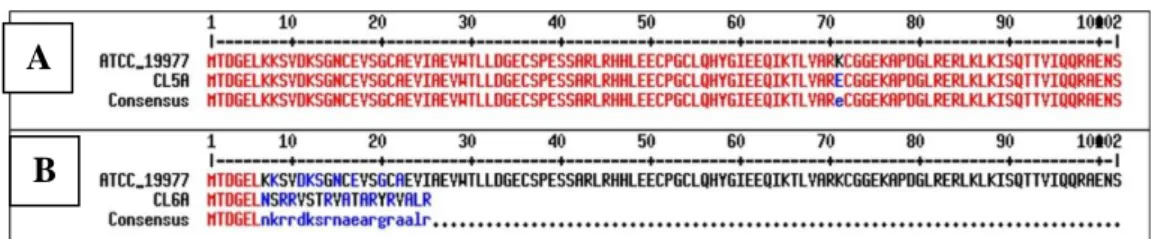
A study by Ng, et al. 2020) described that the mutation in the conserved HXXXCXXC motif of RshA was also associated with the overexpression of the sigH gene, which was verified by RT-qPCR analysis. With further experimental evolution work, Lee, et al. 2021) identified two additional rshA mutations, namely K71E mutation in CL5A and an insertion mutation in CL6A, that were responsible for tigecycline resistance in the two mutants.
Efflux pumps in M. abscessus
- Stokes disk diffusion
- Etest
- Preparation of nucleic acid
- End point PCR
- RT-qPCR
- Bacterial Adenylate Cyclase Two Hybrid (BACTH) assay
- Screening
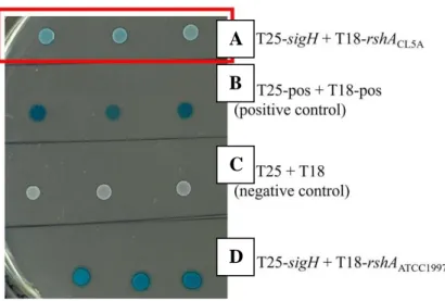
The MIC was read as the lowest concentration of antibiotic that inhibits the visible growth of the bacterium. Essentially, the BACTH kit provided one plasmid carrying the T25 subunit (pKT25) and another carrying the T18 subunit (pUT18) of the adenylate cyclase. In-frame cloning of the rshA gene (amplified from the wt ATCC 19977 or CL5A) into the pUT18 plasmid and the sigH gene (amplified from the wt ATCC 19977) into the pKT25 plasmid was performed.
After purification of the amplicons with the QIAquick PCR purification kit (Qiagen), the amplicons and corresponding plasmids were double digested with the appropriate restriction enzymes described in Table 3.2. The development of the blue color of the BTH101 reporter strain was observed every two hours and compared with three other control strains.
CGTGCGTGCGCTG AGCA
TCAGGCAGCGACA CGATTTC
CTAATCTTCGACG GAGACC
TGATTGCCCGTCT GCG
TTAGCCGGTGGAC ACCG
GGCGTGCCTCGAAC GTCC
CGTGCCCGGCCCTCC GC
GACCGACGGTGAAC TCAAGAAGA
CGGGAGTTCTCGGCC CGCTG
Cloning and transformation in efflux gene dosage studies
- Molecular cloning of target genes
- Transformation of the recombinant plasmids into M. abscessus
- Preparation of electro-competent M. abscessus cells
- Electroporation of the recombinant plasmids into the electro- competent M. abscessus cells
Double digestion was then performed using the appropriate restriction enzymes (Table 3.2) and CutSmart buffer (NEB). Purified fragments, purified with the QIAquick PCR purification kit, were then ligated at a 5:1 insert:vector molar ratio with T4 DNA ligase (NEB). Transformed cells were plated on Luria-Bertani (LB) agar (first base) supplemented with 30 mg/L kanamycin (Gold Biotechnology) and incubated overnight at 37.
The positive colonies were selected after colony PCR screening (see section 3.3.2) and the recombinant plasmids were expanded and purified from the LB broth (first base) cultures supplemented with 30 mg/l kanamycin containing the DNA spin plasmid. Purification kit (iNtRON Biotechnology). In a 0.2 cm electroporation cuvette (Bio-Rad), the recombinant plasmids were electroporated into the electro-competent M.
BLAST analyses
Quantitative experiments were all performed in triplicate and the data obtained were recorded in mean ± one standard deviation. The difference in means between two groups was analyzed using the two-sample t-test and p-value < 0.05 as a cut-off value for statistical significance, with the GraphPad Prism 5 software.
Characterisation of mutants
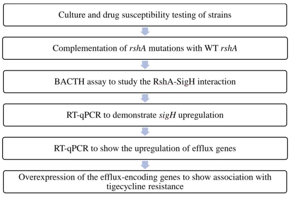
Complementation of CL5A and CL6A with wt rshA
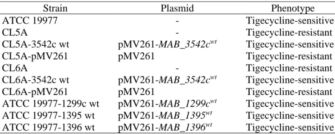
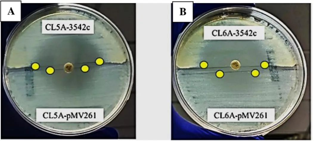
Screened and selected by colony PCR, the PCR products were sent for Sanger sequencing to verify that the mutants were supplemented with wt rshA. As expected, complementation of the tigecycline-resistant mutants with the wt rshA successfully restored their sensitivity to tigecycline. Using disk diffusion from Stokes agar plate, the sizes of the tigecycline inhibition zones of the strains supplemented with the wt rshA were remarkably larger compared to the mutants with the empty plasmid (Figure 4.2).
This was then confirmed using the Etest in which the MIC of the complemented strains decreased to 0.25 mg/L while the mutants with the empty plasmid maintained the MIC of 2 mg/L (Table 4.2). These results indicated that the rshA mutations possessed by CL5A and CL6A were responsible for the tigecycline-resistant phenotype seen in the mutants.

BACTH assay to demonstrate the disruption of RshA-SigH interaction
RT-qPCR to demonstrate sigH upregulation
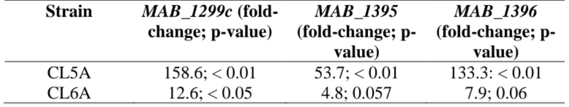
Efflux-encoding genes in CL5A and CL6A
The purpose of this experiment was to determine whether the efflux pumps were also upregulated in the mutants with rshA mutations. The RT-qPCR results suggested that efflux genes might have played a role in the tigecycline resistance seen in mutants CL5A and CL6A.
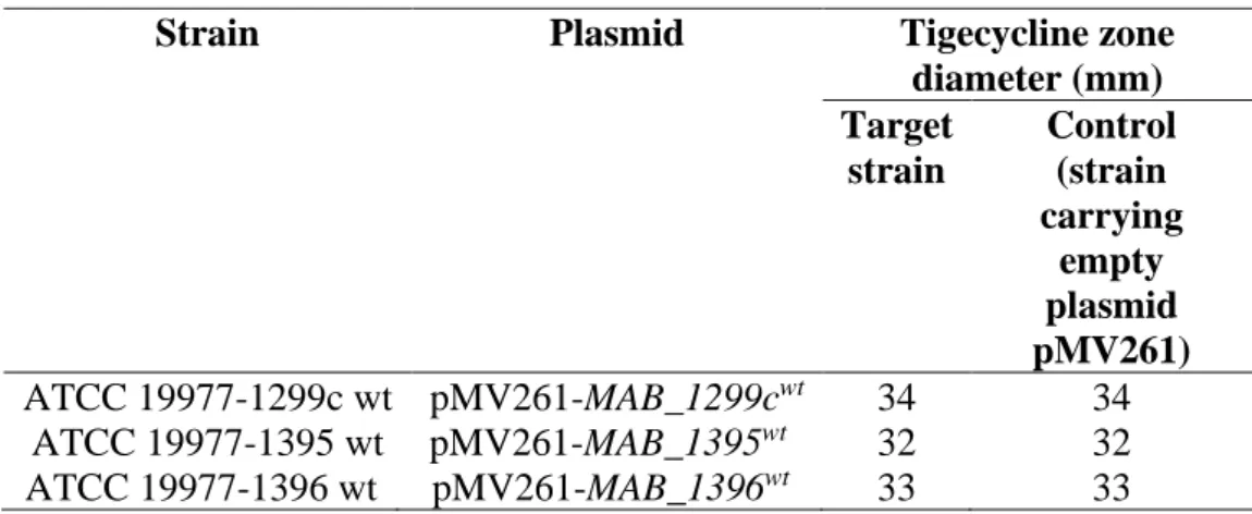
Overexpression of efflux-encoding genes in wt ATCC 19977
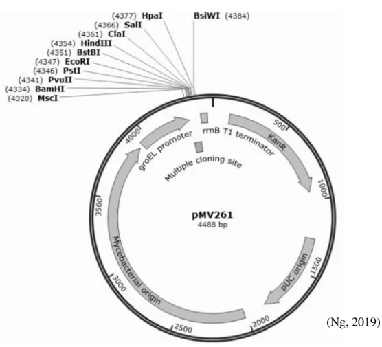
Using the mycobacterial origin of replication, the plasmids carrying the efflux genes (pMV261-MAB_1299cwt, pMV261-MAB_1395wt and pMV261- MAB_1396wt) were multiplied in the mycobacterial host M. This increased the copy number of the target causative efflux genes . transcription from the promoter, groEL. Disappointingly, the overexpression of the three efflux-encoding genes in ATCC 19977 did not show an increase in tigecycline resistance as demonstrated by the zones of inhibition in Stokes disc diffusion assay (Table 4.5).
Currently, it is considered one of the most effective treatment options for patients with M. In this study, rshA mutations and efflux pumps were further investigated to have a better understanding of their role in tirecyclin resistance in M.
Causal relationship of the rshA mutations and tigecycline resistance in M. abscessus established using gene complementation
Complementation of CL5A and CL6A with the pMV261 plasmid carrying the wt rshA gene efficiently restored the tigecycline sensitivity phenotype (Figure 4.2) in the mutants. The results suggested that the rshA mutations were responsible for the tigecycline resistance phenotypes in both CL5A and CL6A. The result of this study was in good agreement with the previous work of Ng, et al. 2018), who showed the T151C → C51R mutation in the rshA gene as a possible tigecycline resistance determinant in M.
The anti-sigma factor, RshA, suppresses SigH, a sigma factor, during non-stressful conditions and both factors play a role in regulating the stress response (Song, et al., 2003). The detailed mechanism on how rshA mutations and its sigma factor, SigH, cause tigecycline resistance in M.
Bacterial Adenylate Cyclase Two Hybrid (BACTH) system demonstrated disrupted RshA-SigH interaction in CL5A
This finding suggested that the mutation of rshA in CL5A could disrupt the interaction between RshA and SigH. RshA acts as an anti-sigma factor that inhibits the sigma factor SigH under normal conditions. The findings in this present study indicated that in the absence of external stressors, the interaction between RshA and SigH could be disrupted by the rshA to CL5A mutation.
RT-qPCR verified the upregulation of sigH in CL5A and CL6A
Thus, if bacterial strains such as these laboratory-derived mutants were present in the environment, a horizontal transfer of the genetic determinant of tigecycline resistance to other bacteria could occur over time, causing widespread tigecycline resistance, as Wintersdorff, et al. 2016) argued that antimicrobial resistance can be acquired through horizontal gene transfer of antibiotic resistance genes.
Efflux-encoding genes in the tigecycline-resistant mutants, CL5A and CL6A
Overexpression of the three efflux-encoding genes failed to induce resistance to tigecycline in M. abscessus
Whole genome sequencing to identify transmission of Mycobacterium abscessus between patients with cystic fibrosis: a retrospective. First US outbreak of Mycobacterium abscessus hand and foot disease in children associated with a wading pool. Selection and characterization of a tigecycline-resistant mutant of Mycobacterium abscessus to identify potential resistance determinants.
A mutation in anti-sigma factor MAB_3542c may be responsible for tigecycline resistance in Mycobacterium abscessus. Antagonism between first-line antibiotics clarithromycin and amikacin in the treatment of Mycobacterium abscessus infections is mediated by the whiB7 gene. High levels of intrinsic tetracycline resistance in Mycobacterium abscessus are conferred by a tetracycline-modifying monooxygenase.
Clinical experience in 52 patients with tigecycline-containing regimens for salvage treatment of Mycobacterium abscessus and Mycobacterium chelonae infections.