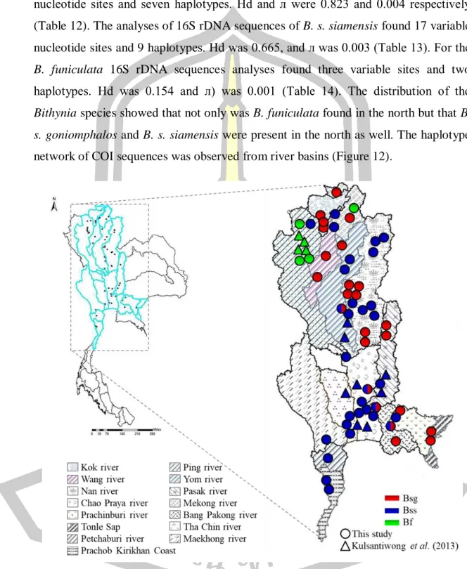
The phylogenetic trees of COI and 16S rDNA reveal that some samples belonged to a cryptic group that was unable to identify to subspecies and clustered between B. 47 Table 10 Analysis of molecular variance (AMOVA) based on COI and 16S rDNA -sequences of seven groups defined by seven different river basins as shown in Figure 7. Bootstrap values for neighbor-joining, maximum likelihood and posterior probability for Bayesian analysis are shown above or near the branches, compared to the median linkage haplotype network of COI and 16S rDNA sequences.

- Background
- Objective of study
- Scopes of study
- Places of study
Mitochondrial cytochrome c oxidase subunit I (COI) gene was applied for differentiation and identification of snails in the family Bithyniidae from Thailand (Kulsantiwong et al., 2013). Thus, this study examined some species of freshwater snails in the family Bithyniidae for cercarial infection, as well as genetic variations using mitochondrial DNA (COI and 16S ribosomal DNA) and nuclear DNA (intron regions of Arginine kinase; AK) sequences as molecular means. markers. The genetic analyzes were mostly performed in the Department of Environmental Medicine, Kochi Medical School, Kochi University, Japan.
Introduction
Material and Methods
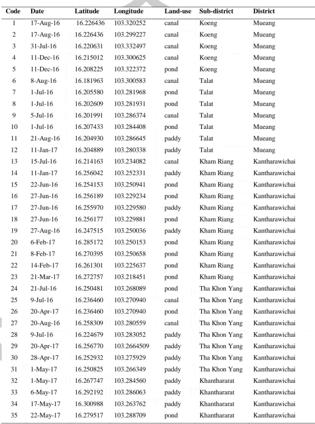
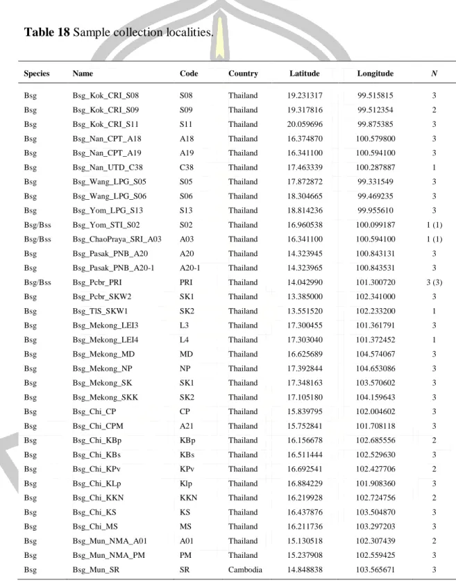
A total of 3,757 individuals of six freshwater snail families namely Bithyniidae, Lymnaeidae, Thiaridae, Planorbidae, Viviparidae and Ampullariidae (Figure 4) were collected from 51 localities of four districts of Maha Sarakham provinces. earlier reports by Brandt (1974), Chitramvong & Upatham (1989) and Chitramvong (1992). After that, place an individual freshwater snail in a small plastic cup (2 oz.) with 5 ml of dechlorinated tap water, exposed to light (1200 lx) for five hours at room temperature (25±2 ºC).

Results
Type of cercariae and metacercariae by crushing method ABCDEFGHL-M% infection ABCGHL-MNOP-QR-ST-UV-WXY% infection 1Bithyniaspp. Type of cercariae and metacercariae by crushing method ABCDEFGHL-M% infection ABCGHL-MNOP-QR-ST-UV-WXY% infection 9Bithyniaspp. Type of cercariae and metacercariae by crushing method ABCDEFGHL-M% infection ABCGHL-MNOP-QR-ST-UV-WXY% infection 39Bithyniaspp.

Discussion
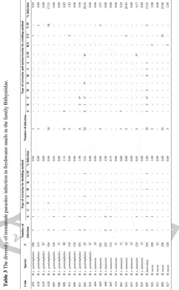
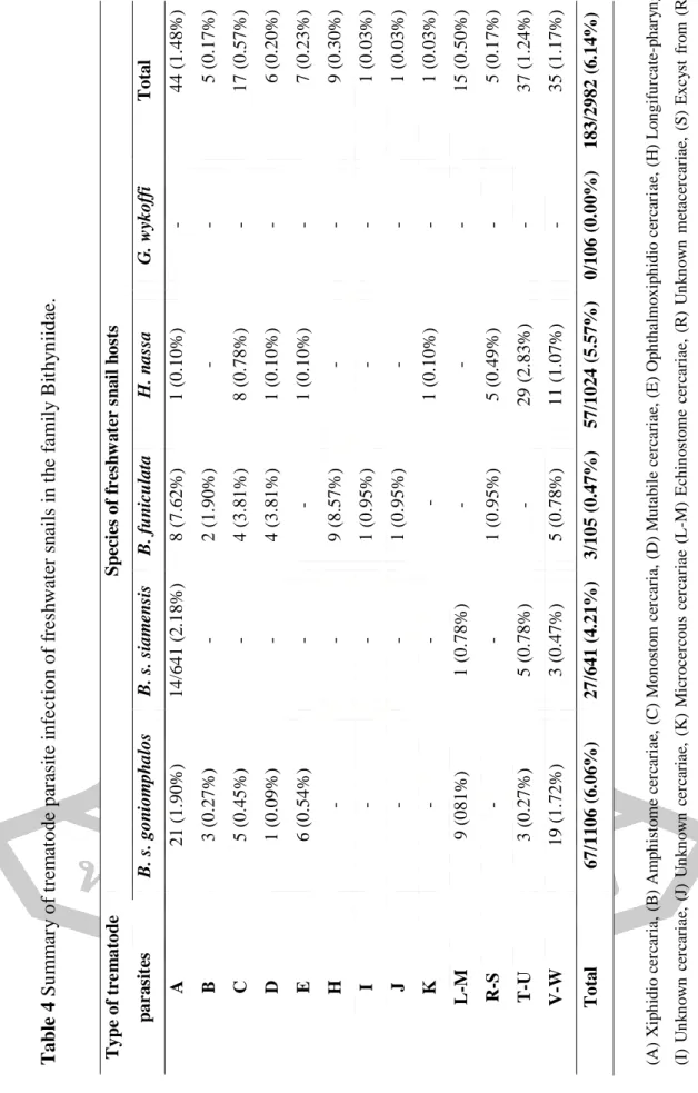
In this study, 13 types of trematode cercariae were found in five freshwater snails, namely B. Of these, five types of cercariae were found in H. nassa, namely Xiphidio, Monostom, Mutabile, Ophthalmoxiphidio, Microcercous cercariae. To my knowledge, this is the first report on the detection of cercarial trematodes in H. On the other hand, Kulsantiwong et al. 2015) studied freshwater snails of the family Bithyniidae in Thailand and found six types of cercariae, viz. according to the previous report, the most common type of trematode infection in snails was Xiphidio cercaria (Kiatsopit et al., 2016; Kulsantiwong et al., 2015; Mohammed et al., 2016), which is similar to my current study that Xiphidio and Echinostome cercariae are the most commonly found parasites in freshwater snails.
Freshwater snails in six families namely Thiaridae, Ampullariidae, Lymnaeidae, Planorbidae, Viviparidae and Bithyniidae collected from Chao-Phraya Basin nine species of cercariae were found viz. hydiocercaria, Virgullate, Monostome, and Echinostome, with an infection rate of 5.90% (Anucherngchai et al., 2016). The present study examined six species of freshwater snails namely; Indoplanorbis, Bithynia, Filopaludina, Thiaridae, Lymnaea and Pila from the Chi River Basin and Maha Sarakham Province and found the infection rate to be 10.17%, with nine species of cercariae and eight species of metacercariae.
- Introduction
- Material and Methods
- Results
- Discussion
The COI marker shows more variation than 16S rDNA, so the COI marker should be used to classify organisms (Elejalde et al., 2008). For example, snails Radix dolgini were screened for COI and nuclear internal transcribed spacer 2 (ITS2) genes to classify within- and between-population diversity (Vinarski et al., 2016). In addition, the ITS2 sequence was also used to identify parasites and freshwater snails from the Chao Praya Basin (Anucherngchai et al., 2016).
According to Kulsantiwong et al. 2013) it is possible to classify snails in the family Bithyniidae from Thailand using COI sequences. PCR product sizes were approximately 650 and 470 bp for the COI and 16S rDNA regions, which were excised and purified using E.Z.NA® Gel. The neighbor-joining tree was constructed using MEGA7 programs ( Kumar et al., 2016 ) based on the Kimura 2-parameter model ( Tamura et al., 2011 ).
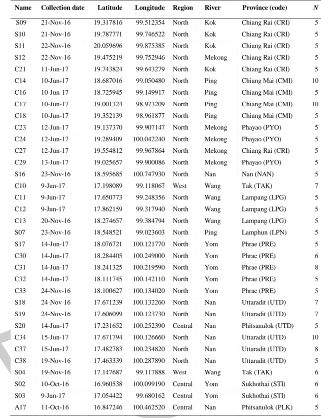
Analysis of the 16S rDNA sequence reveals that the mutation parameter before expansion (θ0) was 1.759 and the mutation parameter after expansion (θ1) was 24.333. Thus, the present study used a molecular marker, COI sequence (Kulsantiwong et al., 2013), for the identification of H. Almost all rivers in these areas drain into the Mekong River (Li et al., 2017; Mohammed et al., 2018 ) because the Mekong River is the main stream on the border between northern Thailand and Lao PDR (Bravard et al., 2014).
Living organisms in this area have always shown genetic differences from others because the Nan and Sa Kaeo sutures in Thailand have traditionally been from the Palaeo-Tethys suture (Barr & Macdonald, 1987), such as the black fly, Simulium weji (Pramual & Pangjanda , 2015 ), catfish, Clarias macrocephalus (Na-Nakorn et al., 2004) and Limnonectes gyldenstolpei and L .
Introduction
The source of infection is the dietary consumption of fish by humans and domestic animals (i.e. cats and dogs) (Aunpromma et al., 2012). This leads to confusion and misidentification in classification by morphology alone (Brandt, 1974; Harinasuta & Harinasuta, 1984; . Chitramvong. The genetic markers have been used for the classification and differentiation of snail species in the family Bithyniidae, such as cytochrome c oxidase subunit I (COI) marker (Kulsantiwong et al., 2013).
Population genetic studies of Bithynia species have been investigated using several genetic markers/techniques such as multilocus enzyme electrophoresis (MEE), random amplified polymorphic DNA (RAPD) and DNA sequencing (Kodcharin, 2006; . Saijuntha et al., 2006; Kulsantiwong et al., 2013; Kiatsopit et al., 2013). Bithynia genetic clusters have been clustered and correlated within wetland systems and possible co-evolution between O. Population genetics of Bithynia snails were analyzed and compared using COI and 16S rDNA sequences.
Materials and Methods
DNA was individually extracted from the head and leg of snails using the Mollusk DNA kit (Omega, USA) according to the manufacturer's protocol. The PCR product sizes were approximately 650 and 470 bp for the COI and 16S rDNA regions, respectively, which were excised and purified using the E.Z.NA® Gel Extraction kit (Omega bio-tek, USA). A neighbor-joining tree was constructed using MEGA7 (Kumar et al., 2016) based on the Kimura 2-parameter model (Tamura et al., 2011).
The population genetic analyzes were performed in Arlequin v.3.5.2.2 (Excoffier & Lischer, 2010), including analysis of molecular variance (AMOVA).

Results
The haplotype and minimum stretch haplotype network was generated using DnaSp v.5.10.01 (Librado & Rozas, 2009) and Network v.5.0 (http://www.fluxus-engineering.com). N = number, s = number of polymorphic sites, H = number of haplotypes, Sh = shared haplotypes, Uh = Unique haplotypes, Hd. Of all variable sites, the 12 positions can be used as diagnostic positions to distinguish the Bithynia species.
Interestingly, there are some sequences of Bithynia siamensis, which were unable to be identified into subgroups and groups separately from B. For 16S rDNA, variance components were contracted and FCT values were obtained for six groups (16S rDNA: FCT not significant ). The source of variation between groups, population within groups and within populations were calculated to be 33.41% respectively.
The source of variation between groups, population within groups and within populations accounted for 27.17%, respectively. The distribution of COI and 16S rDNA mismatches revealed a multimodal mismatch pattern (Figure 17). Figure 17 Distribution of mismatches across COI and 16S rDNA sequences We observed and expected pairwise differences based on the predictions of the sudden population expansion model: A-B) B.
Discussion
Introduction
Materials and methods
DNA was extracted from the cephalopods of each individual snail using the Mollusk DNA kit protocol (Omega, USA). The second PCR, the PCR mixer used the same ratio as the first PCR and 1 µl of the first PCR product. PCR products were excised and purified using Nucleospin® Gel and PCR purification kit (MACHEREY- .. NAGEL GmbH & Co., Germany).
Ligation was used with DynaExpress TA PCR Cloning Kit (pTAC) (BioDynamics Laboratory Inc., Japan) and purified PCR product. The solution was mixed by gently tapping the tube and then the tube containing the sample was turned down. Jet competent cells (DH5α) (BioDynamics Laboratory Inc., Japan) were used in the transformation step. First take 15 µl of competent cells from the -80°C freezer.
The transformation solution is then incubated at 43°C in a water bath for 45 seconds, and then quickly placed in ice water for 2 minutes. Then we pick up a small part of the colony with a toothpick and place it in a suitable test tube. Plasmid was extracted using FAST GENE Plasmid mini kit 300 preps (NIPPON Genetics Co., Ltd., Japan).
The population genetics analyzes were performed in Arlequin v.3.5.2.2 (Excoffier & Lischer, 2010), including analysis of molecular variance (AMOVA) among groups, among populations within groups, and within populations.
Results
Intron-long pattern indicated that 96 variable positions were observed, which were classified into 17 haplotypes. Based on intron-long region analyses, haplotype diversity=0.982, nucleotide diversity=0.041, by Fu's Fs test, Tajima's D= -1.568 and -2, respectively -1.568 and -2) revealed. Percentage of variation within populations was among groups, and 70.59% of among populations within groups. Percentage of variation within populations was among groups, and 89.74% of among populations within groups.
The percentage of variation within populations was between groups, and 91.53% among populations within groups. The percentage of variation within populations was between groups, and 55.10% among populations within groups. The percentage of variation within populations was between groups, and 59.79% among populations within groups.
Discussion
Tatamsa’ina olaanaa infekshinii Opisthorchis viverrini keessummeessitoota kuusa bishaanii keessatti aanaalee afur Bulchiinsa Khon Kaen, naannoo Taayilaandi keessatti dhukkuba opisthorchiasis keessatti mul’atu. Mallattoolee jeneetikii qorannoo sirnaa fi jeneetiksii baay’ina ummataa Bithynia spp., keessummeessitoota gidduugaleessaa jalqabaa Opisthorchis viverrini Taayilaanditti. Lafa jiidhina adda addaa Taayilaandi fi Laa’oo PDR keessatti baay’inni infekshinii Bithynia siamensis goniomphalos Opisthorchis viverrini cercariae garmalee ol’aanaadha.
Seasonal patterns of cercarial emergence of Opisthorchis viverrini infecting Bithynia siamensis goniomphalos from Vientiane Province, Lao PDR. Genetic variation of Bithynia siamensis goniomphalos the first intermediate host of Opisthorchis viverrini in the Moon and Chi River Basin, Northeast, Thailand by Random Amplification of Polymorphic DNA - Polymerase Chain Reaction (RAPD-PCR). Cercarial emergence patterns for Opisthorchis viverrini sensu lato infecting Bithynia siamensis goniomphalos from Sakon Nakhon Province, Thailand.
Daddarbiinsa waqtii Opisthorchis viverrini sensu lato fi gosa Lecithodendriid trematode ilbiisota Bithynia siamensis goniomphalos kaaba baha Taayilaanditti argaman keessatti. Jijjiirama tartiiba DNA maaytookondriyaa adda baafamtoota teessuma lafaa Opisthorchis viverrini Taayilaandi fi Lao PDR keessatti, fi hariiroo faayiloojenetikii trematodes biroo waliin. Sirnaafi jeneetiksii baay’ina ummataa Opisthorchis viverrini sensu lato: Dhiibbaa dhibee paraasitii fi kaansarii ujummoo dhiigaa irratti qabu.
Opisthorchis viverrini: intensity and rates of infection in cyprinoid fishes from an endemic hotspot in northeastern Thailand.