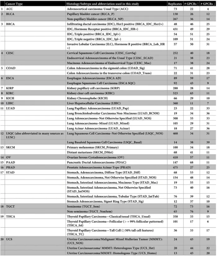
Therefore, we tested the pharmacological effect of carnosol on the following receptors: Insulin Receptor (IR), Chemokine CXCL12 Receptor (CXCR4), Angiotensin II (AngII) Receptor (AT1R), Glucagon-like Peptide-1 Receptor (GLP-1R), Thrombin /Protease-activated receptor (PAR1), and the vasopressin receptor (V2R). The effect of carnosol was investigated on the activation of G-proteins and the recruitment of β-arrestin or Insulin Receptor Substrate 1 (IRS1) by the receptors. This may have implications in the anti-diabetic effects of carnosol as reported in previous studies and supported by our glucose uptake assay showing carnosol promoted glucose uptake in HepG2 cells.
Introduction
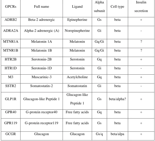
Carnosol and its Structural and Functional Properties
These two compounds are thought to contribute to approximately 90% of the antioxidant properties of many of the plants that contain them. In fact, carnosol is usually extracted by converting the majority of carnosic acid in the plant's extract to carnosol through a series of procedures, which ultimately lead to the oxidation of carnosic acid (Cao et al, 2021; Selamoglu, 2017).
Carnosol Therapeutic Potential in Diabetes and Cancer
The ability of ROS to contribute to carcinogenesis is mainly through their ability to cause DNA damage, such as in their ability to form DNA adducts (Ren et al., 2022). Some of the receptors studied in diabetes and cancer research include GPCRs such as the protease-activated receptor-1 (PAR-1), glucagon-like peptide receptor (GLP-1R), Vasopressin V2 receptor (V2R), and C-X-C chemokine receptor type 4 (CXCR4), as well as RTKs such as the insulin receptor (IR) (Chen et al., 2019;.
GPCRs and RTKs, Their Signaling and Their Role in Cancer and Diabetes
The binding of the agonist causes the physical association with and the activation of several heterotrimeric Gαβγ proteins (Gs, Gi, Gq and G12/13) leading to the dissociation of Gα and Gβγ, each to promote specific downstream signaling pathways that control various cell responses . V2R is primarily known for its important role in regulating salt and water levels in the body. In addition, GLP-1R plays a key role in the regulation of glucose-mediated insulin secretion by the beta cells.
The specificity of downstream signals transmitted through different families of RTKs is determined in part by the variations found in C-. IRS/PI3K/Akt and Grb2/Ras/ERK, controlling multiple cell responses in different insulin-sensitive tissues. RTKs are very important for maintaining normal β-cell development and function, and they also play a crucial role in regulating insulin granule exocytosis (Oakie & Wang, 2018).
In turn, blood glucose levels are the main molecular switch that controls insulin secretion and effects. It serves as an important growth factor; directly via the insulin receptor and indirectly via its role in the IGF/IGFR system on the majority of cells.

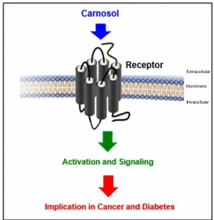
BRET Technology, Principle and Applications
These two fusion proteins are then transfected into cells followed by the addition of coelenterazine substrate (Rluc substrate). The BRET signal is measured through the intensity of light emitted by YFP and Rluc (acceptor/donor ratio). This technique is superior to other commonly used techniques for protein-protein interactions, such as enzyme-linked immunosorbent assay (ELISA) or co-immunoprecipitation (Co-IP) as these techniques are unable to provide temporally or spatially resolved results. as they depend on an endpoint measurement (De et al., 2013).
Also, BRET can be used in live cells in vitro without the need to fix or lyse the cells, which is a requirement of ELISA and Co-IP, or possibly add any chemicals that can alter the structural integrity of cellular proteins. which is one. of the many reasons why this technique is invaluable in studying proteins in their native environment, especially in cases such as signal transduction, cellular transport, cell division, and others. In addition to its in vitro use, this technique has also been used in vivo, in studies involving non-invasive imaging in living subjects and examination of pharmacological interactions in tissue-scale studies. In this work, we used BRET to study certain GPCRs and RTKs since both of these receptor superfamilies rely on downstream proteins to transmit cellular signals upon binding of their ligands.
This may have implications in drug development, for example, where it is important to know which cellular mechanisms should be targeted by certain drugs and which particular members of these mechanisms are pharmacologically beneficial or not. Also, basic research can benefit significantly from this technique in cases such as the discovery of ligands that bind a particular receptor, protein modifications such as phosphorylation and protease function, to name a few of a long list of applications (Bacart et al., 2008; De et al., 2013).
Statement of the Problem and Hypothesis
Research Objectives
Materials and Methods
- Chemicals and Reagents
- Cell Culture and Transfection
- Preparation of Hormone and Chemical Solutions
- BRET Assays
- Glucose Uptake
- SDS-PAGE and Western Blot
- Data Analysis and Statistics
Coelenterazine H was used as a substrate for the donor molecule, a Renilla Luciferase (Rluc) enzyme conjugated to the protein/receptor of interest. 25 cells were seeded overnight in 96-well plates (50,000 cells/well); then the cells were starved in phenol red-free, glucose-free and serum-free medium (DMEM containing 0.01 mM HEPES, 0.3 mg/ml glutamine, 100 IU/ml penicillin and 100 µg/ml streptomycin). After this, the cells were treated or not with insulin or carnosol, as indicated, for 60 minutes at 37°C.
After incubation, the treatment was removed, and the cells were treated with freshly prepared 2-deoxyglucose (500 μM) (50 μL/well) for 10 min at RT. This luminescence, which is proportional to the amount of 2-deoxyglucose transported into the cells during treatment, was then measured using a 0.3- to 1-s integration on GloMax Discover luminometer. HepG2 cells were maintained in DMEM medium in 5% CO2 at 37°C and then seeded in 6-well plate at 106 cells/well for the protein phosphorylation studies.
Before cell treatment, cells were washed in serum-free DMEM for at least 3 h and then treated with hormone (Insulin 1uM) and carnosol (50uM) and both respectively, as indicated in the figure legends. After treatment, cells were harvested by scraping into RIPA lysis buffer, and the resulting cell lysate was centrifuged at 13,000xg for 20 min at 4°C, and finally the supernatant was collected and assayed for protein concentration by bicinchoninic acid protein assay. , as previously reported (Ali et al., 2019) and further analyzed through Western blotting.
Results and Discussion
Results
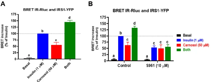
Data are mean ± SEM of BRET increase (normalized as % of insulin signal) of five to seven independent experiments performed in triplicate measurements. Data are mean ± SEM of BRET increase (normalized as % of thrombin's signal) of three to seven independent experiments performed in triplicate measurements. Data are mean ± SEM of BRET increase (normalized as % of thrombin's signal at 1 U/ml) of three to five independent experiments performed in triplicate measurements.
Data are means ± SEM of BRET increase (normalized as % of signal from CXCL12) from seven independent experiments performed in triplicate measurements. Similarly, Exendin abolished the positive effect of carnosol and the combined treatment on the BRET increase between GLP1R-Rluc and Venus-β-arrestin (Figure 15C). 37 Figure 15: Effect of carnosol on the activation of the glucagon-like peptide receptor (GLP1R) in HEK293 cells.
Data are means ± SEM of BRET increase (normalized as % of GLP-1 signal) from six independent experiments performed in triplicate measurements. Data are means ± SEM of BRET increase (normalized as % of AngII's signal) from seven independent experiments performed in triplicate measurements.
Discussion
This is very interesting in the context of GPCR-biased signaling, and suggests that carnosol is a -arrestin-biased bioactive compound that could be of great interest for the administration of carnosol in the disorders involving these two receptors, such as diabetes (for GLP1R) and cancer or viral infections (for CXCR4) (Britton et al., 2021; Kim et al., 2022; Seyedabadi et al., 2019; Seyedabadi et al., 2022; Yokono et al., 2020) . In the context of diabetes, our data on the positive effect of carnosol on both IR and GLP1R may be promising due to the crucial role of these two receptors in the control of glucose transport, glucose metabolism and insulin secretion (Deng et al., 2019) ; Tekin et al., 2020). Indeed, carnosol has been shown to promote anti-hyperglycemic effects similar to the reference drug Glibenclamide in streptozotocin-induced diabetic rats (Khan et al., 2016; . Samarghandian et al., 2017).
In addition, carnosol was able to suppress the upregulation of glucose-6-phosphatase (G6PC) and phosphoenolpyruvate carboxykinase 1 (PCK1), rate-limiting enzymes of hepatic gluconeogenesis (Hasei et al., 2021). In addition, in streptozotocin-induced diabetic Sprague Dawley rats, carnosol reduced the concentration of triglycerides, total cholesterol and Low-Density Lipoproteins (LDL) (Tekin et al., 2020). Strikingly, AMPK activator drugs are often used to counteract the effects of insulin resistance, and carnosol has shown similar activity (Vlavcheski et al., 2018).
On the other hand, the anti-cancer properties of carnosol have been demonstrated in breast cancer cells, ovarian cancer cells, colorectal cells, B-cell leukemia cells and many others (Cao et al., 2021). Furthermore, it was demonstrated that carnosol caused triple negative breast cancer cells to undergo autophagy, as well as apoptosis, through an increase in ROS in these cells (Al Dhaheri, 2014; Cao et al., 2021).
Conclusion
Carnosic acid and carnosol, diterpene phenolic compounds of the labiate plants rosemary and sage, are activators of the human peroxisome proliferator-activated receptor. Potential therapeutic use of the rosemary diterpene carnosic acid for Alzheimer's disease, Parkinson's disease, and chronic COVID through NRF2 activation to combat the NLRP3 inflammasome. Changing cancer burden profiles worldwide and in China: a secondary analysis of global cancer statistics 2020.
Evaluation of antidiabetic activity of carnosol (phenolic diterpene in rosemary) in streptozotocin-induced diabetic rats. Bioluminescence resonance energy transfer methods for studying G protein-coupled receptor-receptor-tyrosine kinase-heteroreceptor complexes. Characterization of five novel vasopressin V2 receptor mutants causing nephrogenic diabetes insipidus reveals role of tolvaptan for M272R-V2R mutation.
Mechanisms of carnosol in chemoprevention of ultraviolet B light-induced nonmelanoma skin carcinogenesis. Functional selectivity profiling of the angiotensin II type 1 receptor using pathway-wide BRET signal sensors. Biased signaling by G protein-coupled receptors (GPCRs): Molecular determinants of GPCR/transducer selectivity and therapeutic potential.
Molecular basis of the antidiabetic properties of camel milk by profiling the bioactive peptides on dipeptidyl peptidase IV (DPP-IV) and insulin receptor activity.