This book also describes the latest developments with future thoughts on wound healing science and research. The effect and contribution of various growth factors and cytokines to normal and abnormal wound healing is discussed in Chapter 6.
Introduction
Various polymers are being used or under investigation with respect to wound healing technology; she. Wound healing and wound care technologies are an ever-expanding field with the advancement of materials science, biomedicine and tissue engineering.
Challenges in Wound Healing
This book discusses the evolution of wound care devices and protocol over the years, and different technologies used in contemporary wound care treatment. Novel strategies involving engineered tissues and drug delivery to mimic the natural wound healing microenvironment will also be discussed.
Classification of Wound Dressing Products
- Nonresorbable Gauze/Sponge Dressing for External Use
- Hydrophilic Wound Dressing
- Occlusive Wound Dressing
- Hydrogel Wound and Burn Dressing
- Interactive Wound and Burn Dressings
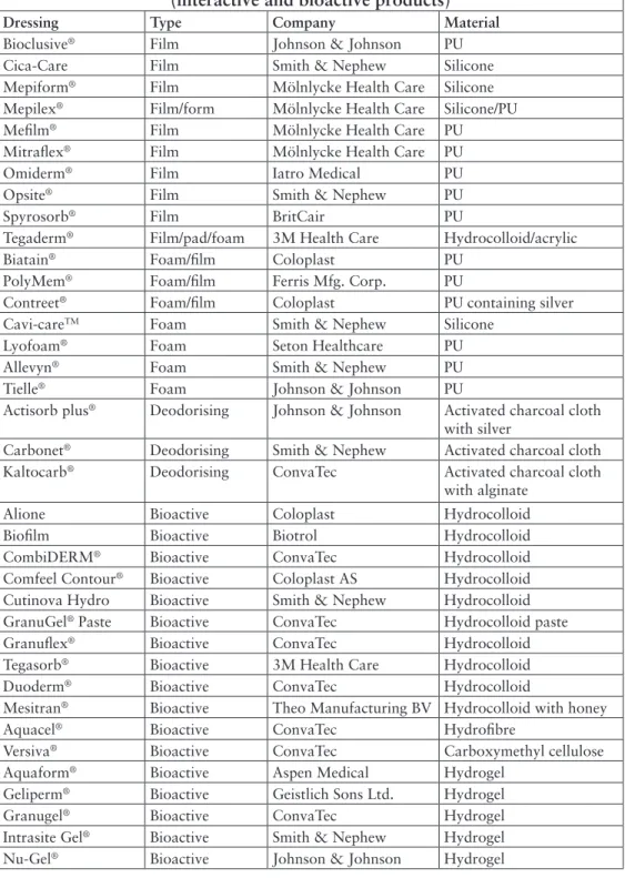
Comfeel Contour® Bioactive Coloplast AS Hydrocolloid Cutinova Hydro Bioactive Smith & Nephew Hydrocolloid GranuGel® Paste Bioactive ConvaTec Hydrocolloid pasta. Acticoat Silver Smith & Nephew High-density polyethylen mesh og nonwoven stof af rayon og polyester.
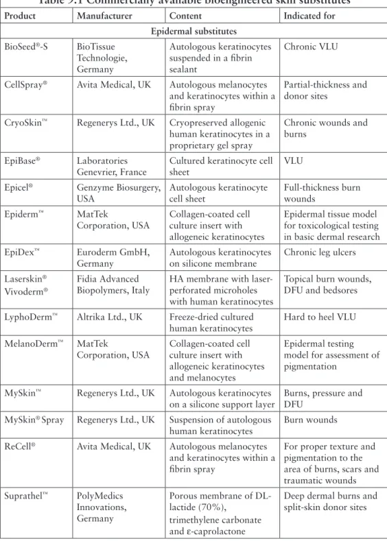
Skin Substitutes
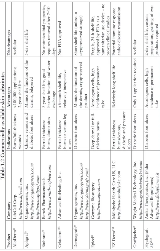
Short shelf life (unless in cryopreserved storage) Epicel®Genzyme Biosurgery http://www.epicel.comDeep dermal or full-thickness burns Autologous cells, high incidence of permanent uptake. Operative removal of silicone layer and autograft requires Laserskin®Anika Therapeutics, Inc. Fidia Advanced Biopolymers) http://www.fidiapharma.it.
Conclusion
The currently available cell-based skin substitutes are effective in accelerating wound healing and their appropriate use can achieve closure in non-healing wounds. The production of specific growth factors and cytokines, depending on the wound nature, by this cell-based bioengineered skin can increase its therapeutic effect [12].
Introduction
Statistics
The Indian advanced wound care market mainly uses collagen technology (32%), hydrogels (22%), foam dressings (17%), film dressings (10%) and calcium alginate dressings (6%). In addition, advanced wound care products are more efficient in wound healing and lead to shorter hospital stays.
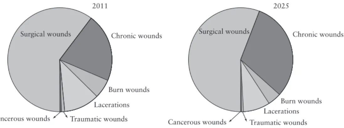
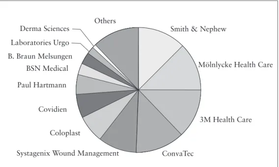
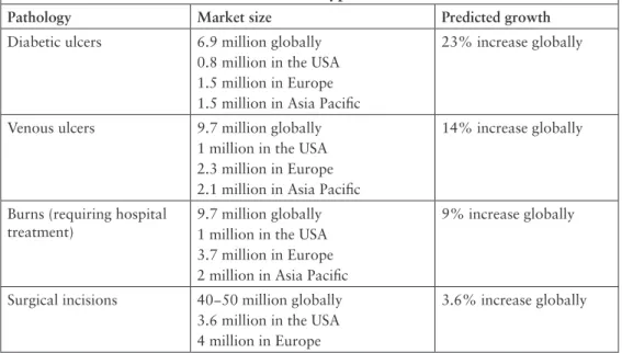
Wound-care Industry
Diabetic wounds 6.9 million globally 0.8 million in the US 1.5 million in Europe 1.5 million in the Asia Pacific region. Venous ulcers 9.7 million globally 1 million in the USA 2.3 million in Europe 2.1 million in the Asia Pacific region.
Textile-based Wound Dressings
KCI Concepts Inc., manufactures Graftjacket®, a regenerative dermal tissue matrix product for the repair or replacement of damaged or inadequate integumentary tissue, such as DFU, venous leg ulcers, pressure ulcers or for other homologous uses for the treatment of damaged human integument. Covidien also concentrates on traditional dressings such as woven and non-woven dressings, and antimicrobial sponge and foam dressings.
Conclusion
Research is underway to develop new cell therapies for wound care based on autologous adipose stem cells. Kalorama Information in Advanced Wound Care Markets Worldwide, Market Research Group LLC, New York, USA, 2014.
Introduction
The Epidermis
The lifespan of an individual keratinocyte cell is believed to be about 6 weeks [4], i.e. the time it takes to renew the entire epidermis [5]. The differentiation of keratinocytes is proportional to the calcium gradient in the epidermal layers, with the stratum basale and stratum corneum having a very low concentration of calcium, while the stratum granulosum contains the highest.
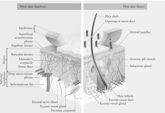
The Dermis
In humans there are two types of sweat glands; the eccrine sweat glands are mainly involved in the regulation of heat and are mostly found on the soles of the feet. The proximal coiled secretory duct and the straight tubular structure in the lower dermis, and the intraepidermal spiral duct opening to the skin, are the three main components of the eccrine sweat gland.
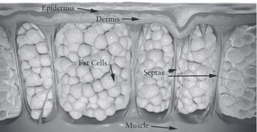
Subcutaneous Fat
Some parts of the skin contain more nerve endings than others, for example the tips of the fingers and toes contain many nerves and are extremely sensitive to touch. Fat cells or lipocytes also produce the hormone leptin, which regulates body weight via the hypothalamus [10].
![Figure 3.3 A normal dermis. Adapted from Kilbad, Normal Epidermis and Dermis with Intradermal Nevus 10x, Wikimedia Commons [7]](https://thumb-ap.123doks.com/thumbv2/1libvncom/9201709.0/41.756.233.502.249.608/figure-adapted-kilbad-normal-epidermis-intradermal-wikimedia-commons.webp)
The Dermo-Epidermal Junction
The subcutaneous fat layer is also a depot that acts as a protective cushion that protects the muscles and bones from bumps and falls; it is a storehouse of energy and is considered an endocrine organ.
Skin Functions
These cells secrete a variety of cytokines that are important in the pathogenesis of contact dermatitis, atopic dermatitis, histiocytosis X, human immunodeficiency virus type 1, and skin graft rejection.
Introduction
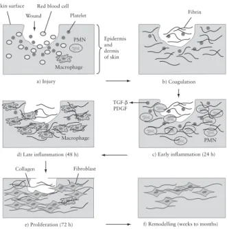
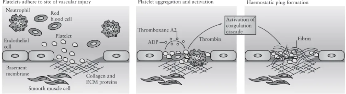
Vascular permeability is temporarily increased to allow neutrophils [polymorphonuclear neutrophils (PMNs)], platelets, and plasma proteins to infiltrate the wound. The proliferation phase begins at approximately 72 hours, when fibroblasts, recruited to the wound by growth factors released by inflammatory cells, begin to synthesize collagen. f) Although the rate of collagen synthesis slows down.
The Biochemical Process of Wound Healing
- Haemostasis
- Inflammation
- Proliferation
- Maturation
Neutrophil infiltration during the time course of wound healing is demonstrated in an animal model shown in Figure 4.3. Macrophages recruit a new cell type, i.e., the fibroblast, which lays down a network of collagen fibers surrounding the neovasculature of the wound.
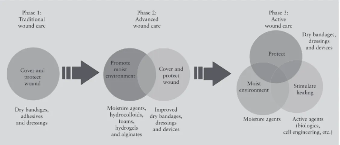
Moist Wound-healing Theory
Some moist wound healing dressings include alginates, foams, hydrogels, hydrocolloids, and some topical treatments and gels. Ease of application, reasonable cost and quality of healing along with a reduced risk of infection are among the main advantages of dressings that support moist wound healing.
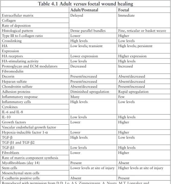
Foetal Wound Healing
This success initiated the commercial production of various polyurethane-based dressings supporting the wet wound theory. Alginate- and chitosan-based dressings were introduced to the market due to their increased ability to absorb fluids and inherent wound healing ability.
Abnormal Wound Healing
Systemic related factors such as age, stress, other medications, obesity and alcohol consumption, smoking and low nutrients can also delay wound healing and make wounds chronic. An altered inflammatory response, such as a delayed T-cell infiltration in the wound area, reduced chemokine production and macrophage phagocytic capacity is due to the age-related factor, which leads to delayed re-epithelialization, collagen synthesis and angiogenesis, and delayed wound healing.
Conclusion
Introduction
Hydrophilic wound dressings are designed to cover the wound and are capable of absorbing exudates; these dressings consist of hydrophilic non-resorbable materials such as cotton, cotton derivatives, alginates, dextran and rayon. Occlusive wound dressings are designed to cover the wound, maintain a moist wound environment, and allow the exchange of gases such as oxygen and water vapor through the device;.
Types of Wound Dressings
Inert or Passive Products
Traditional dressings such as fiber and gauze have been used for many years and are called inert or passive dressings. Absorbent pad dressings help absorb excess exudates from the wound and should only be used on wounds with minimal to low exudation.
Interactive and Bioactive Products
They are dry, do not provide a moist wound environment, and are commonly used to plug and conceal wounds. However, these dressings have low water vapor permeability and exudate can become trapped in the wound, which can cause maceration.
Classification of Wounds
In addition to these four grades, some ulcers that show full-thickness loss of skin or tissue are unclassified because the actual depth of the ulcer is completely obscured by ulceration and/or oozing at the base of the wound. If only a part of the epidermis or the entire depth of the epidermis is damaged, it regenerates to the state before the injury and these wounds are called partial thickness wounds.
Classification of Wound Closure
These wounds are usually caused by surgery and are closed in the operating room itself. If the wound is left to heal on its own, this is called secondary intention healing.
Skin Equivalents
Chitosan is a natural hemostat which interacts with red blood cells and aggregates [6], thus aiding in wound healing. From studies reported in the literature, cell-seeded chitosan sponges appear to be an excellent candidate material for wound healing applications in chronic wounds.
Conclusion
Introduction
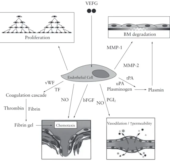
The Role of Growth Factors in Wound Healing
FGF-1 and FGF-2 have been shown to stimulate angiogenesis and are present in the early stages of wound healing. It has been reported that stress, depression and a hostile marital environment have a possible role in the modulation of MMP and in the expression of TIMP [13, 14], thereby delaying wound healing.
The Role of Matrix Metalloproteinases in Wound Healing
MMP-28 is a 59 kDa protein that is expressed in basal keratinocytes at the wound edge. TIMP-3 binds strongly to ECM and is a good inhibitor of TNF-α converting enzyme.
![Table 6.2 lists examples of MMP actions that affect cell migration, differentiation, growth, the inflammatory process, neovascularisation, apoptosis and so on [17]](https://thumb-ap.123doks.com/thumbv2/1libvncom/9201709.0/79.756.87.652.443.942/examples-actions-migration-differentiation-inflammatory-process-neovascularisation-apoptosis.webp)
The Role of Matrix Metalloproteinases in Abnormal Wound
Wound healing Wound dressings for chronic non-healing wounds should absorb the heavy exudate, relieve pain and control infection, in addition to normalizing wound healing for better regeneration. Currently, only collagen-based wound dressings are indicated for diabetic non-healing wounds; such wounds also require a secondary dressing and growth factor supplementation.
Nutritional Support
Conclusion
Introduction
Classes of Wound Dressings
- Paraffin Gauze Dressing
- Hydrogel Dressings
- Hydrocolloid Dressings
- Alginates
- Foam Dressings
- Composite Dressings
- Activated Charcoal Dressings
- Transparent Film Dressings
- Antimicrobial Dressings
- Honey-based Dressings
- Iodine-based Dressings
- Polyhexamethylene Biguanide Antimicrobial Dressings
Hydrogel dressings are occlusive and therefore provide a moist wound healing environment that promotes autolytic debridement and isolation of the wound. Alginate dressings are available in the form of freeze-dried porous sheets or foam, or as flexible fibers.
Haemostasis and Haemostatic Agents in Wound Healing
- Fibrin Sealants
- Gelatin Haemostats
- QuickClot ®
- WoundStat TM
Greater knowledge of the hemostasis process was developed by the Egyptians based on the study of the mummification process. It is a cross-linked gelatin granular product which reduces the swelling of the granules in vivo.
Chitosan-based Haemostats
HemCon ®
Upon contact with anionic erythrocytes, the chitosan salts rapidly 'crosslink' and adhere strongly to the wound surface. Improved HemCon® dressings are now in production that are thinner and more flexible than the original product, and are designed to allow better conformation to the wound surface and easier handling.
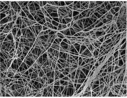
Celox™
Chitoflex® is made from the same material as HemCon® and offers the same life-saving technology, but is available in strip form and can be used to pack wounds. This free-flowing powder should be poured onto the wound area and pressure applied for up to 5 minutes to stop bleeding.
Hemogrip TM
Drug Delivery in Wound Healing
Conclusion
Introduction
Alginates
Physicochemical Properties of Alginate
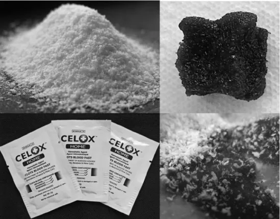
The gel strength will depend on the guluronic acid content and also on the average number of G units in the G blocks. When gelation occurs in the presence of excess calcium, a modified egg-crate model has been proposed involving more than two alginate chains in the gelation zone, which may have an impact on gel porosity.
Alginate Wound Dressings
It has been demonstrated in a study that calcium alginate increased the proliferation of fibroblasts but decreased the proliferation of human microvascular endothelial cells and keratinocytes. The effects of calcium alginate on cell proliferation and migration have been reported to be mediated by released calcium ions.
Calcium Alginate as a Haemostat
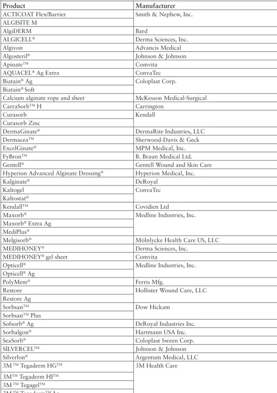
Calcium alginate wound dressings have been indicated for leg and pressure ulcers, burn wounds and donor sites, surgical wounds, and as hemostatic dressings; therefore, products such as SorbsanTM, TegagelTM, Kaltostat® and so on have been subjected to many controlled clinical trials, which have shown their significant and beneficial effect on leg ulcers, SorbsanTM, Kaltostat®, Seasorb® on burns and donor sites, Algosteril ® , SorbsanTM, Kaltostat® and so on, for pressure ulcers and surgical wounds. A combination of alginate and chitosan (alchite) has been investigated to form fibers that could be developed for use in wound care; a study on its antibacterial properties suggested its suitability in the development of wound dressings [27].
Types of Dressings
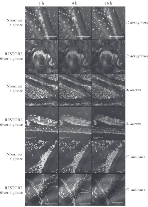
Silver in Alginate Wound Dressings
A silver ion release rate of at least 1 part per million (ppm) is required to provide effective antimicrobial efficacy. Exposure to wound exudate dissolves the silver and stimulates the release of silver ions, creating a controlled-release antimicrobial effect.
Honey in Alginate Wound Dressings
Chitin and Chitosan
Chemical Modifications
Chitin and chitosan are interesting polysaccharides due to the presence of the amino group functionality, which can be suitably modified to confer desired properties and characteristic biological functions, including solubility. The hydroxyl functional groups also enable various reactions such as o-acetylation, H-bonding with polar atoms, grafting and so on.
Action of Chitosan
Chitosan Derivatives
Partially deacetylated chitin hydrochloride showed hemostatic properties similar to collagen hydrochloride and has a positive effect on wound healing [71]. Phosphorylated chitin and chitosan have also been reported to have beneficial application in wound healing [89].
Skin Tissue Engineering
Forms of chitosan used for tissue engineering include porous scaffolds, which help increase cell penetration and adhesion, rod-shaped chitosan, commonly used in bone tissue engineering, and hydrogel, which is for tissue engineering applications. soft tissues [94]. The interaction of various cell types with chitosan scaffolds, alone or in combination with other biomolecules, has been extensively examined and these show very promising results for tissue engineering applications, and include: neuronal differentiation of stem cells derived from muscle. in chitosan-based hydrogel scaffolds [107], neural regeneration with stem cells along the chitosan channel was observed [108] and co-delivery of adipose stem cells and growth factors with an RGD sequence was attempted with N-methacrylate-glycol - modified chitosan gel for chondral repair [109].
Conclusion
Millions of these droplets store oxygen and carry it to the wound, which is difficult to heal due to the low oxygen concentration at the wound site (diabetic ulcers and so on). Depending on the location of the carboxymethyl group attachment, CMCh can be referred to as 'N' when the carboxymethyl group attaches to the amine, 'O' when attached to the primary hydroxyl group, or NOCC when attached to both [117].
Introduction
Biopolymers, Italy HA ester with laser perforated micro holes containing human keratinocytes and fibroblasts in combination with Hyalograft 3D™ and Laserskin.
Biobrane ®
Chitoderm ®
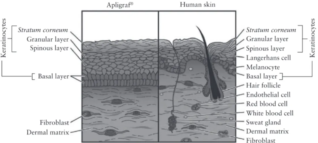
Apligraf ®
During the 6-day incubation period, fibroblasts divide and multiply, move through the collagen and rearrange the matrix by contracting the collagen filaments, producing human matrix proteins to form a dermal layer. During the 4-day incubation period, these cells attach and multiply on top of the dermal layer to form an epidermal layer.
OrCel ®
Initially, fibroblast and keratinocyte cells are obtained from the patient via a biopsy; this biopsy is first digested and the fibroblast and keratinocyte cells are extracted and cultured before being seeded onto a collagen scaffold. The packaged 75 mm diameter double layer disc has a shelf life of 10 days if stored in a polyethylene bag containing 10%.
Dermagraft ®
VLU treated with Apligraf® healed in 99 days and DFU healed in 65 days compared to 184 days and 90 days for conventional treatment, respectively [13, 14]. As cells divide and proliferate, they cover the scaffold to create a three-dimensional (3D) bioengineered dermal replacement containing metabolically active living cells, which is indicated for DFU.
Epicel ®
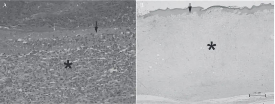
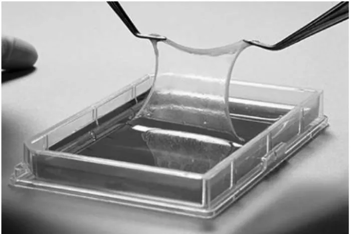
The size and shape of the graft will be approximately the size of B8 paper, as shown in Figure 9.3. In addition to having a high utility value, Epicel® can be a life-saving method in the treatment of patients with full-thickness burns covering >60% of the total body surface area.
MySkin ®
TransCyte ®
Hyalograft 3D TM and Hyalomatrix ® PA
Hyalomatrix® PA closely matched the hydration, transepidermal water loss, and skin surface 3D pattern of normal skin, showing better cell regulation, ECM regeneration, and neoangiogenesis. Hyalomatrix® PA has also been reported to provide beneficial results in the treatment of deep burns in children.
Laserskin ®
3DTM grafting increased keratinocyte proliferation and reduced the rate of wound hypertrophy and contraction when compared with exclusive application of keratinocyte cultures [29].
Bioseed ® -S
Integra TM
TeruDermis TM
Collagen is replaced by the organically produced new dermal skin cells. e) Day 21+: When a new dermal layer is formed, the silicone layer is removed. f) Day 21+: a thin epidermal autograft is applied over the new dermal skin and (g).
Conclusion
Even today, collagen is used in several skin substitutes, although HA derivatives are also on the market, mainly due to the cost factor. The advent of nanotechnology and commercially successful skin substitutes in combination can help manipulate a precise microenvironment, leading to accelerated and near-natural wound healing.
Introduction
Scarless Wound Healing
Keloid scars are cosmetically unpleasant and are mostly associated with pain and itching. Fetal wounds heal rapidly with complete regeneration of the dermal and epithelial tissue without any scar tissue formation.
Gene Therapy in Wound Healing
Usually, superficial partial-thickness burns that heal in less than 10 days are less prone to scar formation. An elevated mast cell histamine level is one of the factors contributing to abnormal growth [3]; Estrogen overproduction and adrenal cortical hypersecretions are also reported to be associated with keloid formation [4].
Stem Cells in Wound Healing
Future Outlook
3D Three-dimensional ADP Adenosine diphosphate AgNP Silver nanoparticle(s) ATP Adenosine triphosphate bFGF Basic fibroblast growth factor CMC Carboxymethylcellulose CMCh Carboxymethylchitosan. NOCC N,O-carboxymethyl chitosan PAR Protease-activated receptor PDGF Platelet-derived growth factor(s) PEG Polyethylene glycol.
![Figure 3.2 Schematic representation of the epidermis. Adapted from BruceBlaus, Epidermis, Wikimedia Commons [3]](https://thumb-ap.123doks.com/thumbv2/1libvncom/9201709.0/39.756.128.602.145.427/schematic-representation-epidermis-adapted-bruceblaus-epidermis-wikimedia-commons.webp)