A PHARMACOLOGICAL STUDY OF SOME NIGERIAN MEDICINAL PLANTS
by
Jude Chinedu Chukwujekwu
Submitted in fulfilment of the requirements for the degree of
Doctor of Philosophy
In the
School of Biological and Conservation Sciences University of Kwazulu-Natal
Pietermaritzburg
2005
"Recall the face of the poorest and weakest man whom you have
seen, and ask yourself, if the steps you contemplate are going to
be any use to him, will he gain anything by it? Will it restore to
him control over his own life and destiny?" Mahatma Ghandi
DECLARATION
I hereby declare that this thesis, unless otherwise acknowledged to the contrary in the text, is the result of my own investigation, under the supervision of Professor J. van Staden, in the Research Centre for Plant Growth and Development, School of Biological and Conservation Sciences, University of KwaZulu-Natal, Pietermaritzburg.
Jude Chinedu Chukwujekwu
I certify that the above statement is correct.
Supervisor: ProfessorJ. van Staden
ACKNOWLEDGEMENTS
What a journey! First and foremost, I give all the glory, honour and adoration to God Almighty for giving me the strength to successfully pull through this journey.
To my supervisor, Prof. J. van Staden, thank you for giving me the opportunity to study at this great Research Centre. Thank you also for your fatherly advice, encouragement and financial support. Iwill always be indebted to you.
To all the Staff and Students at the Research Centre for Plant Growth and Development, thanks for all your assistance especially Dr. Esam Elgorashi, Dr.
Geogina Arthur, Ibrahim, Usa, Zama, Antoine and Noluyolo, you guys were great to me.
To Professor D.A. Mulholland and Dr. Phil Coombes (University of KwaZulu- Natal, Durban), thank you for helping me with the structural elucidation of isolated compounds.
To Professor Peter Smith (University of Cape Town), thank you for helping me with the antiplasmodial bioassay.
To my family, dad, mum, Chinyere, Emma, Dr. Emeka, Nnamdi and most especially Dr. Ifeanyi, thank you for your prayers, encouragement and financial
support. May God shower you all with his blessings. I love you all and will always do.
ABSTRACT
Petroleum ether, dichloromethane, and 80% ethanol extracts of 15 plant species collected in Nigeria were screened for in vitro antibacterial, anti-inflammatory and antimalarial activities. Antibacterial activity was tested using the agar diffusion method, while the minimum inhibitory concentrations (MIC) of the active extracts were determined using the microtitre serial dilution method. Most antibacterial activity detected was against Gram-positive bacteria with Staphylococcus aureus being the most susceptible. The highest activity was found in petroleum ether and dichloromethane leaf extracts of Mallotus oppositifolius; petroleum ether, dichloromethane and ethanolic root extracts of Newbouldia laevis; and ethanolic root extracts of Morinda lucida and Canthium subcordatum. Against the Gram- negative bacterium Escherichia coli, the highest activity was found in dichloromethane leaf extracts of Newbouldia laevis, ethanolic root extracts of Phyllanthus amarus, Mallotus oppositifolius, and Canthium subcordatum. A total of 60 plant extracts were screened for antiplasmodial activity. A chloroquine sensitive strain of Plasmodium falciparum (D10) was used. In the assay, the parasite lactate dehydrogenase (pLDH) activity was used to measure parasite viability. About 11 extracts showed promising activity with an ICso ranging from 2.5 to 13.4 J..lg/ml. The petroleum ether leaf extract of Hyptis suaveolens had the highest activity (ICso
=
2.5 J..lg/ml). The cyclooxygenase (COX-1 and COX-2) assays were used to test for anti-inflammatory activity. All the plant species, with the exception of Hedranthera barteri and Picralima nitida showed anti- inflammatory activity. Apart for a few ethanolic extracts, all the activities wererecorded with petroleum ether and dichloromethane extracts. Employing bioassay-guided activity fractionation, an antibacterial anthraquinone identified as emodin was isolated from ethanolic root extract of Senna occidentalis. Although this compound had been isolated from other sources, this was the first report of isolation from Senna occidentalis. Using a similar approach a novel antimalarial diterpenoid was isolated from the petroleum ether leaves extract of Hyptis suaveolens. It had ICso of 0.1 I-lg/ml. This new compound is worthy of further investigation and may act as an important lead compound for future antimalarial drugs.
PAPERS PUBLISHED FROM THIS THESIS
CHUKWUJEKWU, J.C., VAN STAOEN, J. and SMITH, P. 2005. Antibacterial, anti-inflammatory and antimalarial activities of some Nigerian medicinal plants.
South African Journalof Botany, 71 (In press).
CHUKWUJEKWU, J.C., VAN STAOEN, J, SMITH, P, MULHOLLANO, 0, and COOMBES, P. A novel antiplasmodial diterpenoid from the leaves of Hyptis suaveolens (Submitted for publication).
CHUKWUJEKWU, J.C., VAN STAOEN, J, MULHOLLANO, 0, and COOMBES, P. Emodin, an antibacterial anthraquinone from the roots of Cassia occidentalis (Submitted for publication).
CONFERENCE PARTICIPATION
Oral presentation
CHUKWUJEKWU, J. C., VAN STAOEN, J. and SMITH, P. 2005. Antimalarial activity of some Nigerian medicinal plants, 31st Annual Congress of SAAB, Bloemfontein, South Africa, 10 - 14 January 2005.
TABLE OF CONTENTS
I . .
Dec aratlon 1
Acknowledgements .i i
Abstract
IVPapers Published from this Thesis vi
Conference Participation vi
Table of Contents vi i
List of Figures
I• • I• • • • • • • • • • • • • • • • • • • • • • • • • • • • • • • • • • • • • • • •xii List of Tables
1 1 • • • • • • • • • • •xiv
List of Abbreviations xvi
CHAPTER 1. LITERATURE REVIEW
1.1 Introduction 1
1.2 Traditional Medicine: A Global View 3
1.3 Traditional Medicine and Primary Health Care (PHC) 7
1.4 Medicinal Plants and Malaria 9
1.5 Drug Discovery and Development. 10
1.6 Aims and Objectives 16
CHAPTER 2. ANTIBACTERIAL SCREENING
2.1 Introduction 17
2.2 Disease Causing Bacteria 17
2.2.1 Escherichia coli
18
2.2.2 Klebsiella pneumoniae
18
2.2.3 Staphylococcus aureus
19
2.2.4 Bacillus subtilis
19
2.2.5 Micrococcus luteus
20
2.3 Antibacterial Agents 20
2.3.1 Resistance to Antibacterial Agents 21
2.4 Antibacterial Assays 22
2.4.1 Diffusion Method 22
2.4.2 Microdilution Method 23
2.4.3 Bioautographic Method 24
2.5 Materials and Methods 25
2.5.1 Plant Collection 25
2.5.2 Plant Extract Preparation 25
2.5.3 Test Organisms 25
2.5.4 Diffusion Method: Disc-diffusion Assay 29 2.5.5 Dilution Method: Microdilution Assay 30
2.6 Results and Discussion 30
2.7 Conclusions 36
CHAPTER 3. ANTI-INFLAMMATORY SCREENING
3.1 Introduction 38
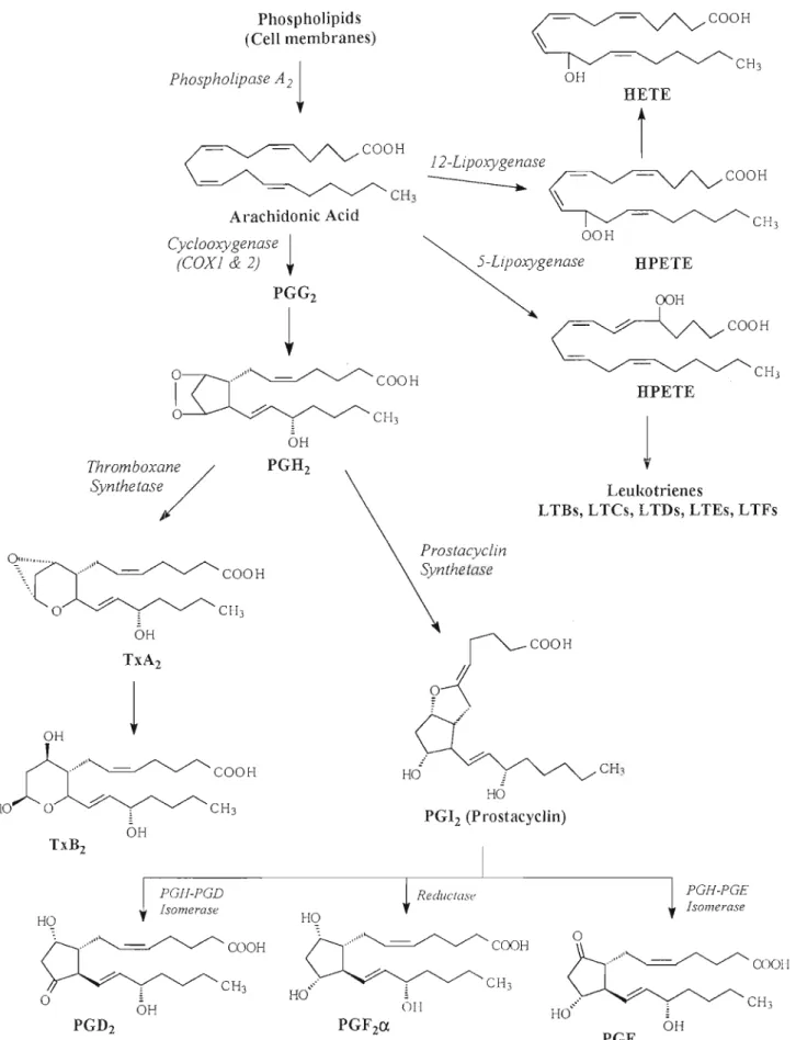
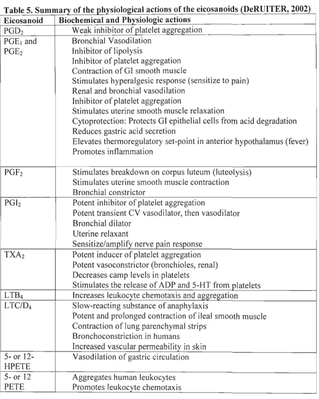
3.2 Cyclooxygenase Isoenzymes (COX-1 and COX-2) 39 3.3 Biosynthesis of Prostaglandins and their Functions .42
3.4 Aims and Objectives .44
3.5 Materials and Methods .47
3.5.2 CQX-2 Bioassay .47
3.5.3 CQX-1 Bioassay .48
3.5.4 Controls 49
3.5.5 Calculation of Inhibition .49
3.5.6 Determination of IC
so50
3.6 Results and Discussion 50
3.7 Conclusions 56
CHAPTER 4. ANTIPLASMODIAL SCREENING
4.1 Introduction 57
4.2 Drug Resistance against Malaria 58
4.3 Plants as Sources of Antimalarial Drugs 58
4.4 Aims 59
4.5 Materials and Methods 60
4.5.1 Plant Collection and Extraction 60
4.5.2 Antiplasmodial Assay 60
4.6 Results and Discussion 60
4.7 Conclusions 66
CHAPTER 5. ISOLATION AND IDENTIFICATION OF ANTIPLASMODIAL COMPOUNDS FROM Hyptis suaveo/ens
5.1 Introduction 67
5.1.1 Description and Traditional Medicinal Uses 67
5.1.2 Chemical Constituents and Biological Activity 68
5.2 Aims 69
5.3 Materials and Methods 69
5.3.1 Plant Extraction 69
5.3.2 Antiplasmodial Assay 69
5.3.3 Bioassay-Guided Fractionation for Isolation of Active
Compound(s) 69
5.3.3.1 Vacuum Liquid Chromatography (VLC) 70 5.3.3.2 Gravity-Assisted Column Chromatography 70 5.3.3.3 Preparative Thin Layer Chromatography 71 5.3.4 Identification of Purified Active Compound(s) 71
5.4 Results and Discussion 72
5.4.1 Plant Extraction 72
5.4.2 Bioassay-Guided Fractionation for Isolation of Active
Compound(s) 72
5.4.3 Identification of the Pure Compound ("Hyptimisin") 74
5.5 Conclusions 76
CHAPTER 6. ISOLATION AND IDENTIFICATION OF EMODIN, AN ANTIBACTERIAL AGENT FROM Senna occidentalis
6.1 Introduction '" 81
6.1.1 Description and Traditional Medicinal Uses 81
6.1.2 Chemical Constituents and Biological Activity 82
6.2 Aims 83
6.3 Materials and Methods 83
6.3.1 Plant Extraction 83
6.3.2 Antibacterial Assay 84
6.3.3 Bioassay-Guided Fractionation for Isolation of Active
Compound(s) 84
6.3.3.1 Vacuum Liquid Chromatography (VLC) 84 6.3.3.2 Gravity-Assisted Column Chromatography 85 6.3.3.3 Preparative Thin Layer Chromatography 86 6.3.4 Identification of Purified Active Compound 86
6.3.5 MIC Determination 87
6.4 Results and Discussion 87
6.4.1 Plant Extraction 87
6.4.2 Bioassay-Guided Fractionation for Isolation of Active
Compound(s) 87
6.4.3 NMR Spectroscopy 89
6.4.4 MIC Determination 91
6.5 Conclusions 92
CHAPTER 7. GENERAL CONCLUSIONS
7.1 Introduction 96
7.2 General Screening of Plants for Biological Activity 97
7.3 Isolation and Identification of Active Compound(s) in Plants 98
REFERENCES , , , , 99
LIST OF FIGURES CHAPTER 3
Figure 1. Biosynthesis of ecosanoids .45
Figure 2a. Inhibition (%) of COX-2 and COX-1 by some crude plant
extracts 51
Figure 2b. Inhibition (%) of COX-2 and COX-1 by some crude plant
extracts 52
CHAPTERS
Figure 3. A picture of
Hyptis suaveolens in the wild67 Figure 4. Chemical structures of (1) 4-epipalustric acid-9a,13a-
endoperoxide, (2) isolated compound 76
Figure 5. Flow diagram of the isolation steps for antiplasmodial
compound(s) using dried
H. suaveolens leaves78 Figure 6. 1H NMR Spectrum of antimalarial compound (F4-33)
isolated from
H. suaveolens leaves79
Figure 7. 13C NMR Spectrum of antimalarial compound (F4-33)
isolated from
H. suaveolens leaves80
CHAPTER 6
Figure 8. Senna occidentalis
L. 81
Figure 9. Chromatogram of ethanolic root extract of Senna
occidentalis showing antibacterial activity (Inhibition zone) of the
active component. 88
Figure 10. Structure of emodin and the antibacterial isolated from
Cassia
occidentalis90
Figure 11. Flow diagram of the isolation steps for antibacterial
compound(s) using dried S.
occidentalis roots93
Figure 12.
1H
NMR Spectrum of antibacterial compound isolatedfrom S.
occidentalis roots94
Figure 13. 13C NMR Spectrum of antibacterial compound isolated
from S.
occidentalis roots95
LIST OF TABLES
CHAPTER 1
Table
1. Some drugs discovered from ethnobotanicalleads 12
CHAPTER 2
Table
2. Selected medicinal plants used and collected in Nigeria investigated for antibacterial, anti-inflammatory and antiplasmodial
activities 26
Table
3. Determination of antibacterial activitya of some medicinal plants used and collected in Nigeria using the disc-diffusion (Dif) and microdilution assays (MIC expressed in mg/ml) 32
CHAPTER 3
Table
4. Differences between COX-1 and COX-2 Isoenzymes ...41
Table
5. Summary of the physiological actions of the eicosanoids . 46
Table
6. COX selectivity of some plant extracts 53
CHAPTER 4
Table 7.
In vitro antiplasmodial activity of some extracts fromNigerian medicinal plants against P.
falciparum 010 (CaS); L, Leaves;R, Roots; PE, Petroleum ether; OM, Oichloromethane; ET, 80%
ethanol. 61
CHAPTER 5
Table 8.
In vitro antiplasmodial activity (%) of a fractionatedpetroleum ether leaf extract of
H. suaveolens againstP.
falciparumstrain 010 73
Table 9.
In vitro antiplasmodial activity (% parasite viability) of F4against P.
falciparum strain 01073
Table 10. Antiplasmodial activity of isolated compounds 74
CHAPTER 6
Table 11. Minimum Inhibition Concentrations (mg/ml) at which the isolated compound from
Senna occidentalis roots showed abacteriostatic effect against test bacteria (average of two
experiments) 91
LIST OF ABBREVIATIONS
AIDS Acquired Immune Deficiency Syndrome CAM Complimentary Alternative Medicine HIV Human Immunodeficiency Virus INT p-iodonitrotetrazolium Violet ISIS Institute of Science in Society
MBC Minimum Bactericidal Concentration
MH Mueller-Hinton
MiC Minimum Inhibitory Concentration
NMR Nuclear Magnetic Resonance
PHC Primary Health Care
R
f ..•...mobility relative to front
TLC Thin Layer Chromatography
UKZN '" University of KwaZulu-Natal
UNESCO United Nations Educational, Scientific and Cultural Organisation
UV ultraviolet
VLC Vacuum Liquid Chromatography
WHO '" World Health Organisation
CHAPTER 1 LITERATURE REVIEW
1.1. Introduction
Nature has always been the mother of all treatments, which provides therapies for all sorts of illnesses and diseases. Medicinal plants, since times immemorial have been used in virtually all cultures as a source of medicine. Then their enormous usefulness in the primary health care system cannot be overemphasized (BALANDRIN et al., 1993).
The widespread use of medicinal plant preparations obtained from commonly used traditional herbs and medicinal plants, has been traced to the occurrence of natural products with medicinal properties (HOAREAU and DA SILVA, 1999). The use of traditional medicine and medicinal plants in most developing countries, as normative basis for the maintenance of good health, has been widely reported (UNESCO, 1996).
There has been an increasing reliance on the use of medicinal plants in Western Societies, which has been traced to the extraction and development of several drugs from these plants as well as from traditionally used rural herbal remedies (UNESCO, 1998). Evidence of the therapeutic effects of medicinal plants is seen in their continued use. Well-known examples are India's traditional medical system, Ayurveda, and the traditional medicine developed by Native American, African, Chinese and South
American Cultures. It is estimated that about 25% of all modern prescription medicines are directly or indirectly derived from plants. Such drugs include quinine, reserpine, ephedrine, ipecac and morphine that have been in widespread use for a long time, and
more recently adopted compounds such as the antimalarial artemisinin (BALANDRIN et al., 1993)
In 2001, the House of Lords in the United Kingdom sanctioned the regulation of acupuncture and herbal medicine. As from August 2002, 25 of WHO's 191 member states have developed a National Traditional and Complimentary Alternative Medicine known as TM/CAM (ISIS, 2002). About 70% of the population in Canada, 48% in Australia, 42% in USA, and 38% in France use CAM. The global market for TM stands at US$60 billion, with the United Kingdom's expenditure at US$2.3 billion per year (ISIS, 2002).
Natural products are typically secondary metabolites, produced by organisms in response to external stimuli such as nutritional changes, infection and competition (CanON, 1996). Natural products produced by plants and fungi have been isolated as biologically active compounds using bio-activity guided separation methods. About 25%
of modern medicines in the world today are higher plant natural products, or are from their derivatives, often with an ethnopharmacological background (lSIS, 2002). They are widely recognized in the pharmaceutical industry for their broad structural diversity as well as their wide range of pharmacological activities. Less than 10% of the estimated 250,000 flowering plant species on earth have been examined for biological activity (VINCENT and FURNHAM, 1997). It can thus be assumed that natural products will continue to offer new leads for novel therapeutic agents once the untapped source of
biodiversity are available for screening. Thus, the future of plants as sources of drugs seems secure (VINCENT and FURNHAM, 1997).
1.2. Traditional Medicine: A Global View
Traditional medicine can be seen as diverse health practices, approaches, knowledge and beliefs incorporating plant and, animal, and/or mineral based medicines, spiritual therapies, manual techniques and exercises applied singularly or in combination to maintain well-being, as well as to treat, diagnose or prevent illness (WHO, 2003). These approaches to health form part of the traditions of each country, and have been handed down from generation to generation (WHO, 1996).
Over the years, the World Health Assembly has adopted a number of resolutions
drawing attention to the fact that most of the populations in developing countries around the world depends on traditional medicine for primary health care. The work force represented by the practitioners of traditional medicine is a potentially important resource for the delivery of health care as medicinal plants are of great importance to the health of individuals and communities. This is being supported by the fact that about 80% of the developing world's population depends on traditional medicine for their primary health care (ABELSON, 1990; CHAUDHURY, 1995). The huge reliance on traditional medicine by most of the developing world's population has been partly attributed to the high cost of modern drugs. However, the use of traditional medicine over the years has gone beyond the borders of the developing world. Western cultures are gradually embracing many aspects of traditional medicine. This can be attributed to
the proven efficacy of some remedies used in traditional medicine. In Europe, North America and other industrialized regions, over 50% of the population have used complementary or alternative medicine at least once (WHO, 2003). Yet, there are still criticism about several aspects of traditional medicine such as the practice, safety, and efficacy of remedies. WHO, through its Traditional Medicine Programme supports member states in their efforts to formulate national policies on traditional medicine, to study the potential usefulness of traditional medicine including evaluation of practices and examination of the safety and efficacy of remedies, to upgrade the knowledge of traditional and modern health practitioners, as well as to educate and inform the general public about proven traditional health practices.
Traditional medicine is more holistic in its approach than western medicine, which has been accused of emphasizing the cure of symptoms rather than underlying causes, and of dividing illness too rigorously into 'physical' and 'mental' categories. Other positive features accredited to traditional medicine include diversity and flexibility, accessibility and affordability in many parts of the world, broad acceptance among many populations in developing countries, increasing popularity in developed countries, comparatively low cost, low levels of technological input, and growing economic importance (WHO, 2001).
Although modern and traditional health care may exist side by side, they seldom cooperate. Herbal medicine no doubt makes obvious and important contributions to primary health care, however it continues to be frowned upon, especially by the majority
scientific basis due to the secrecy of most traditional doctors with regards to methods, techniques, medicines and even training. In addition they also make highly contentious claims (ADDAE-MENSAH, 1992). It is frequently argued that herbalists are not
competent to diagnose, especially diagnosing chronic illness in time; no data on long- term toxicity of herbs have been documented, absence of standard dosages can lead to overdosage or underdosage: herbal preparations are generally unhygienic and poorly packaged, and dosages are usually large and difficult to cope with (ADDAE-MENSAH, 1992). SIDINGA (1995) indicated that the rational use of traditional medicine has not been well defined as it relies on mysticism and intangible forces, for example witchcraft.
Herbal medicine has existed for a long time and continues to grow in popularity.
According to VINCENT and FURNHAM (1997) the upsurge of interest in plant-based medicines has several explanations that include the success of plant derived anti- cancer drugs and other bioactive compounds. Plants, while not clinically effective in themselves, can provide molecular templates for the design of more effective drugs.
Within contemporary rural Africa, there is no doubt about the efficacy of herbal
medicine. Many Africans, especially rural people and the urban poor, rely on the use of herbal medicine when they are ill. In fact, many rural communities in Africa still have areas where traditional herbal medicine is the major, and in some cases, the only source of health care available.
Ethnobotanical researches have proved the efficacy of many herbal medicines.
Recently a Chinese herbal remedy Artemisia annua was found to be efficacious against
strains of drug-resistant malaria (ISIS, 2002). The fact that many valuable drugs such as quinine, codeine and some anti-cancer drugs originate from herbal medicine gives some credence to the efficacy of some herbal medicines. However, in as much as some herbal medicines are effective, one should not turn a blind eye to some dangers
associated with some herbal remedies. Like drugs, some herbal preparations clearly have effects on the body and so have a potential for harm as well as therapeutic benefit (VINCENT and FURNHAM, 1997). Herbs are potentially dangerous if misused. The particular danger of herbal preparations amongst its users stems from the belief that they are natural and therefore necessarily safe because they act in harmony with the body's own functions (VINCENT and FURNHAM, 1997).
Besides efficacy, accessibility plays a huge role in the use of traditional medicine in developing Africa. The relative ratios of traditional practitioners and orthodox doctors in Africa are revealing. In Ghana, for example, there are 224 people for every traditional practitioner, compared to nearly 21,000 people for one orthodox doctor. The same applies to Swaziland where the ratio is 110 people for every traditional doctor and 10,000 people for every orthodox doctor (RUKANGIRA, 2002). In Tanzania, Uganda, and Zambia, researchers have also found a ratio of traditional doctors to population of 1:200 - 1:400. This contrasts starkly with the availability of orthodox doctors where the ratio is typically 1:20,000 or less (WHO, 2002).
1.3. Traditional Medicine and Primary Health Care (PHC)
The importance of traditional medicine as a source of Primary Health Care (PH C) was first officially recognized by WHO in the PHC Declaration of Alma Ata 1978 and has been globally addressed since 1976 by the Traditional Medicine Programme of the World Health Organisation. Since then many researchers in this field have been advocating the promotion and integration of traditional medicine in the PHC system.
According to DAVIES (1995), PHC is health care based on practical, scientific, and socially acceptable methods and technology. Its main priorities include making health care accessible, acceptable, and affordable; gaining maximum self-reliance; and
encouraging community participation. The traditional medical practitioners' approach to health care is, in general, holistic, considering environmental, social, psychological, and spiritual factors. Like PHC, traditional medicine sees poor health as not simply resulting from biological malfunctions, but also relating to living conditions (DAVIES, 1995). On the basis of the trust they engender, their well-established customers, and cultural acceptance, traditional healers could contribute to PHC programs if recognized as legitimate caregivers and are trained in the use of the modern instruments of PHC.
In Africa, up to 80% of all health care is traditional medicine, and 60% of children in the region with malaria are treated with herbal preparations at home (WHO, 2003). This highlights the need for reliable, accessible, and affordable herbal medicines that are locally available. Greater accessibility to traditional medicine and confidence in their ability to manage debilitating and incurable diseases probably explain why most
Africans living with HIV/AIDS use traditional herbal medicine to obtain symptomatic relief and to manage opportunistic infections (WHO, 2002).
For a better healthcare, the integration of traditional medicine into the PHC system is desirable. Unlike ethnomedicine, biomedicine is inaccessible both in physical distance and cost, shortage of drugs and equipment, inability to treat and cure certain diseases and impersonal care like distance between medical staff and patients (SIDINGA, 1995).
Its numerous benefits notwithstanding, traditional medicine can be dangerous. For its full integration into the PHC, a lot of work needs to be done. Governments need to establish the necessary institutional financial support to promote the potential role of herbal medicine in PHC delivery. In doing this, priority should be given to listing and documenting the various medicinal plants and herbs, which are used to treat common diseases in each country. Moreover, establishing local botanical gardens for the preservation of essential medicinal herbal plants, and most importantly setting up of testing laboratories with adequate facilities for the assessment of the efficacy of medicinal herbs. Essential also is investigating possible long-term deleterious effects, and establishing dosage norms for the most efficacious of herbal extracts. Finally, laying a more pragmatic and scientific basis for the practice is paramount (ADDE-MENSAH, 1992).
For too long, traditional and modern medicine have followed their separate paths in mutual antipathy. Their aims are surely identical - the improvement of human health
participate and contribute to health, well-being and healing. Each area of knowledge and expertise has something to offer, and healing potential is expanded by the synergy of all methods joining in a common interest determined to serve a healing purpose. It has a greater effect when people work together, rather than competing.
1.4. Medicinal Plants and Malaria
Malaria is a disease caused by Apicomplex protozoans, represented by 150 species ofPlasmodium, transmitted by the bites of mosquito vectors to man, simians, rodents, birds, and reptiles (KRETTLI et al., 2001). Over 90
%
of cases occur in sub-Saharan Africa, causing over two million deaths each year with high mortality among children (see KRETTLI et al., 2001). Typically, malaria produces fever, headache, vomiting and other flu-like symptoms (RBM, 2001). Malaria, together with HIV/AIDS and TB, is one of the major public health challenges undermining development in the poorest countries of the world (RBM, 2001).Employing medicinal plants, African traditional healers have been trying the much they can to combat malaria. However, resistance ofPlasmodium falciparum to currently used antimalarials have rendered these drugs almost ineffective. Increases in the incidences of malaria due to drug-resistant parasites and the need for more effective and safer drugs have necessitated the search for new malaria agents. With the isolation of artemisinin from Artemisia annua a compound very active against drug resistant malaria parasites (KLAYMANN, 1985), there has been growing interest in medicinal plants as a source of new
antimalarials. By screening some selected medicinal plants for anti plasmodial activity, one would be able to advice the traditional healers on the plants to use.
This process will also afford us the opportunity to select plants for further studies.
1.5. Drug Discovery and Development
Historically, plants have provided a source of inspiration for novel drug compounds, as plant derived medicine have made large contributions to human health and well-being.
Cardiac glycosides from some Digitalis species are almost certainly the only major discovery of the 18th Century, followed later by morphine from Papaver somniferum, quinine from Cinchona species, atropine from Atropa belladonna (NJAU, 1991). Natural products from plants may become the base for the development of a medicine, a natural blueprint or template for the development of new drugs, or a phytomedicine to be used for the treatment of diseases. It is estimated that plant materials are present in, or have provided the models for, 50% of Western drugs (ROBBERS et al., 1996). Many
commercially proven drugs used in modern medicine were initially used in crude form in traditional or folk healing practices, or for other purposes that suggested potentially useful biological activity (IWU et al., 1999). An estimated 250,000 higher plants have been described and only a small number have been exhaustively studied for their potential value as a source of drugs. In other words, tested for several bioactivities instead of only one bioactivity (FARNSWORTH, 1988). There are at least 120 distinct chemical substances derived from plants that are considered as important drugs currently in use in one or more countries of the world (TAYLOR, 2000). Several of the
substances. Some of these drugs/chemicals include anti bacteriaIs, antiinflammatories, and antimalarials (Table 1).
The objectives of research in the field of drug development are the identification of the bioactive agents in medicinal plants, and investigation of the extracts to ensure that they are safe, effective, and of constant activity. Ultimately the isolation of the bioactive agents, and determination of their structure in order that they may be synthesized, structurally modified or simply extracted more efficiently are of major concern (CAVE, 1986). There are many approaches to the search for new biologically active principles in higher plants, but experimental evidence has proved the 'ethnopharmacological
approach' the best (PEI SHENG, 2001). Other approaches include the
'chemotaxonomical approach' that relies on correlations between plant taxonomy and the occurrence of specific chemical constituents (RASOANAIVO and
RATSIMAMANGA-URVERG, 1993). In the 'random selection approach', a variety of plants (and plant parts) are subjected to routine extraction and bioassay without
preconceived selection on the basis of ethnobotanical knowledge or chemotaxonomical data (RASOANAIVO and RATSIMAMANGA-URVERG, 1993). Ethnopharmacological studies have been used for the discovery of new drugs and new drug development (PE I SHENG, 2001). Therefore the role of ethnopharmacology in drug discovery cannot be questioned (Table 1). According to CANIGUERAL (1999) 75% of drugs used in pharmacies that were obtained from higher plants had originally been isolated from species used in traditional medicine.
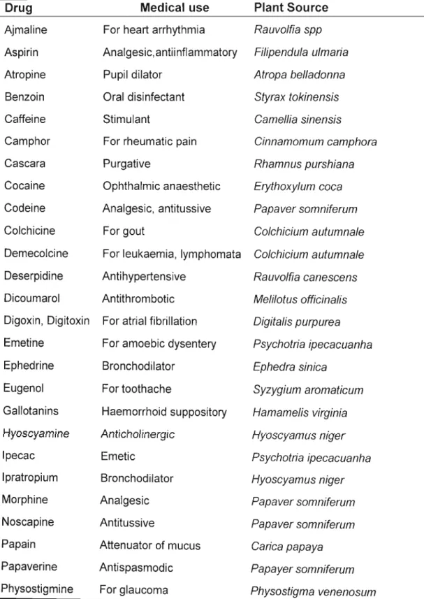
Table 1. Some drugs discovered from ethnobotanicalleads (COX, 1994)
Drug Medical use Plant Source
Ajmaline For heart arrhythmia Rauvolfia spp Aspirin Analgesic,antiinflammatory Filipendula ulmaria
Atropine Pupil dilator Atropa belladonna
Benzoin Oral disinfectant Styrax tokinensis
Caffeine Stimulant Camellia sinensis
Camphor For rheumatic pain Cinnamomum camphora
Cascara Purgative Rhamnus purshiana
Cocaine Ophthalmic anaesthetic Erythoxylum coca Codeine Analgesic, antitussive Papaver somniferum
Colchicine For gout Colchicium autumnale
Demecolcine For leukaemia, lymphomata Colchicium autumnale Deserpidine Antihypertensive Rauvolfia canescens Dicoumarol Antithrombotic Melilotus officinalis Digoxin, Digitoxin For atrial fibrillation Digitalis purpurea Emetine For amoebic dysentery Psychotria ipecacuanha
Ephedrine Bronchodilator Ephedra sinica
Eugenol For toothache Syzygium aromaticum
Gallotanins Haemorrhoid suppository Hamamelis virginia Hyoscyamine Anticholinergic Hyoscyamus niger
Ipecac Emetic Psychotria ipecacuanha
Ipratropium Bronchodilator Hyoscyamus niger
Morphine Analgesic Papaver somniferum
Noscapine Antitussive Papaver somniferum
Papain Attenuator of mucus Carica papaya
Papaverine Antispasmodic Papayer somniferum
Physostigmine For glaucoma Physostigma venenosum
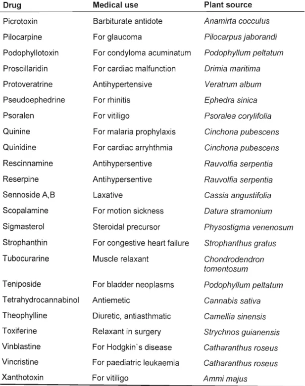
Table 1. continued
Drug Medical use Plant source
Picrotoxin Barbiturate antidote Anamirta coccu/us
Pilocarpine For glaucoma Pilocarpus jaborandi
Podophyllotoxin For condyloma acuminatum Podophyllum pe/tatum Proscillaridin For cardiac malfunction Drimia maritima
Protoveratrine Antihypertensive Veratrum a/bum
Pseudoephedrine For rhinitis Ephedra sinica
Psoralen For vitiligo Psora/ea cory/ifo/ia
Quinine For malaria prophylaxis Cinchona pubescens Quinidine For cardiac arryhthmia Cinchona pubescens Rescinnamine Antihypersentive Rauvolfia serpentia
Reserpine Antihypersentive Rauvo/fia serpentia
Sennoside A,B Laxative Cassia angustifo/ia
Scopalamine For motion sickness Datura stramonium Sigmasterol Steroidal precursor Physostigma venenosum Strophanthin For congestive heart failure Strophanthus gratus
Tubocurarine Muscle relaxant Chondrodendron
tomentosum
Teniposide For bladder neoplasms Podophyllum pe/tatum Tetrahydrocannabinol Antiemetic Cannabis sativa Theophylline Diuretic, antiasthmatic Camellia sinensis Toxiferine Relaxant in surgery Strychnos guianensis Vinblastine For Hodgkin's disease Catharanthus roseus Vincristine For paediatric leukaemia Catharanthus roseus
Xanthotoxin For vitiligo Ammi majus
Several studies have also shown that the rate of discovery of active compounds was higher when plants were selected according to traditional use rather than at random (CANIGUERAL, 1999). In the field of anticancer activity, a correlation between biological activity and use in traditional medicine has been demonstrated (HOSTETTMANN et al., 1996).
Drug discovery and development is a time-consuming and lengthy process and it takes about 12 years at a cost of around US$231 million to develop a drug (FARNSWORTH, 1994). The sequence usually begins with the identification of active lead molecules, detailed biological assays, and formulation of dosage forms in that order. This is
followed by several phases of clinical studies designed to establish safety, efficacy and the pharmacokinetic profile of the new drug (IWU et al., 1999).
The use of ethnobotanical data provides a valuable short cut by indicating plants with specific folk-medicinal uses which might be likely sources of biologically active
chemicals (GENTRY, 1993). Historically, ethnobotanicalleads have resulted in three different types of drug discovery: unmodified natural plant products where ethnomedical use suggested clinical efficacy (e.g. digitalis); unmodified natural products of which the therapeutic efficacy was only remotely suggested by indigenous plant use (e.g.
vincristine); and modified natural or synthetic substances based on a natural product used in folk medicine (e.g. aspirin) (COX, 1994). In as much as this approach has a great potential for discovering potent new compounds, it may be limited in the type of
Crucial to any investigation of plants with biological activities is the availability of suitable bioassays for monitoring the required effects (HOSTETTMANN et al., 1996).
This has lead to the development and introduction of high-throughput tests for biological activity (HOUGHTON, 1999). The test systems should ideally be simple, rapid,
reproducible and inexpensive, and the number of false positives should be reduced to a minimum. These tests have the advantage that a large number of samples can be investigated in a relatively short time and the amount of sample required is usually quite small. The process lends itself, not only to the investigation of a large number of plants obtained, either through random collection or selected according to
ethnopharmacological information, but also to fractions from individual plant extracts so that the chemical compounds responsible can be determined (HOUGHTON, 1999).
In as much as drug development requires an enormous time frame and money - higher plants still remain chemical factories that are capable of synthesizing significant
numbers of highly complex and unusual chemical substances whose structures could escape the imagination of synthetic chemists (FARNSWORTH, 1988). Tropical forests represent nature's main storehouse of raw materials for modern medicine. There is a need for continued search for new drugs or blueprints for new drugs in plants for better treatment of diseases that has plagued mankind for centuries.
1.6. Aims and Objectives
Infectious diseases account for approximately one-half of all deaths in tropical countries (IWU et al., 1999). Malaria is the most widespread insect-borne disease. It kills between one and two million people every year, with as many as 300-500 million people being infected (SAXENA et al., 2003). Therefore the aims when embarking on this project were to screen a number of medicinal plants collected, for antiplasmodial activity. The infection leads to loss of resistance and greater susceptibility to bacteria, fungi and other disease causing microorganisms. For this reason attention was also given to screening for antibacterial and anti-inflammatory activity. Attempts were also made towards isolating the most bioactive constituent(s). Validation of the traditional use of these plants was a prime consideration.
CHAPTER 2
ANTIBACTERIAL SCREENING
2.1. Introduction
Bacteria have been on the earth for millions of years yet it was not until the late 1600s that scientists discovered them. They are prokaryotic and they come in a variety of sizes and shapes that include spherical, rodlike and spiral forms. Most bacterial cells have a rigid cell wall made of peptidoglycan. Since they do not posses a nucleus, the genetic material simply floats around in the cytoplasm (BLACK, 2002). Besides being pathogenic, bacteria play a very vital role in our ecosystem. The actinomycetes produce antibiotics such as streptomycin and nocardicin; others live symbiotically in the gut of animals including humans, or on the roots of some legumes converting nitrogen into a usable form. They also help to break down dead organic matter and form the base of the food web in our environment (BLACK, 2002).
2.2. Disease Causing Bacteria
Infectious diseases account for approximately one-half of all deaths in tropical countries (IWU et al., 1999). Mortality rates due to infectious diseases are actually increasing in developed countries such as the United States (IWU et al., 1999). Death from infectious diseases, which ranked fifth in 1981, has become the third leading cause of death in 1992, an increase of 58% (pINNER et al., 1996). As examples a number of bacteria that can cause major health problems are discussed.
2.2.1. Escherichia coli
This is a Gram-negative, rod-shaped bacterium. It belongs to a group of bacteria known as enterobacteria, so called because they inhabit the intestines of humans and animals.
It is a very common, but certainly not the most abundant bacterium. Some strains of this bacterium cause enteritis in young infants and the young of farm animals, where it can cause diarrhoea and fatal dehydration. It is a common infectant of the urinary tract and bladder in humans (HUGO, 1992).
2.2.2. Klebsiella pneumonia
Klebsiella pneumonia is a Gram-negative, facultative anaerobic, rod-shaped and non- motile bacterium. It is a member of the bacteria family known as Enterobacteriaceae. It is commonly found in water, soil, and occasionally food and can form part of the intestinal flora of humans and animals. Distinguished by the presence of a capsular polysaccharide, Klebsiella pneumonia colonies are larger than those of other bacteria and are highly mucoid. Although it can cause severe pneumonia, it is most commonly the cause of hospital-acquired urinary tract infections or burn wound infections. The autoimmune disease, ankylosing spondylitis is thought to be a possible sequel of Klebsiella infection (HUGO, 1992). The virulence of Klebsiella is not well understood, but its antiphagocytic capsule plays a role in the lung infections by preventing phagocytosis (HUGO, 1992).
2.2.3. Staphylococcus aureus
It is a Gram-positive, spherical bacterium that occurs in microscopic clusters resembling grapes. Staphylococcus aureus forms a fairly large yellow colony on rich medium, often hemolytic on blood agar. It can grow at a temperature range of 15 to 45°C and at NaCI concentrations as high as 15%. Staphylococcus aureus is a normal pathogen of humans, found in nasal passages, on skin and mucous membranes. Some strains are capable of producing a highly heat-stable protein toxin that causes illness in humans.
Enterotoxins produced by some strains of this bacterium cause staphylococcal food poisoning. It also causes a wide range of suppurative (pus-forming) infections and a toxic shock syndrome. It is also a major cause of hospital acquired (nosocomial) infections of surgical wounds and infections associated with indwelling medical devices.
Today in excess of 95% of this bacterium worldwide is resistant to penicilin, ampicilin, and the antipseudomonas penicillin (NEU, 1992).
2.2.4. Bacillus subtilis
It belongs to the family Bacillaceae. Bacillus subtilis is a Gram-positive rod, endospore- forming, motile and obligate aerobe. It is common in soil and dust. Harmless as it might be, Bacillus subtilis may occasionally cause human eye infections. Bacillus subtilis is a source of the antibiotic bacitracin. The only species within this family Bacillaceae that regularly causes serious infection is Bacillus anthracis. It causes a disease called anthrax in farm animals and humans (FUERST, 1978).
2.2.5. Micrococcus luteus
This is an aerobic Gram-positive coccus and a pigment (yellow to cream-white) producer. It is a member of the family Micrococcaceae. Micrococcus luteus is a common inhabitant of human skin, meat, and dairy products. It is non-pathogenic, but may be opportunistic in immunosuppressed individuals (FUERST, 1978).
2.3. Antibacterial Agents
An antibiotic was originally defined as a substance produced by one microorganism, which kills or inhibits the growth of other microorganisms (RUSSELL, 1992). This has since being modified considering the fact that microorganisms are not the only source of antibiotics. Most people now define it as drugs that help the host fight bacteria, viruses and other microorganisms, either by directly killing the causative microorganism, inhibiting its growth, or weakening it so that the host's immune system can fight and kill it more easily (RUSSELL, 1992). Besides microorganisms, plants are also good sources of antibiotics. An ideal antibiotic must display selective toxicity. In other words, it is harmful to the microbe without being harmful to the host.
As early as the 1940s, the quest for chemotherapeutic agents active against pathogenic microbes had already begun. This was when Howard Florey and his colleagues seized upon Alexander Fleming's penicillin and turned it into a major therapeutic compound (GREENWOOD, 1995a). Ever since then many more antibiotics have emerged. Most antibiotics over the years have been discovered by screening of soil samples for such
natural products that kill bacteria including known pathogens, first on culture plates and then in animal systems (WALSH, 2000).
There are three proven targets for the main antibacterial drugs - bacterial cell wall biosynthesis; bacterial protein synthesis; and bacterial DNA replication and repair. The fact that mammalian cells lack cell walls makes the bacterial cell wall a good target.
Although not all the bacterial cell walls are the same, in general they all possess a crossed-linked chain of peptidoglycan, which gives the cell its strength. Cell-wall-active antibiotics act by interfering with the biosynthesis of this structure (GREENWOOD, 1995b and c). Despite the fact that the general mechanism of protein synthesis is thought to be universal, the process as it occurs in bacterial cells is sufficiently different from mammalian protein synthesis to offer scope for the selective toxicity required of therapeutically useful antibacterial agents. Besides, some antibacterial agents kill bacteria by targeting the enzyme DNA gyrase (SHEN, 1993). This is the enzyme responsible for uncoiling the intertwined circles of double-stranded bacterial DNA that arise after each round of DNA replication (WALSH, 2000).
2.3.1. Resistance to Antibacteria Agents
Resistance of bacterial strains to antibiotics is one of the biggest problems faced when combating infectious diseases. According to WALSH (2000), once an antibiotic is proven to be effective and enters widespread human therapeutic use, its days are numbered. A strain of bacteria becomes resistant to an antibiotic by making a protein that its susceptible ancestors could not make. This protein, usually an enzyme, may
inactivate incoming molecules of the antibiotic, as ~-Iactamases do. Alternatively, it may somehow protect the bacterial function targeted by the molecules for sabotage, as does a methylase found in many erythromycin-resistant bacteria (0' BRIEN, 1992). The enzyme methylates, and thus, shields from erythromycin, the specific site on the bacterial ribosome to which the drug would bind to stop protein synthesis and the growth of the bacteria (WEISBLUM, 1984). The resistance of some bacterial strains to antibiotics has contributed substantially to the high mortality rates recorded with infectious diseases. According to FAUCI (1998), a multi-pronged approach that includes prevention, improves monitoring, and allows for the development of new treatments should be employed.
2.4. Antibacterial Assays
Antibacterial sensitivity testing is usually employed to establish the degree and spectrum of in vitro activity of new antibacterial agents or to screen for a new antibacterial agent. This can be done in a variety of ways, and most methods fall into one of three main categories.
·2.4.1. Diffusion Method
This was originally designed to monitor the amount of antibiotic substances in crude extracts (RIOS et al., 1988). Agar diffusion tests, in which the sample to be tested is allowed to diffuse from a point source (disc, hole, or cylinder), commonly in the form of an impregnated filter paper disc, into an agar medium that has been seeded with the
the filter paper disc (inhibition diameter) is measured. In most studies, inhibition zones or diameters are compared with those obtained for antibiotics that are normally used as the positive control. This is useful in establishing the sensitivity of the test organism, but the comparison of the antibacterial potency of the samples and antibiotics (positive control) cannot be determined from this (RIOS
et
al., 1988). The size of the inhibition zone or diameter may be profoundly influenced by the physio-chemical characteristics of the sample such as solubility, ionic charge, and molecular size. Growth rate of the bacterial cell, for example slow-growing organisms give rise to large zones (GREENWOOD, 1995d). It is widely used for reasons of speed, simplicity, cost, as up to six samples can be tested on one culture plate (RIOSet
al., 1988; GREENWOOD, 1995d). In addition, it is also well suited for preliminary screening of pure substances (RIOSet
al., 1988).2.4.2. Microdilution Method
Dilution techniques are those, which require a homogenous dispersion of the sample in water (RIOS,
et
al., 1988). They are used when only a few strains of bacteria need to be tested or when accurate minimum inhibition concentration (MIC) values are estimated for an extract, essential oil or pure compound(s) (RIOSet
al., 1988; GREENWOOD, 1995d). They can also be used in the preliminary screening of antimicrobial activity (RIOSet
al., 1988). A series of two-fold dilutions of the sample under study is prepared in a suitable broth medium and a standard inoculum of the test strain (commonly 105 bacteria) is introduced into each tube. The test is incubated at 370C overnight and the end point is read as that concentration of the sample in which turbidity can be seen.Uninoculated tubes containing broth (or water) plus antibiotic and broth alone act as the sterility controls. An antibiotic free tube inoculated with the test organism serves to indicate that the organism is alive and well in case the end point is missed (RIOS et al., 1988; GREENWOOD, 1995d). This is the only method for determining minimum bactericidal concentrations (MBC). It is done by subculture of the tube with inhibition in an agar plate or liquid medium. When the bacterium does not grow, the sample is a microbicide (RIOS et al., 1988; GREENWOOD, 1995d).
Microdilution method is a dilution method using a 96-well microtitre plate developed by ELOFF (1998). In this method, a two-fold serial dilution of sample to be tested is prepared in the wells of the microplate, and bacterial culture added afterwards. It is incubated overnight at 37°C like other dilution methods. After incubation, p- iodonitrotetrazolium violet is added, and bacterial growth is indicated by a deep red colour, while wells with antibacterial activity stay clear. This technique requires only a small amount of test sample, and is quick, sensitive and reproducible (ELOFF, 1998).
2.4.3. Bioautographic Method
This is the most important detection method for new or unidentified antimicrobial compounds (RIOS et al., 1988). It is based on the biological (eg. antibacterial) effects of the substances under study. The method makes it possible to localize antimicrobial activity on a chromatogram. Inhibition zones are visualised by a dehydrogenase-activity- detecting reagent (RIOS et al., 1988).
However, since some factors like inoculum size, composition of culture medium, extraction method, pH and solubility of the sample in the culture medium can influence results, it is difficult using these methods, to standardize a procedure for the study of antimicrobial plants (RIOS et al., 1988; GREENWOOD, 1995d).
2.5. Materials and Methods 2.5.1. Plant Collection
After consultation with various traditional healers who advised as to the uses of various plants, plant material was collected between May and June 2002 from Oraifite in Anambra state of southeastern Nigeria. Prof. Jonathan G. Okafor of Tree Crops and Tropical Ecology Consultancy, 3 Kingsway Road, Enugu, Nigeria confirmed their identity. Voucher specimens were deposited in the Enugu State Herbarium (Table 2).
2.5.2. Plant Extract Preparation
Plant parts were air-dried for two weeks and then powdered. Plant material (10 g) was extracted sequentially with 150 ml each of petroleum ether (PE), dichloromethane (DCM) and then with 80% ethanol (ET) using a sonication bath for 1 h. Crude extracts obtained were filtered and dried under reduced pressure at 30°C.
2.5.3. Test Organisms
Test organisms used were from The American Type Culture Collection (ATCC):
Staphylococcus aureus(ATCC 12600), Micrococcus luteus(ATCC 4698), Escherichia
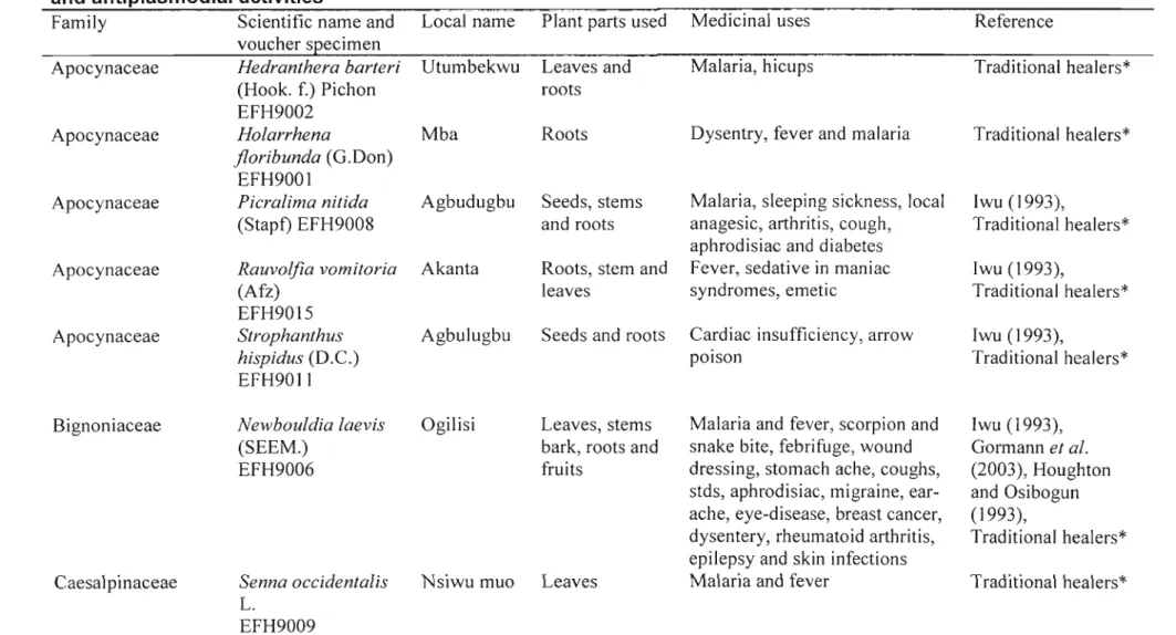
Table 2. Selected medicinal plants used and collected in Nigeria investigated for antibacterial, anti-inflammatory and anti plasmodial activities
Family Scientific name and Local name Plant parts used Medicinal uses voucher specimen
Traditional healers*
Traditional healers*
Iwu (1993),
Traditional healers*
Reference
Iwu (1993),
Traditional healers*
Iwu (1993),
Traditional healers*
Dysentry, fever and malaria Malaria, hicups
Cardiac insufficiency, arrow poison
Malaria, sleeping sickness, local anagesic, arthritis, cough, aphrodisiac and diabetes Fever, sedative in maniac syndromes, emetic Roots
Seeds and roots Roots, stem and leaves
Seeds, stems and roots Mba
Akanta Agbudugbu
Agbulugbu Rauvolfia vomitoria
(Afz) EFH9015 Strophanthus hispidus (D.C.) EFH9011
Hedranthera barteri Utumbekwu Leaves and
(Hook. f.) Pichon roots
EFH9002 Holarrhena
floribunda (G.Don) EFH9001
Picralima nitida (Stapf) EFH9008
Apocynaceae Apocynaceae
Apocynaceae Apocynaceae Apocynaceae
Bignoniaceae
Caesal pinaceae
Newbouldia laevis (SEEM.)
EFH9006
Senna occidentalis L.
EFH9009
Ogilisi Leaves, stems bark, roots and fruits
Nsiwu muo Leaves
Malaria and fever, scorpion and snake bite, febrifuge, wound dressing, stomach ache, coughs, stds, aphrodisiac, migraine, ear- ache, eye-disease, breast cancer, dysentery, rheumatoid arthritis, epilepsy and skin infections Malaria and fever
Iwu (1993), Gormannet al.
(2003), Houghton and Osibogun (1993),
Traditional healers*
Traditional healers*
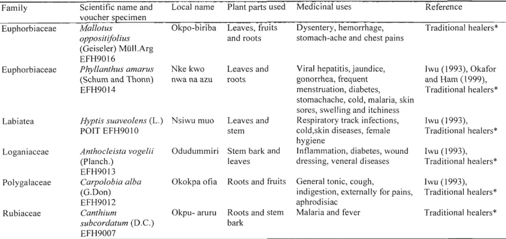
Table 2. Continued
Family Scientific name and Local name Plant parts used Medicinal uses Reference voucher specimen
Euphorbiaceae Mallotus Okpo-biriba Leaves, fruits Dysentery, hemorrhage, Traditional healers*
oppositijolius and roots stomach-ache and chest pains
(Geiseler) Mlill.Arg EFH9016
Euphorbiaceae Phyllanthus amarus Nke kwo Leaves and Viral hepatitis, jaundice, Iwu (1993), Okafor (Schum and Thonn) nwa na azu roots gonorrhea, frequent and Ham (1999),
EFH9014 menstruation, diabetes, Traditional healers*
stomachache, cold, malaria, skin sores, swelling and itchiness
Labiatea Hyptis suaveolens(L.) Nsiwu muo Leaves and Respiratory track infections, Iwu (1993),
POIT EFH9010 stem cold,skin diseases, female Traditional healers*
hygiene
Loganiaceae Anthocleista vogelii Odudummiri Stem bark and Inflammation, diabetes, wound Iwu (1993),
(Planch.) leaves dressing, veneral diseases Traditional healers*
EFH9013
Polygalaceae Carpolobia alba Okokpa ofia Roots and fruits General tonic, cough, Iwu (1993),
(G.Don) indigestion, externally for pains, Traditional healers*
EFH9012 aphrodisiac
Rubiaceae Canthium Okpu- aruru Roots and stem Malaria and fever Traditional healers*
subcordatum (D.e.) bark
EFH9007
Table 2. Continued
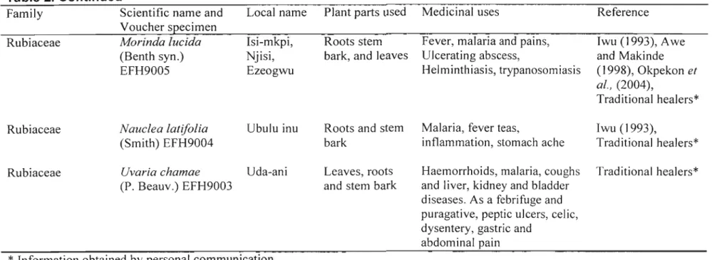
Family Scientific name and Voucher specimen Rubiaceae Morinda lucida
(Benth syn.) EFH9005
Rubiaceae Nauclea latifolia (Smith) EFH9004
Local name Plant parts used Medicinal uses Reference Isi-mkpi, Roots stem Fever, malaria and pains, Iwu (1993), Awe Njisi, bark, and leaves Ulcerating abscess, and Makinde Ezeogwu Helminthiasis, trypanosomiasis (1998), Okpekon et
aI., (2004),
Traditional healers*
Ubulu inu Roots and stem Malaria, fever teas, Iwu (1993),
bark inflammation, stomach ache Traditional healers*
Rubiaceae Uvaria chamae Uda-ani
(P. Beauv.) EFH9003
Leaves, roots and stem bark
Haemorrhoids, malaria, coughs Traditional healers*
and liver, kidney and bladder diseases. As a febrifuge and puragative, peptic ulcers, celic, dysentery, gastric and
abdominal pain
* Information obtained by personal communication.
coli (ATCC 11775), Klebsiella pneumoniae (ATCC 13883), and Bacillus subtilis (ATCC 6051). They were maintained at 4°C on nutrient agar plates.
2.5.4. Diffusion Method: Disc-diffusion Assay
The disc-diffusion assay (RASOANAIVO and RATSIMAMANGA-URVERG, 1993) was employed to investigate the inhibition of bacterial growth by plant extracts. Plants assayed for antibacterial activity and what they are used for traditionally are listed in Table 2. Plant extracts were resuspended in extracting solvent at a concentration of 100 mg/ml. Base plates were prepared by pouring 10 ml Mueller-Hinton (MH) agar into sterile 90 mm Petri dishes. MH agar maintained at 48°C was inoculated with a MH broth (106-108bacteria per ml) of each of the test organisms and poured over the base plates to form a homogenous layer. Filter paper discs (6 mm diameter, Whatman no. 3) were sterilized by autoclaving. Plant extract (10 I-II) was dispensed on each filter paper disc.
This gave a final concentration of 1 mg plant extract per disc. Discs with plant extracts were air-dried and each placed onto the seeded MH agar plates. Each extract was tested in quadruplicate (4 discs per plate) with a neomycin disc (500 I-Ig) as a positive control. Air-dried solvent (80% ethanol, petroleum ether, and dichloromethane) discs were used as negative controls. The plates were incubated overnight at 37°C.
Antibacterial activity was expressed as a ratio of the inhibition zone produced by the plant extract and the inhibition zone produced by the positive control (neomycin).
2.5.5. Dilution Method: Microdilution Assay
The microtitre bioassay (ELOFF, 1998) was used to determine the Minimal Inhibitory Concentration (MIC) for the plant extracts that inhibited bacterial growth or showed a bacteriostatic effect in the disc-diffusion assay. Plant extracts (both polar and non-polar) were made up to 50 mg/ml with 25% ethanol. Plant extracts (100 Ill) were two-fold serially diluted with distilled water in 96-well microplates to give concentrations from 12.5 - 0.098 mg/ml. Overnight MH broth cultures (grown at 370C in a water bath with continuous shaking) of the test organisms were diluted 100 fold with MH broth, and 100 III of the resulting bacterial culture were added to each well. Neomycin (100 Ilg/ml) was used as a positive control for each bacterium, with solvent and bacteria free wells being included as negative controls. Microplates were covered and incubated overnight at 370C. To indicate bacterial growth, 40 III of 0.2 mg/ml p-indonitritetrazolium violet (INT) were added to each well and incubated at 37 QC for 30 min. The colourless tetrazolium salts act as an electron acceptor and it is reduced to a red-coloured formazan product by biologically active organisms (ELOFF, 1998). Clear wells with INT after incubation indicate inhibition of bacterial growth. MIC values were recorded as the lowest concentration of extract that completely inhibited bacterial growth.
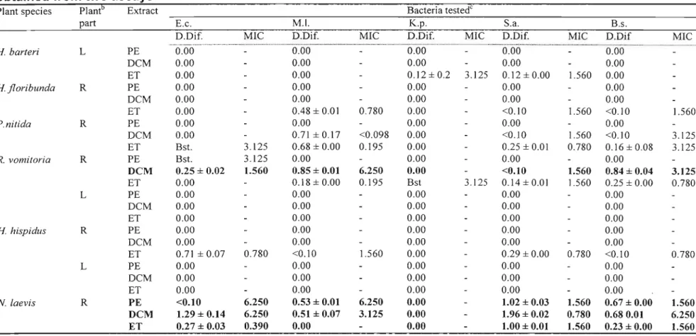
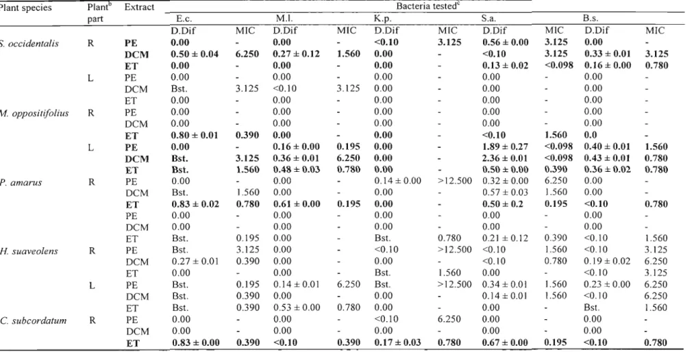
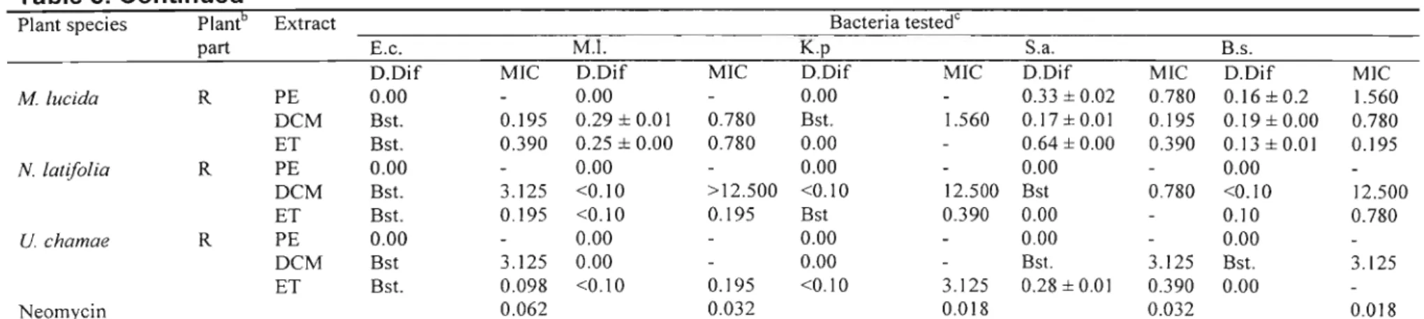
2.6. Results and Discussion
Antibacterial activity detected using the disc diffusion assay and MIC values of crude extracts of different plant parts of different species are presented in Table 3. Plant extracts with high antibacterial activity are highlighted in bold. Most activity was
activity was detected in the petroleum ether extracts. Of the 57 extracts screened, 27 were active against E. coli; 22 against M. luteus; 14 against K. pneumonae; 32 against S. aureus and 29 against B. subtilis. The Gram-positive bacteria, especially S. aureus, were the most susceptible. Few extracts showed activity against the Gram-negative bacteria used. Gram-negative bacteria are generally more difficult to inhibit due to the presence of the thick murin layer that tends to prevent the entry of inhibitors (MARTIN, 1995; VLlETINCKet al., 1995).
As earlier stated, the microtitre bioassay (ELOFF, 1998) was used to determine the MIC for the plant extracts that inhibited bacterial growth or showed a bacteriostatic effect in the disc-diffusion assay. In as much as this assay records the minimum inhibitory concentration, it can also indicate a bacteriocidal effect. Since there were no further tests carried out to actually determine if it was inhibition of the bacterial growth or a bacteriocidal effect detected, results are recorded as MIC. One would expect those extracts that had high inhibition zones when compared to the inhibition zones of the reference (neomycin) ratio in the disc diffusion assay to show much lower MIC values, but this was not the case. Some did exhibit much lower MIC values (dichloromethane leaf extract ofM. oppositifolius against S. aureus) while some did not (dichloromethane root extract ofN. laevisagainst S. aureus).
In the disc-diffusion assay, petroleum ether, dichloromethane and 80 %ethanol extracts ofN. laevisall exhibited broad-spectrum activity with the dichloromethane extract
Table 3. Determination of antibacterial activitya of some medicinal plants used and collected in Nigeria using the disc-diffusion (Dif) and microdilution assays (MIC expressed in mg/ml). Values are the mean ±S.E.M. of results obtained from two assays
Plant species Plantb Extract Bacteria testedC
part E.c. M.l. K.p. S.a. B.s.
D.Dif. MIC D.Dif. MIC D.Dif. MIC D.Dif. MIC D.Dif MIC
H barteri L PE.-6:o-6.·- ·..··
-=.. -....-..O:OO-- ---.-.·--·=---·-··---0:00··· ·..··..
·=·-···---·--·-6:00..·..·---·=--···..··..
·0:00··· - .DCM 0.00 - 0.00 - 0.00 - 0.00 - 0.00
ET 0.00 - 0.00 - 0.12±0.2 3.125 0.12±0.00 1.5600.00
H floribunda R PE 0.00 - 0.00 - 0.00 - 0.00 - 0.00
DCM 0.00 - 0.00 - 0.00 - 0.00 - 0.00
ET 0.00 - 0.48 ± 0.01 0.780 0.00 - <0.10 1.560 <0.10 1.560
P.nitida R PE 0.00 - 0.00 .. 0.00 .. 0.00 - 0.00
DCM 0.00 .. 0.71 ±0.17 <0.098 0.00 - <0.10 1.560 <0.10 3.125
ET Bst. 3.125 0.68 ± 0.00 0.195 0.00 .. 0.25±0.01 0.780 0.16±0.08 3.125
R. vomitoria R PE Bst. 3.125 0.00 .. 0.00 .. 0.00 - 0.00
DCM 0.25 ± 0.02 1.560 0.85 ± 0.01 6.250 0.00 - <0.10 1.560 0.84 ± 0.04 3.125
ET 0.00 - 0.18 ± 0.00 0.195 Bst 3.125 0.14 ± 0.01 1.560 0.25 ± 0.00 0.780
L PE 0.00 - 0.00 - 0.00 - 0.00 .. 0.00
DCM 0.00 - 0.00 - 0.00 - 0.00 - 0.00
ET 0.00 - 0.00 - 0.00 - 0.00 - 0.00
H hispidus R PE 0.00 - 0.00 - 0.00 .. 0.00 - 0.00
DCM 0.00 - 0.00 .. 0.00 .. 0.00 - 0.00
ET 0.71±0.07 0.780 <0.10 1.560 0.00 .. 0.29±0.00 0.780 <0.10 0.780
L PE 0.00 - 0.00 - 0.00 - 0.00 - 0.00
DCM 0.00 - 0.00 - 0.00 - 0.00 .. 0.00
ET 0.00 - 0.00 - 0.00 .. 0.00 - 0.00
N. laevis R PE <0.10 6.250 0.53 ± 0.01 6.250 0.00 - 1.02 ± 0.03 1.560 0.67 ± 0.00 1.560
DCM 1.29 ± 0.14 6.250 0.51 ± 0.07 3.125 0.00 - 1.96 ± 0.02 0.780 0.680.01 6.250
ET 0.27 ± 0.03 0.390 0.00 - 0.00 - 1.00 ± 0.01 1.560 0.23 ± 0.00 1.560
Table 3. Continued
Plant species Plantb Extract Bacteria testedC
part E.c. M.1. K.p.