For Peer Review
Manuscript ID: JAM-2008-0805.R1
Journal Name: 1 Journal of Applied Microbiology - JAM Manuscript Type: JAM - Review
Date Submitted by the Author: n/a
Complete List of Authors: Thuku, Robert; University of Cape Town, Electron Microscope Unit Brady, Dean; CSIR, Biosciences
Benedik, Michael; Texas A&M University, Biology
Sewell, Bryan; University of Cape Town, Electron Microscope Unit
Key Words: Biotechnology, Molecular genetic, Enzyme activity, Protein Engineering, Structural Biology
For Peer Review
Microbial nitrilases: versatile, spiral forming, industrial enzymes
R. Ndoria Thuku1,4, Dean Brady2, Michael J. Benedik3and B. Trevor Sewell1,5
1Electron Microscope Unit, University of Cape Town, South Africa.
2CSIR Biosciences, Modderfontein 1645, Private Bag X2, Johannesburg, South Africa
3Department of Biology, Texas A&M University, Texas, USA
4Department of Molecular and Cell Biology, University of Cape Town, South Africa
5Institute of Infectious Disease and Molecular Medicine, University of Cape Town, South Africa
Running title: The spiral forming nitrilase structures Subdivision: Review
Corresponding author: B. Trevor Sewell
Electron Microscope Unit, University of Cape Town, Private Bag, Rondebosch 7700, South Africa.
Telephone: +27216502817 Fax: +27216891528
Email: [email protected] Enzyme: EC 3.5.5.1 nitrilase
Keywords: nitrilase, oligomer, spiral enzymes, nitrilase superfamily, helical enzymes Word Count: 15855
3 4 5 6 7 8 9 10 11 12 13 14 15 16 17 18 19 20 21 22 23 24 25 26 27 28 29 30 31 32 33 34 35 36 37 38 39 40 41 42 43 44 45 46 47 48 49 50 51 52 53 54 55 56 57 58 59 60
For Peer Review
1. Summary 2. Introduction
3. Enzyme structure and homology
4. Substrate specificity and enzyme engineering 5. Enzyme engineering
6. The reaction mechanism
7. Spiral formation among microbial nitrilases 7.1 The ‘A’ surface
7.2 The ‘C’ surface
7.3 The ‘D’ and ‘F’ surface 7.4 The ‘E’ surface
8. The role of the extended C-terminal region 9. Conclusion
10. References
3 4 5 6 7 8 9 10 11 12 13 14 15 16 17 18 19 20 21 22 23 24 25 26 27 28 29 30 31 32 33 34 35 36 37 38 39 40 41 42 43 44 45 46 47 48 49 50 51 52 53 54 55 56 57 58 59 60
For Peer Review
1. Summary
The nitrilases are enzymes that convert nitriles to the corresponding acid and ammonia. They are members of a superfamily, which includes amidases and occur in both prokaryotes and eukaryotes. The superfamily is characterized by having a homodimeric building block with a - sandwich fold and an active site containing four positionally conserved residues:
cys, glu, glu and lys. Their high chemical specificity and frequent enantioselectivity makes them attractive biocatalysts for the production of fine chemicals and pharmaceutical intermediates. Nitrilases are also used in the treatment of toxic industrial effluent and cyanide remediation. The superfamily enzymes have been visualized as dimers, tetramers, hexamers, octamers, tetradecamers, octadecamers and variable length helices, but all nitrilase oligomers have the same basic dimer interface. Moreover, in the case of the octamers, tetradecamers, octadecamers and the helices, common principles of subunit association apply. While the range of industrially interesting reactions catalysed by this enzyme class continues to increase, research efforts are still hampered by the lack of a high resolution microbial nitrilase structure which can provide insights into their specificity, enantioselectivity and the mechanism of catalysis. This review provides an overview of the current progress in elucidation of structure and function in this enzyme class and emphasizes insights that may lead to further biotechnological applications.
2. Introduction
The nitrilases (EC 3.5.5.1) are an important class of industrial enzymes belonging to the nitrilase superfamily (Pace and Brenner, 2001), and are expressed widely in both prokaryotes and eukaryotes. Nitrilases hydrolyze various nitriles to the corresponding acid and ammonia, although they occasionally release an amide product (Fernandes et al., 2006). The nitrilase superfamily comprises thirteen different enzyme classes that share significant structural homology despite varying sequence conservation. In general, the superfamily members include the microbial nitrilases (nitrilases, cyanide dihydratases and cyanide hydratases), aliphatic amidases, amidohydrolases and acyl transferases of differing specificities (Brenner, 2002). The majority of the known enzymes were obtained from bacteria, fungi and plants by a variety of selection methods on media containing nitriles as nitrogen sources (O’Reilly and Turner, 2003;
Martinkova et al., 2008) or through direct cloning and expression. Although the physiological role of nitrilases is still not clear, they may play a variety of diverse roles in the cell. The production of metabolites is exemplified by the synthesis of indole acetic acid (Bartel and Fink, 1994; Bartling et al., 1992, 1994), whereas their role in detoxification has been shown for
3 4 5 6 7 8 9 10 11 12 13 14 15 16 17 18 19 20 21 22 23 24 25 26 27 28 29 30 31 32 33 34 35 36 37 38 39 40 41 42 43 44 45 46 47 48 49 50 51 52 53 54 55 56 57 58 59 60
For Peer Review
cyanide in plants (Piotrowski et al., 2001), the degradation of glucosinolates (Bestwick et al.
1993), and the aldoxime degrading pathway (Kato et al. 2000).
Some information regarding their natural role can be surmised from studying the control of nitrilase gene expression. While bacteria such as Bacillus subtilis ZJB-063, constitutively express nitrilases (Zheng et al., 2007), the majority of the characterized nitrilases are inducible by the presence of nitriles in the growth media (Banerjee et al., 2002) which would seem to indicate that they play a role in detoxification or utilization. In a variety of fungi, expression of the cyanide degrading nitrilase is specifically induced by the presence of cyanide in the growth medium. In contrast, the expression of the CynD from B. pumilus was not found to be regulated by cyanide, but is controlled by the sporulation cascade. It is induced by Mn2+
(Meyers et al., 1993), a well-known inducer of sporulation, and is abolished in spoOA mutants.
CynD has not been specifically linked to any biological role in bacteria, but nonetheless has been exploited for biotechnological purposes.
In seven classes of the nitrilase superfamily, the nitrilase domain is fused to another domain (Brenner, 2002). One such case is the NAD+ synthetase from Mycobacterium tuberculosis (Bellinzoni et al., 2005). This enzyme relies on an associated amino-terminal amidase domain in order to utilize glutamine as a source of nitrogen and liberate ammonia which is required for the synthesis of NAD+. The inactivation of the amidase domain by mutation of the catalytic cysteine inactivated the associated NAD+ synthetase, and suggested that NAD+ synthetase is a potential drug target (Bellinzoni et al., 2004). Nitrilases are attractive biocatalysts in the fine chemicals and pharmaceutical industry because of their high specificity and chemo-, regio- and enantio-selectivity (Brady et al., 2004). They are also used in the detoxification of industrial waste and herbicide degradation (Banerjee at al., 2002). In general, the nitrile biocatalysts operate in aqueous solutions at moderate temperatures and pH, and this minimizes the costs of chemical processes and the negative impact of industry on the environment (Singh et al., 2006;
Brady et al., 2006).
There are over 200 known nitrilase sequences (Robertson et al., 2004). A recent study identified nitrile-hydrolyzing activities in 40 distinct species of bacterial and yeast isolates (Brady et al., 2006). While our knowledge of their sequence information, environmental distribution and substrate specificity continues to increase, the crystal structure of a microbial nitrilase remains elusive. Consequently, the lack of specific structural information on these enzymes hinders the correlation of sequence, activity and specificity (Podar et al., 2005).
3 4 5 6 7 8 9 10 11 12 13 14 15 16 17 18 19 20 21 22 23 24 25 26 27 28 29 30 31 32 33 34 35 36 37 38 39 40 41 42 43 44 45 46 47 48 49 50 51 52 53 54 55 56 57 58 59 60
For Peer Review
Nevertheless, there are eleven atomic structures of distant homologous enzymes in the nitrilase superfamily which provide key insights into the enzyme structure. These include the Nit domain of the NitFhit fusion protein (PDB code 1ems), three N-carbamoyl-D-amino acid amidohydrolases from Agrobacterium sp. Strain KNK712 and Agrobacterium radiobacter (PDB codes 1erz, 1fo6 and 1uf5), the putative CN hydrolase from yeast (PDB code 1f89), the hypothetical protein PH0642 from Pyrococcus horikoshii (PDB code 1j31), XC1258, a putative prokaryotic Nit protein from Xanthomonas campestris (PDB code 2e11), the -alanine synthase from Drosophila melanogaster (PDB code 2vhi and 2vhh), the AmiF formamidase from Helicobacter pylori (PDB codes 2dyu, 2e2k and 2e2l), and two aliphatic amidases from Pseudomonas aeruginosa and Geobacillus pallidus RAPc8 (PDB codes 2uxy and 2plq, respectively) (Pace et al., 2000; Nakai et al., 2000; Wang et al., 2001; Hashimoto et al., 2004;
Kumaran et al., 2003; Sakai et al., 2004; Chin et al., 2007; Lundgren et al., 2008; Hung et al., 2007; Andrade et al., 2007 and Kimani et al., 2007, respectively). Despite varying sequence identity (~20%), all the structures share a characteristic monomer fold and conserve two glutamates, lysine and cysteine in their catalytic site. The monomers associate in a common manner to form an - sandwich.
The microbial nitrilases have been shown to form homo-oligomeric spirals using a combination of negative stain electron microscopy and the docking of homology models (Sewell et al., 2003; Thuku et al., 2007; Woodward et al., 2008; Dent et al., 2008; Vejvoda et al., 2008). The best characterized enzyme is the nitrilase from Rhodococcus rhodochrous J1 which is capable of converting acrylonitrile and 3-cyanopyridine to acrylic acid and nicotinic acid, respectively (Kobayashi et al., 1988, Mathew et al., 1988). This enzyme exists as an inactive dimer which oligomerizes to form active spirals in the presence of benzonitrile or an autolytic cleavage of the C-terminus (Nagasawa et al., 2000, Thuku et al, 2007). This phenomenon has most frequently been described in those nitrilases arising from the genus Rhodococcus (Harper, 1977b, 1985; Hoyle et al., 1988; Stevenson et al., 1992). The monomer association occurs via two interfaces, namely the ‘A’ and ‘C’ surfaces and forms a one-start, left-handed spiral (Sewell et al., 2003). The modification of residues within these surfaces inactivates the enzyme and possibly suggests that a link exists between oligomerization at these surfaces and the active site (Sewell et al., 2005). In order to fully understand the spiral structures, it is therefore necessary to characterize these interfaces and their mechanism of activation.
3 4 5 6 7 8 9 10 11 12 13 14 15 16 17 18 19 20 21 22 23 24 25 26 27 28 29 30 31 32 33 34 35 36 37 38 39 40 41 42 43 44 45 46 47 48 49 50 51 52 53 54 55 56 57 58 59 60
For Peer Review
In this review, we describe the structural insights we have gained from studying the nitrilase from Rhodococcus rhodochrous J1 and other closely related enzymes, namely the cyanide dihydratases, the cyanide hydratases and an amidase. We also incorporate structural insights from other nitrilase homologues whose structures have been determined at atomic resolution.
Further biotechnological advances involving these enzymes make it important to expand on their structure, the details of the interacting surfaces, and their reaction mechanism. This will lead to the development of improved biocatalysts (more stable, specific and versatile enzymes) and additional processes using nitrile-hydrolyzing enzymes.
3. Enzyme structure and homology
The microbial nitrilases are generally known to exist as inactive dimers in solution except the active dimeric enzyme from Pyrococcus abyssi (Mueller et al., 2006). The majority of these enzymes have a subunit size of between 30-45 kDa (Banerjee et al., 2002; O’Reilly and Turner, 2003), which self associate to form active oligomers having between 4 – 22 subunits (Table 1), or active spirals of variable length (Sewell et al., 2005; Thuku et al., 2007; Woodward et al., 2008; Vejvoda et al., 2008; Dent et al., 2008). The exceptions include the 76 kDa nitrilase from Fusarium solani (Harper, 1977a), and the monomeric enzymes from Arthrobacter sp. strain J1 (Bandyopadhyay et al., 1986) and Rhodococcus rhodochrous PA-34 (Bhalla et al., 1992).
However, as none of these atypical nitrilases have been sequenced, their homology to the nitrilase superfamily enzymes remains unknown. The molecular mass of the enzyme subunit and the active complex is usually determined in the absence of the substrate by size exclusion chromatography, native- and SDS-PAGE, mass spectroscopy, light scattering and electron microscopy.
Despite varying sequence conservation and differing substrate affinities, all the superfamily enzymes demonstrate significant structural homology and can be aligned with the crystal structures of 1ems, 1erz, 1uf5, 1fo6, 1f89, 1j31, 2e11, 2vhi, 2plq, 2dyu and 2uxy using a program such as mGenTHREADER (Jones, 1999; McGuffin and Jones, 2003). Each enzyme monomer has an -fold which associates to form an 8-layered - dimer (Figure 1) across the ‘A’ surface (Sewell et al., 2003). While the solved structures form dimers, tetramers, hexamers or octamers, the microbial nitrilases form larger homo-oligomeric spirals with a varying number of subunits. The dimer is the primary building block for oligomerization and their association occurs in a variety of ways among the superfamily enzymes. In particular, the Rhodococcal nitrilases (Harper 1977b, 1985; Stevenson et al., 1992; Nagawasa et al.; 2000)
3 4 5 6 7 8 9 10 11 12 13 14 15 16 17 18 19 20 21 22 23 24 25 26 27 28 29 30 31 32 33 34 35 36 37 38 39 40 41 42 43 44 45 46 47 48 49 50 51 52 53 54 55 56 57 58 59 60
For Peer Review
could form a complex estimated at having 10-12 subunits in the presence of substrate, on heat treatment, or addition of ammonium sulphate or organic solvent. We recently reported a long regular helix of the nitrilase from R. rhodochrous J1 in which the short active ‘c’ shaped oligomers undergo an autolysis removing 39 C-terminal amino acids and causing them to form long regular helices (Thuku et al., 2007). Active ‘c’ shaped homo-octamers (Figure 2) which appear superficially similar have recently been visualized at atomic resolution in the nitrilase- related -alanine synthase from Drosophila melanogaster (Lundgren et al., 2008). This enzyme catalyzes the removal of the N-carbamyl group of N-carbamyl- -alanine and N-carbamyl- - aminoisobutyrate to form -alanine and -aminoisobutyrate, respectively, with the release of carbon dioxide and ammonia (Schnackerz and Dobritzsch, 2008). The correlation of the shape of the fruit fly enzyme to the ‘c’ shape of the R. rhodochrous J1 nitrilase oligomers (Thuku et al., 2007) and the fact that there are approximately 10 subunits per turn of helix, suggests that the Rhodococcal enzyme could also form an octamer.
The cyanide dihydratase from B. pumilus C1 was also found as both a short spiral and a long helix but differed in that it showed a reversible pH-dependent switching between an 18-subunit terminating spiral to a variable length helix (Jandhyala et al., 2003) at pH5.4. Other known spiral structures include the 14-subunit self-terminating cyanide dihydratase from P. stutzeri AK61 (Sewell et al., 2003), and the long regular helices of the cyanide hydratases from G.
sorghi (Woodward et al., 2008) and N. crassa (Dent et al., 2008), and the nitrilase from Aspergillus niger K10 (Vejvoda et al., 2008).
There are numerous suggestions of the functional significance of the oligomerization.
Jandhyala et al (2005) observed an increase in activity relative to a homologue in which the pH dependent switching does not occur at the pH at which the size of the cyanide dihydratase increased in the B. pumilus C1. The increase in activity accompanying an increase in complex size is commonly observed in Rhodococcal species where inactive dimers and active higher oligomers are found (Harper 1977b, 1985; Nagasawa et al., 2000; Stevenson et al., 1992). The functional complexity of subunit association is nowhere better illustrated than in the case of the plant nitrilases which are known to possess two or three nitrilase isoforms in their tissues.
Recent reports suggest that these enzymes could either have a dual biological function or broaden their substrate spectrum through the formation of higher heteromeric complexes (Jenrich et al., 2007; Kriechbaumer et al., 2007). In particular, the ZmNit1 and ZmNit2 nitrilases from maize were observed to form a complex that could synthesize auxin (indole-3-
3 4 5 6 7 8 9 10 11 12 13 14 15 16 17 18 19 20 21 22 23 24 25 26 27 28 29 30 31 32 33 34 35 36 37 38 39 40 41 42 43 44 45 46 47 48 49 50 51 52 53 54 55 56 57 58 59 60
For Peer Review
acetic acid) as well as hydrolyse -cyanoalanine, an intermediate in cyanide detoxification (Kriechbaumer et al., 2007). In the plant Sorghum bicolor, the individual nitrilase isoforms are inactive but upon heteromeric assembly, they acquired activity and could hydrolyse - cyanoalanine and other substrates (Jenrich et al., 2007).
Multiple alignments of the helix or spiral forming nitrilase sequences showed that these proteins have two significant insertions (12-16 amino acids) in their sequences and an extended C-terminus (Figure 3) relative to the sequences of the first two crystallographically determined structures (1erz and 1ems). Because of the multiple forms of association of the nitrilase monomers, it was necessary to label the areas of association. The residue insertions correspond to the ‘C’ surface and this is required to form the one-start left-handed spiral (Sewell et al., 2003). It has been shown that the modification of the residues in the ‘C’ surface by mutation destroyed the activity of the cyanide dihydratases from B. pumilus C1 and P. stutzeri AK61 (Sewell et al., 2005). As seen in Figure 1, all superfamily enzymes conserve the catalytic residues, namely a glutamic acid, a lysine and a cysteine in the active site (Brenner, 2002). In addition, a glutamic acid (corresponding to glu 142 in the G. pallidus amidase structure), which has been implicated in the nitrilase reaction mechanism is also highly conserved (Kimani et al., 2007). It is interesting that apart from the active site residues, only four glycines are absolutely conserved throughout the known superfamily sequences.
The docking of a homology model of the P. stutzeri AK61 enzyme into the negative stain density of its spiral structure located the extended C-terminal tail facing the center of the spiral (Sewell et al., 2003). This situation is similar to that of the crystalline -alanine synthase (Lundgren et al., 2008), but different in the homologous crystalline amidases which have D3 symmetry and in which the C-terminal tail is located on the outside of the hexamer (Andrade et al., 2007; Hung et al., 2007; Kimani et al., 2007). In the majority of the nitrilase atomic homologues, this part of the molecule is seen to interact at the ‘A’ surface with its equivalent from another monomer. Even though the aliphatic amidases have an extended C-terminal tail similar to that of the microbial nitrilases, the lack of sequence and structural homology suggests there is high variability in this region. We have previously shown that the docking of homology models in the three-dimensional reconstructions of these enzymes, can provide a framework by which the basis for oligomerization and stabilization can be explained (Sewell et al., 2003; Thuku et al., 2007; Woodward et al., 2008; Dent et al., 2008). In addition, the
3 4 5 6 7 8 9 10 11 12 13 14 15 16 17 18 19 20 21 22 23 24 25 26 27 28 29 30 31 32 33 34 35 36 37 38 39 40 41 42 43 44 45 46 47 48 49 50 51 52 53 54 55 56 57 58 59 60
For Peer Review
alignment presented in Figure 3 provides a basis for homology modeling from which the enzyme structure and the identification of the interfacial residues can be inferred.
4. Substrate specificity
The nitrilases have previously been classified on the basis of their substrate affinity (Kobayashi and Shimizu, 1994; Banerjee et al., 2002; O’Reilly and Turner, 2003). While most nitrilases are specific for aromatic nitriles, others have preference for only arylacetonitriles, aliphatic nitriles, bromoxynil or cyanide. We continue to use this broad classification of the biochemically characterized nitrilases and highlight their physical properties, substrate specificity and activity in Table 1 even though there are examples of enzymes that fall into two different categories, for instance arylacetonitrilases that convert aliphatic substrates (Heinemann et al., 2003).
The aromatic nitrilases are highly specific for aromatic and heterocyclic nitriles. The best characterized enzyme is the nitrilase from Rhodococcus rhodochrous J1 (Kobayashi et al., 1989). However, there is a degree of flexibility in the nitrilase substrate range. For instance, the enzymes from R. rhodochrous J1 and Rhodococcus (formerly Nocardia) NCIMB 11216 could hydrolyse acrylonitrile and propionitrile respectively, following activation in the presence of benzonitrile (Nagasawa et al., 2000; Hoyle et al., 1998). Recent reports suggest that the occurrence of aromatic nitrilases in filamentous fungi is common (Kato et al., 2000; Kaplan et al 2006a-c; Vejvoda et al., 2006a-b). Four aromatic fungal nitrilases have been characterized to date, namely those from Fusarium solani IMI196840, Fusarium oxysporum f.sp. melonis, Aspergillus niger K10 and Fusarium solani O1 (Harper, 1977; Goldlust and Bohak, 1989;
Kaplan et al., 2006c; Vejvoda et al., 2008, respectively). These enzymes share high specificity for aromatic substrates and good thermostability. The enzymes from A. niger K10 and F.
solani O1 were shown by electron microscopy (Vejvoda et al., 2008) to form spiral structures similar to those previously reported (Sewell et al., 2005; Thuku et al., 2007).
Although aliphatic nitrilases are capable of hydrolyzing benzonitrile, the rate of hydrolysis is highest with the aliphatic nitriles. In general, these enzymes occur in plants and bacteria (Table 1). There are several reports of homologous nitrilases in plants (Dohmoto et al., 1999, 2000;
Park et al., 2003), however, the AtNIT1 enzyme is the only biochemically characterized plant nitrilase to date. The recombinant NIT1 nitrilase from Arabidopsis thaliana (AtNIT1) had 270
3 4 5 6 7 8 9 10 11 12 13 14 15 16 17 18 19 20 21 22 23 24 25 26 27 28 29 30 31 32 33 34 35 36 37 38 39 40 41 42 43 44 45 46 47 48 49 50 51 52 53 54 55 56 57 58 59 60
For Peer Review
times more activity with 3-phenylpropionitrile than that observed with benzonitrile (Osswald et al., 2002).
The heterologous expression of bacterial nitrilases in plants has also been achieved. The nitrilase of Klebsiella pneumoniae subsp. ozaenae is highly specific for the herbicide bromoxynil (3,5-dibromo-4-hydroxybenzonitrile). The native and recombinant enzymes completely converted bromoxynil to the acid which enabled the bacterium to use the liberated ammonia as the sole source of nitrogen (McBride et al., 1986; Stalker et al., 1988a). This enzyme was observed to be active in vitro as a dimer and showed no activity with benzonitrile (Stalker et al., 1988a). The incorporation of the bacterial gene (bxn) encoding the bromoxynil- specific nitrilase into the leaves of transgenic tobacco plants was reported to confer resistance to high levels of the herbicide Buctril® (or commercial bromoxynil) (Stalker et al., 1988b).
The bacterial gene was spliced to plant-promoters and the genes expressing the bromoxynil- specific nitrilase were introduced into cotton varieties via Agrobacterium transformation with the same effects (Stalker et al., 1996). This discovery has led to the development of Bromoxynil-resistant cotton (BXNTM) which is widely grown in the USA and indeed, this represents one of the most successful biotechnological applications of a nitrilase.
The arylacetonitrilases are generally enantioselective enzymes that display activity with benzonitrile and sometimes the aliphatic nitriles. In particular, the native and recombinantly purified enzymes from Pseudomonas fluorescens EBC191 could convert 2- acetoxybutenenitrile to the corresponding acid with higher specificity than the enzyme from Synechocystis spp. 6803, an aliphatic nitrilase (Heinemann et al., 2003). These observations suggest that the substrate specificities of the microbial nitrilases is wider than is generally assumed. The nitrilases from Alcaligenes faecalis ATCC 8750 (Yamamoto et al., 1991) and P.
fluorescens EBC191 (Kiziak et al., 2005) could convert (R,S)-mandelonitrile to (R)-(- )mandelic acid, an important intermediate in the pharmaceutical industry. While the majority of the arylacetonitrilases could hydrolyse mandelonitrile at a lower rate compared to that of phenylacetonitrile, the mandelonitrile hydrolase from Bradyrhizobium japonicus USDA110 is highly specific for mandelonitrile (Zhu et al., 2007, 2008). The mandelonitrile hydrolase genes have been found to occur on the same operon that encodes other enzymes involved in the mandelonitrile metabolic pathway. For this reason, it was suggested that these enzymes could play a role in the detoxification of harmful nitriles during the metabolism of cyanogenic glycosides (Kiziak et al., 2005; Zhu et al., 2007).
3 4 5 6 7 8 9 10 11 12 13 14 15 16 17 18 19 20 21 22 23 24 25 26 27 28 29 30 31 32 33 34 35 36 37 38 39 40 41 42 43 44 45 46 47 48 49 50 51 52 53 54 55 56 57 58 59 60
For Peer Review
The cyanide dihydratases and cyanide hydratases catalyze the hydrolysis of cyanide (HCN) with high specificity to formate and formamide, respectively. In particular, the cyanide hydratase from the fungus Fusarium lateritium had a 3000-fold higher activity towards KCN compared to benzonitrile (Nolan et al., 2003). The cyanide dihydratases occur mostly in bacteria whereas the cyanide hydratases occur in filamentous fungi (O’Reilly and Turner, 2003). The structures of the enzymes from B. pumilus, P. stutzeri AK61, N. crassa and G.
sorghi are spirals which conserve the oligomeric principles previously reported (Sewell et al., 2005). While the details of the mechanism of cyanide degradation are not clear, it was proposed that subtle differences in the active site configuration of these enzymes dictate whether ammonia or formamide is the better leaving group (Jandhyala et al., 2005). The fungal cyanide hydratases have been shown to be capable of degrading metal cyanides and these enzymes could have potential applications in the treatment of waste from the metal plating industry (Yanase et al., 2000; Barclay et al., 1998). However, the microbial treatment of toxic industrial effluent is often hindered by varying levels of pH and temperature which often inhibits microbial growth (Baxter and Cummings, 2006). Nevertheless, the potential exists for re-engineering these enzymes to tolerate harsh reaction conditions and use them for on-site cyanide remediation (Jandhyala et al., 2003). Mutants of B. pumilus with improved pH tolerance have been identified. However substrate inhibition remains a problem with these enzymes and this limits their industrial application under conditions where high substrate concentrations would occur.
In general, the natural substrates for the majority of the nitrilases are not known. Structural and modeling studies in other homologous superfamily enzymes, namely DCase from Agrobacterium sp (Nakai et al., 2000; Chen et al., 2003; Hashimoto et al., 2004), the CN hydrolase from yeast (Kumaran et al., 2003), and several aliphatic amidases (Andrade et al., 2007; Hung et al., 2007; Kimani et al., 2007) with bound substrates have suggested that the residues lining the active site cavity and the volume of the cavity determine the type and size of substrate that is hydrolysed. Kimani et al. (2007) also suggested that the enantioselective properties for the D- and not the L-enantiomer of lactamide in the enzyme from G. pallidus RAPc8 was due to the steric clash between the L-lactamide hydroxyl group and the glu142 carboxylate. Some insights into the specificity of the amidases, which provide an explanation for the results of Karmali et al. (2001) and Makhongela et al. (2007), were derived from the crystal structures. The location of Trp138, obstructing the opening of the active site, explains
3 4 5 6 7 8 9 10 11 12 13 14 15 16 17 18 19 20 21 22 23 24 25 26 27 28 29 30 31 32 33 34 35 36 37 38 39 40 41 42 43 44 45 46 47 48 49 50 51 52 53 54 55 56 57 58 59 60
For Peer Review
the normal preference for small substrates and explains why its mutation to a glycine allows hydrolysis of phenylamide and p-nitrophenylamide. Clearly, the insights from the nitrilase homologous structures can be applied in the modeling of the substrate preference and the enantioselectivity of the microbial nitrilases.
5. Enzyme engineering
The creation of enzymes catalyzing reactions of use for industrial applications has been successful with nitrilases. Insight into the determinants of substrate specificity or activity of the nitrilases has been obtained through an analysis of random mutagenesis studies (Table 2) in which modification of specific residues has changed either the substrate profile or the activity.
At present, the structural reason for the activity changes can only be inferred from the study of nitrilase homologues, in particular the DCase and the amidases. For example, in the aliphatic amidase from Pseudomonas aeruginosa, the modification of tryptophan 138 (which is located in the substrate binding pocket) to glycine allowed this enzyme to act on aliphatic and aromatic amides (Karmali et al., 2001). This residue is structurally conserved in the other aliphatic amidases whose structures have been determined. The mutant W138G allowed the hydrolysis of bulky aromatic substrates such as phenylacetamide and p-nitrophenylacetamide. In the R.
rhodochrous J1 nitrilase, a threonine occurs at the equivalent position suggesting that a modification of substrate specificity could be achieved in the microbial enzymes.
Gene site saturation mutagenesis (a high throughput screening technique in which all possible point mutations are explored) was applied by DeSantis et al (2002, 2003) to an aliphatic nitrilase isolated from the environment. This resulted in an improved enzyme that could hydrolyse 3-hydroxyglutaryl nitrile to (R)-4-cyano-3-hydroxybutyric acid, an important pharmaceutical intermediate for synthesis of the cholesterol-lowering drug Avorastatin (Lipitor). In particular, a single residue change (alanine 190) to histidine, threonine or serine resulted in an enzyme that could convert the substrate at high concentrations to the (R)-acid with a higher enantiomeric excess than the wild type enzyme. The “enantio-selectivity hot- spots” at 190 and 191 are situated in the loop between 8 and 6. This region is difficult to model based on known homologous structures but probably forms a side of the opening to the active site. Similarly, the enzyme from Acidovorax facilis 72W has recently been engineered for the commercial production of 3-hydroxyvaleric acid (Wu et al., 2007) and glycolic acid (Panova et al., 2007; Wu et al., 2008), using selected mutagenesis, over-expression in E. coli and the immobilization of whole cells in recyclable beads. The specific activity of the mutants
3 4 5 6 7 8 9 10 11 12 13 14 15 16 17 18 19 20 21 22 23 24 25 26 27 28 29 30 31 32 33 34 35 36 37 38 39 40 41 42 43 44 45 46 47 48 49 50 51 52 53 54 55 56 57 58 59 60
For Peer Review
T210A and F168V/L201N clones was approximately 7- and 15-fold higher, respectively, compared to the wild-type enzyme. On the basis of structural alignment, the modified residues are located in the 6 helix (threonine 210), substrate binding pocket and close to active site cysteine (phenylalanine 168), and in the loop between strand 8 and 6 helix (leucine 201). It is not structurally clear why these modifications resulted in a significantly improved biocatalyst. We speculate that the increased activity per cell of the recombinant enzyme (that is, 125-fold higher than the native organism) is mostly due to over-expression.
6. The reaction mechanism
All the superfamily enzymes conserve the three catalytic residues, namely a cysteine which acts as a nucleophile, a glutamate that acts as a general base catalyst and a lysine which stabilizes the tetrahedral intermediate (Brenner, 2002; Wang et al., 2001; Andrade et al., 2007;
Hung et al., 2007; Nakai et al., 2000). While the nitrilase reaction mechanism is not well defined, considerable information can be inferred from structural studies of other members of the superfamily, in particular DCase and amidase. Structures of mutants of both of these enzymes in which the active site cysteine has been modified (Chen et al., 2003; Hung et al., 2007) have enabled the visualization of the bound substrate. In both these enzymes, the amide nitrogen of the substrate is within hydrogen bonding distance of two glutamates in the active site (47 and 146 in DCase). The positions of both of these glutamates are conserved in the known structures and it has been suggested that these interactions play an important role in positioning the substrate. It is thought that the active site cysteine initiates a nucleophilic attack on the substrate to form a tetrahedral intermediate as originally proposed by Stevenson et al (1992). One of the two glutamates (Glu47) increases the nucleophilicity of the cysteine and participates in the proton transfer giving rise to ammonia. The tetrahedral intermediate is thought to be stabilized by the conserved lysine and by the backbone amino group of the residue following the cysteine in the case of the amidase. The thioester intermediate formed after the release of the ammonia then undergoes a general base catalysed nucleophilic attack by a water forming a second stabilized tetrahedral intermediate. This tetrahedral intermediate formed following nucleophilic attack on an acetyl intermediate by hydroxylamine has actually been visualized (Andrade et al, 2007). The tetrahedral intermediate then breaks down with the release of the acid product (in the case of hydrolysis) and the restoration of the enzyme.
The observation that the acyl intermediate would prevent access of a water to the vicinity of the primary active site glutamate (E59 in the Geobacillus pallidus amidase) led to the suggestion
3 4 5 6 7 8 9 10 11 12 13 14 15 16 17 18 19 20 21 22 23 24 25 26 27 28 29 30 31 32 33 34 35 36 37 38 39 40 41 42 43 44 45 46 47 48 49 50 51 52 53 54 55 56 57 58 59 60
For Peer Review
that the hydrolysis could be catalysed by the second glutamate residue (E142 in the Geobacillus pallidus amidase) whose side chain is in a suitable position to act as the general base catalyst (Kimani et al, 2007). It has been observed that the G. pallidus amidase E142L mutant is inactive and leads to the formation of a covalently modified cysteine with certain substrates (Brandon Weber, personal communication). Furthermore, the location of E142 on a loop which in the nitrilases is directed into the ‘C’ surface, has led to the suggestion that the residue is moved into position by oligomerization (Kimani et al., 2007), and therefore explains the inactivity of nitrilase dimers, particularly in the well characterized Rhodococcal nitrilases.
It is suggested (Brenner, 2002; Jandhyala et al., 2005) that in the case of a nitrile substrate, a thioimidate is formed after the first nucleophilic attack which is then hydrolysed with the release of ammonia and an acyl intermediate in the case of the nitrilases and the cyanide dihydratases, or formamide in the case of the cyanide hydratases. In the former case, the acyl intermediate is then subjected to a second hydrolysis step which is similar to that postulated for the amidases and DCases. As seen in Table 1, some significant amide concentration is detected among the nitrilase-catalyzed reaction products, suggesting that the scissile bond in the thioimidate tetrahedral intermediate is not well determined for these substrates. The plant nitrilases, in particular, produce a high proportion of amide by-product (Hook and Robinson, 1964; Robinson and Hook, 1964; Effenberger and Osswald, 2001; Osswald et al., 2002;
Piotrowski et al., 2001). In addition, Kiziak et al (2005) reported amide formation for the arylacetonitrilase from Pseudomonas fluorescens EBC191 in the range 8-89% depending on substrate. In this enzyme, a positive correlation was observed between the amount of amide produced and the electron-deficiency of the -substituent (Fernandes et al., 2006), suggesting a subtle interaction between the active site and substrate.
An interesting question is what distinguishes an enzyme having nitrilase activity from one having amidase activity but since no active nitrilase structures have been solved the question remains unanswered. A close homologue of the Pyrococcus horikoshii enzyme (1j31) has been observed to have activity on fumaronitrile and malononitrile (Mueller et al., 2006) and this suggests a possible fruitful area for future research.
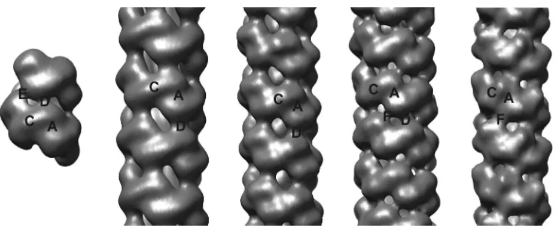
7. Spiral formation among microbial nitrilases
All the nitrilases, cyanide dihydratases and cyanide hydratases studied by us (Figure 3) form a spiral quaternary structure. While some of these enzymes form helices of variable length,
3 4 5 6 7 8 9 10 11 12 13 14 15 16 17 18 19 20 21 22 23 24 25 26 27 28 29 30 31 32 33 34 35 36 37 38 39 40 41 42 43 44 45 46 47 48 49 50 51 52 53 54 55 56 57 58 59 60
For Peer Review
others form short, terminating spirals which have a specific number of subunits (Figure 4). In particular, the cyanide dihydratase from P.stutzeri AK61 forms a 14-subunit spiral (Sewell et al., 2003), whereas the homologous enzyme from B. pumilus C1 and 8A3, form an 18- and a 22-subunit spiral, respectively (Jandhyala et al., 2003; Johann Eicher, personal communication). The nitrilases from Rhodococcus rhodochrous J1 (Thuku et al., 2007) and Fusarium solani O1 (Vejvoda et al., 2008), are also seen as short spirals by electron microscopy, however, the exact number of subunits is yet to be determined. The cyanide hydratase enzymes from G. sorghi (Woodward et al., 2008), N. crassa (Dent et al., 2008) and A. niger K10 (Vejvoda et al., 2008), occur as long, variable length helices.
There is growing evidence that the principles governing the association of monomers to form oligomers (Sewell et al., 2005) are common among this class of enzymes. As seen in Figure 4, the shape of the density which encloses the dimer, the conserved dyadic axes and the connectivity between the dimers which extends the assembly can be clearly discerned. The spiral oligomers are generally left-handed following handedness determination in other homologous enzymes (Jandhyala et al. 2003; Woodward et al., 2008). In addition, the homo- octameric spiral of the homologous, crystallized fruit fly beta alanine synthase is left-handed (Lundgren et al., 2008). Structural studies involving three-dimensional reconstruction and modeling based on the available nitrilase atomic homologues have enabled the identification of interacting regions between the subunits, which have been labeled ‘A’, ‘B’, ‘C’, ‘D’, ‘E’ and
‘F’ surfaces (Figure 4). The ‘A’, ‘C’, ‘D’ and ‘F’ surfaces are related by two-fold axes whereas the ‘E’ surface is asymmetric. Even though these regions of interactions appear to be common, the association of subunits at the different interfaces leads to the variety of oligomeric shapes that are seen in these enzymes. While the ‘A’, ‘C’, ‘D’ and ‘F’ surfaces participate in spiral formation, the ‘B’ surface does not. This surface occurs only in three crystal forms, namely the NitFhit protein (Pace et al., 2000), N-carbamyl-D-amino acid amidohydrolase (DCase, Nakai et al., 2000) and the prokaryotic XC1258 Nit protein (Chin et al., 2007), in which the subunits form tetramers with 222 point group symmetry. Use of the ‘B’ surface results in a closed point group and the basis of these interactions is hydrogen bonding along the exposed side of a beta sheet between subunits. On the basis of sequence alignment, model building and three- dimensional electron microscopy, it can be inferred that the interactions at the surfaces are generally electrostatic. The only surfaces that have been visualized at atomic resolution in the nitrilase homologues are the ‘A’ and ‘C’ surfaces. Details of other surfaces arise from docking
3 4 5 6 7 8 9 10 11 12 13 14 15 16 17 18 19 20 21 22 23 24 25 26 27 28 29 30 31 32 33 34 35 36 37 38 39 40 41 42 43 44 45 46 47 48 49 50 51 52 53 54 55 56 57 58 59 60
For Peer Review
of homology models into their three-dimensional electron microscopic reconstructions and must therefore be regarded as suggestions until they are verified by mutational analysis.
7.1 The ‘A’ surface
The association at the ‘A’ surface, involving helices 5 and 6, occurs in all crystalline and spiral structures. The surface has been frequently visualized at atomic resolution and the interactions across this surface are the basis for dimerization. The details of the intermolecular salt bridges and hydrophobic interactions in the high resolution structures vary. Thus, for example, in the G. pallidus amidase, interactions in 6 comprise a series of interdigitated methionines (M202, M203 and M207) and 5 has a pair of salt bridges formed by E173 and R176, in the case of NitFhit there are salt bridges in 6 between R211 and E214, in the Pyrococcus hypothetical protein (1j31) there are salt bridges between both 5 (E153 and R156) and 6 (R184 and E187) ( all the salt bridges are highlighted in Figure 3) and in DCase the 336 intersubunit interactions are mostly hydrophobic (Wang et al., 2001). In the prokaryotic XC1258 Nit protein (Chin et al., 2007), the 5 helix is slightly longer. An inspection of the sequences in the 5 and 6 helices of the spiral forming enzymes shows there is possible substitution of the salt bridges with hydrophobic residues. This situation is not yet clear due to lack of atomic detail in these enzymes. Additional structural elements sometimes interact across the ‘A’ surface and ‘strengthen’ it. In the G. pallidus amidase, a region of the C-terminal tail comprising four helices ( 8, 9, 10 and 11) form an ‘interlock’ with the equivalent region from the two-fold related monomer (Kimani et al., 2007). There is considerable sequence and structural variability in the tail region and generalization about the details is not possible, however, interactions in the tail which contribute to the ‘A’ surface do occur in all the known structures where the tail exists. In the case of the -alanine synthase from D.
melanogaster (Lundgren et al., 2008), the ‘A’ surface is strengthened by interactions between the two additional N-terminal helices and a pair of C-terminal helices similar to those seen in the crystalline amidases ( 9). In addition, the carboxy-terminal end of the tightly associated dimer is held in place by a parallel interaction between a pair of strands, one located in the tail region ( 16) and the other on the surface of the partner subunit ( 9). In general, the interactions at the ‘A’ surface not only hold the subunits together, but are also necessary for positioning the catalytic cysteine residue within the active site pocket. This residue is located on a 310 helix which is part of the -strand-turn-helix structural motif referred to as the ‘nucleophilic elbow’
(Kumaran et al., 2003). This motif is generally conserved in all the superfamily enzymes and is formed by the strand 7 and the 5 helix. This also provides an explanation for the loss of
3 4 5 6 7 8 9 10 11 12 13 14 15 16 17 18 19 20 21 22 23 24 25 26 27 28 29 30 31 32 33 34 35 36 37 38 39 40 41 42 43 44 45 46 47 48 49 50 51 52 53 54 55 56 57 58 59 60
For Peer Review
activity that was reported in these enzymes after modification of the interfacial residues (Sewell et al., 2005). Although the structural elements at the interface appear to be common in the solved structures, there is very little sequence conservation either in the interacting helices or in the linking regions.
7.2 The ‘C’ surface
The association at the ‘C’ surface is the key to the formation of the spiral quaternary structures.
It is located approximately at right angles to the ‘A’ surface and thus it is easy to see how the association at the ‘A’ and ‘C’ surfaces leads to an extended assembly. The details of the angular relationship between the two surfaces clearly determine the nature of the spiral which the extended assembly will form. As seen in Figure 3, there is no obvious conservation of amino acids occurring in this region. There is some evidence of conformational flexibility in this region from the structures of the fungal cyanide hydratases (Dent et al., 2008a; Woodward et al., 2008), the fruit fly -alanine synthase (Lundgren et al., 2008) and several aliphatic amidases (Andrade et al., 2007; Hung et al., 2007; Kimani et al., 2007), which leads to a variety of oligomeric structures. When homology models are fitted into the low-resolution spiral structures, there is some vacant density visible at the ‘C’ surface which suggests these residues are absent in the solved structures (Figure 5, Thuku et al., 2007). The two insertions commonly occurring in the 2 helix and the beta bend between 10 and 11 are strongly implicated in this region. Predictions of secondary structure using PSIPRED (Bryson et al., 2005) showed that the residues inserted at 2 simply extend the helix in its amino-terminal while those located in the bend between 10 and 11 form a coil which makes it difficult to make a reasonable statement about the path of these residues or their possible contribution in the ‘C’ surface. The first detailed visualization of the ‘C’ surface has come recently from the crystal structure of the -alanine synthase from Drosophila melanogaster (Lundgren et al., 2008), which clearly identifies the the major contributors to this surface that arise from three loop regions in each subunit, namely the residues between 2 and 2, 5 and 6, and 10 and 11, respectively. In particular, the residues between 10 and 11 in each subunit are seen to extend into a long coil which runs approximately at right angles to the ‘A’ surface, and interacts across the interface via two, symmetric salt bridges (E298 and K306). This region is disordered in the terminal subunits and was suggested to cause the octameric spiral assembly to terminate (Lundgren et al., 2008). Interestingly, charged residues are present in the region between 10 and 11 in the microbial spiral or helix forming enzymes, but not in identical locations according to our alignment. Nevertheless, the structure of the fruit fly beta alanine
3 4 5 6 7 8 9 10 11 12 13 14 15 16 17 18 19 20 21 22 23 24 25 26 27 28 29 30 31 32 33 34 35 36 37 38 39 40 41 42 43 44 45 46 47 48 49 50 51 52 53 54 55 56 57 58 59 60
For Peer Review
synthase provides a basis for modeling these residue insertions in the microbial nitrilases and the structural alignment (Figure 3) can be used to design experiments which explore interacting residues in this region. The interactions in this region are both electrostatic and hydrophobic and between residues which are mostly buried. Apart from the residue insertions, there is involvement of other residues arising from different parts of the molecule which possibly confer stability to the ‘C’ surface (Lundgren et al., 2008). Similarly, the docking in the helical structure of the Rhodococcus rhodochrous nitrilase places an aspartate (residue 108) located in the bend between beta sheets 3 and 4 in close proximity to a lysine (residue 289) located on 7 suggesting the possibility of a stabilizing interaction across the interface (Thuku et al., 2007). The disruption of this surface by mutation abolished the activity in the cyanide dihydratases (Sewell et al., 2005). Therefore, a study of this region will definitely reveal how steric changes are transmitted to the active site so that the enzyme is activated.
7.3 The ‘D’ and ‘F’ surfaces
The ‘D’ surface association occurs only when the spiral completes one turn. The interactions at this surface occur across the groove of the spiral or helix (Figure 4 and 5). The docking of homology models into the spiral structures unambiguously locates the helices 1 and 3 in this region (Sewell et al., 2003). These two long helices are located on the same two-fold axis as the ‘C’ surface but on the opposite side of the spiral. An interesting feature of the helices in this region is that they comprise a mixture of positively and negatively charged residues, and we postulate that two-fold symmetric, electrostatic interactions maintain the elongating assembly (Sewell et al., 2005; Thuku et al., 2007). The location of charged residues in the ‘D’ surface is generally not conserved (Figure 3), and mutating much of the region made little difference to activity in the case of the cyanide dihydratases (Sewell et al., 2005). Interestingly, crystal packing interactions in the G. pallidus amidase structure (Kimani et al., 2007) involve residues K36 in 1 and E82 in 3 helices, which interact across a two-fold axis. A further stabilizing two-fold symmetric interaction is present in the cyanide hydratases from G. sorghi (Woodward et al., 2008) and N. crassa (Dent et al., 2008). This interaction, which we have called the ‘F’
surface, occurs close to the ‘D’ surface in a region where there is a hole in other homologous enzymes (Figure 4). We have seen helical fibres in which the interactions across the groove are at the D surface only (B. pumilus CynD and R. rhodochrous J1 nitrilase), both D and F surfaces (N. crassa CHT) and the F surface only (G. sorghi CHT). The interactions occurring at the ‘C’,
‘D’ or ‘F’ surfaces dictate the helical symmetry (the rotation ( ) and axial rise ( z) of each subunit along the helical axis) and consequently, the number of dimers per turn of the helix.
3 4 5 6 7 8 9 10 11 12 13 14 15 16 17 18 19 20 21 22 23 24 25 26 27 28 29 30 31 32 33 34 35 36 37 38 39 40 41 42 43 44 45 46 47 48 49 50 51 52 53 54 55 56 57 58 59 60
For Peer Review
The enzymes with the ‘F’ surface interaction are seen to have an increased helical twist (Dent et al., 2008; Woodward et al., 2008). The atomic details of the location of the charged residues in these surfaces are yet to be visualized.
7.4 The ‘E’ surface
The ‘E’ surface interaction differs from other surfaces because its contributors are asymmetric and arise from different regions of the subunit. Interpretation of the docked three-dimensional map of the P. stutzeri AK61 enzyme (Sewell et al., 2003), suggested that spiral elongation occurs via interactions at the ‘A’ and ‘C’ surfaces until an opportunity for the ‘E’ interaction occurs across the groove of the helix. The putative electrostatic interactions which result in helix termination are between residues 266EID268 and 92RKNK95 in the P. stutzeri CynD and these may well be unique to this enzyme. The positive residues implicated in this region are located in the carboxy-terminal end of the 3 helix while the negative residues occur at the end of strand 14 (Figure 3). We speculate that the ‘E’ surface interaction generally involves the conserved negative cluster E/DID and the positive cluster R/K-R/K-X-E/K/X which is not conserved. This interaction distorts the spiral by tilting the terminal dimer such that the diameter is reduced preventing further addition of subunits and causing the cyanide dihydratse spiral to terminate (Sewell et al., 2003). In contrast, the enzyme from D. melanogaster does not form a complete spiral due to disorder in the terminal subunits (Lundgren et al., 2008). While the P. stutzeri and G. pallidus DAC521 enzymes are seen to form short terminating spiral structures, the R. rhodochrous J1 nitrilase (Thuku et al., 2007) and the cyanide dihydratase from B. pumilus C1 (Jandhyala et al., 2003) occur either as a short or long spirals. Details of the interactions which limit helix formation remain speculative and their proper elucidation is an obvious direction for future research.
8. The role of the extended C-terminal region
All the microbial nitrilases for which sequence data are available and the related crystalline amidases are seen to have an extended C-terminal sequence about 40-100 amino acids longer than that of the other homologous structures (Figure 3). The C-terminal region is located on the inside of the spiral. Several amidase structures have allowed visualization of structure in this region but the alpha helical content of the amidases seems to be far greater that of the nitrilases (as predicted by PSIPRED) and the structures may not be transferable (Andrade et al., 2007;
Hung et al., 2007; Kimani et al., 2007). Furthermore, the C-terminus in these structures lies on the outside of the hexamer. The G. pallidus amidase has 66 amino acids in its carboxy-
3 4 5 6 7 8 9 10 11 12 13 14 15 16 17 18 19 20 21 22 23 24 25 26 27 28 29 30 31 32 33 34 35 36 37 38 39 40 41 42 43 44 45 46 47 48 49 50 51 52 53 54 55 56 57 58 59 60