Their total number is 95 of which a clearly largest group (41) measures 3 to 5 mm., the rest gradmg down to less than 1 mm. Of the older corpora lutea there are a total of 6, up to 3 mm. in diameter and of an ivory-yellow colour. Eleven of the older corpora lutea can be counted, 3 to 4 mm. in diameter and of a creamy yellow colour.
Of the older corpora lutea only whitish remains, about 1 :mm. in diameter, can be found. Of the older corpora lutea there are 3 in each ovary, all 3 mm. in diameter and of a deep creamy yellow color. The color of the corpus is pale purplish wine-like. the shape is spherical and the consistency soft.
The color of the body is pale purple wine-like. the shape is spherical and the vascularization is very clear. corpora rupture points still show a raised papillae. Older corpora lutea are only found in the left ovary, up to 2 mm. in diameter and of a yellowish color. Of the follicles there are only a few under 1 mm., the largest showing brightly colored capillaries scattered over the surfaces.
Some of the older corpora lutea are up to 3 mm in diameter, the color is deep yellow, most are present as remnants.
F. BURGER
On section, all cavities are found closed in with fibrous tissue and the cut surfaces are flesh-coloured. An atrophic embryo appears on the right side with a diameter of 7 mm., the whole body is greatly congested. Only a few of the older corpora lutea are found, the largest is 2 mm., their color is yellowish.
The follicles are shell pink in the largest group, while the smaller ones are opaque white. With only three corpuscles in the right ovary, this is a clear case of embryo migration. On the right side, there is a further resorbed embryo, of which there are only whitish remains in the embryonic envelopes.
On section, all cavities are found closed with fibrous tissue and the cut surfaces are light flesh-colored. The largest follicles are seashell pink in color and the capillaries are only visible at their bases.
F. BURGER
A correct count is not possible due to the large number of small follicles of less than 1 mm.;. the largest have diameters of 5 to 6 mm. There are a total of 74 follicles, the smallest group is under 1 mm. large and the largest 5 mm. The left side also contains one atrophic embryo, 8 mm. long and without any appendages.
The older corpora lutea range in size from mere remnants to 3 mm, and their color is yellow. Only one of the older corpora lutea, 2 mm. in diameter, are found in the two ovaries, while others are visible only as small spots. The older corpora lutea are still clearly visible, up to 3 mm in size, their color is creamy yellow.
The left side of the uterus carries 7 and the right side 4 apparently normal embryos, all 2·1 em. The left side of the uterus contains 2 and the right side 1 normal embryos, measuring 2 to 2·1 em. The total number is 121, of which 26 have a diameter varying from 7 to 9 mm., which is nearly that of the mature follicles; however, they are not as blunt and glistening as they are at maturity.
Mere traces of older corpora lutea can be seen, but some reach up to 2 mm; the color is deep yellow. A number of yellow corpora are still clearly visible about 2 mm in diameter, the rest are mere remnants and difficult to count. The left side of the uterus carries 8 and the right side 7 normal embryos, all of which are 2·1 em.
Only remnants of older corpora lutea are seen, with a diameter of up to almost 3 mm; the color is orange-yellow. In addition, the left side carries 1 clearly atrophic embryo, a whitish mass of 4 x 2 mm, located in a clearly pregnant part of the cornu. A largest group of twenty are all about 6 mm in diameter, being sharply defined from the rest; the number of smaller groups is exceptionally low and there are none below 1 mm.
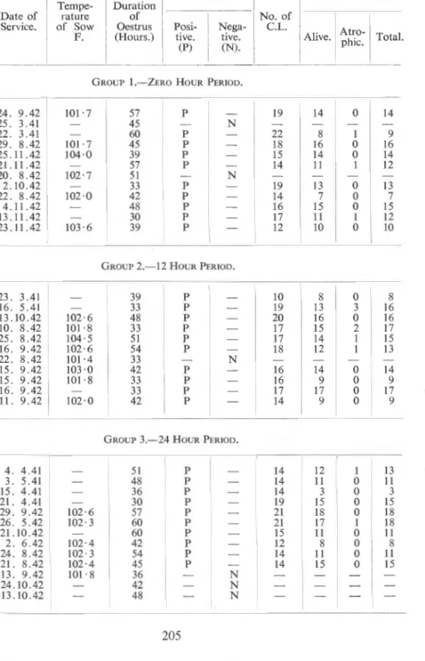
GROUP 1.-ZERO HOUR
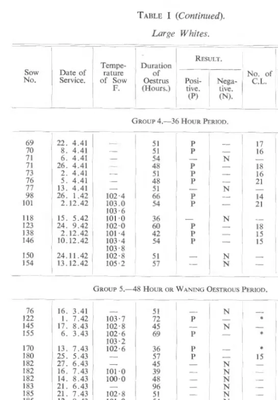
GROUP 3.- 24 HOUR PERIOD
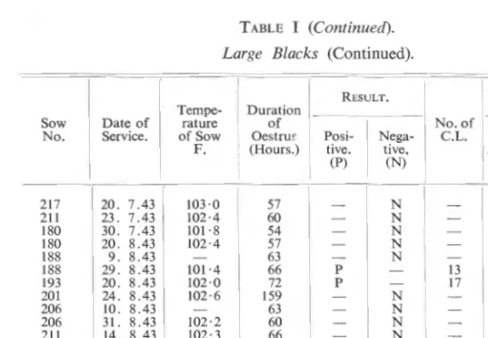
GROUP 5.-48 HOUR OR WANING OESTROUS PERIOD
F. )3URGER
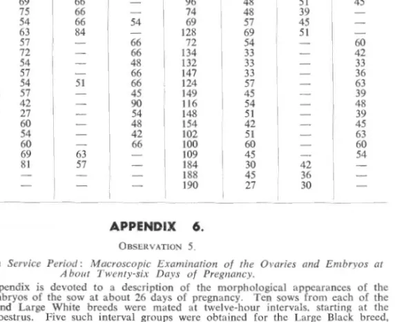
The deposition of semen by the pig in the genital tract of the sow Durin!. To determine in which compartment of the genital tract semen is deposited by the bear during coitus, four sows were serviced in oestrus and killed immediately after 1>ervice. On cutting the cervix, an uninterrupted gelatinous plug (bouchon vaginale) was exposed, firmly stuck between the rugae, measuring 16 x 2 · 5 ern.
Scrapings from the walls of the fallopian tubes were not examined microscopically for the presence of spermatozoa. Scrapings from the ovarian extremities of the fallopian tubes were examined under the microscope, revealing numerous motile spermatozoa. A gelatinous plug was found, broken into two parts, a part in the vagina, measuring 3.5 em., and a section in the cervix, measuring 5 · 5 em.
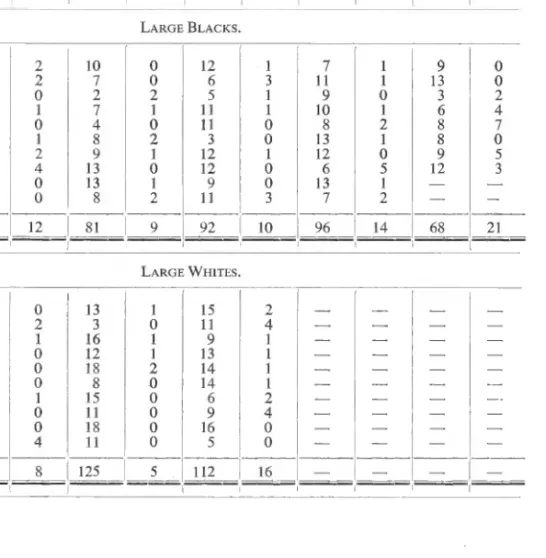
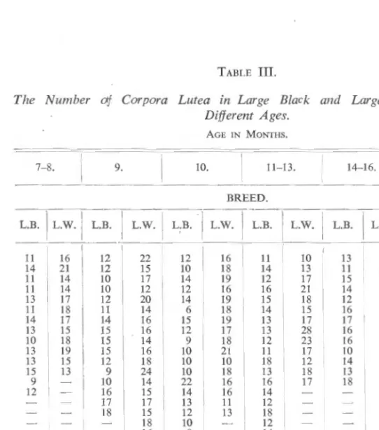
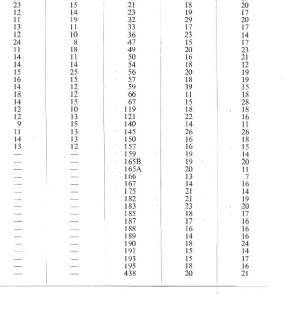
Microscopic examination of scrapings from the walls of the fallopian tubes revealed the presence of motile spermatozoa at the ends of their ovaries. Differences in basic fertility between groups of daughters of different boars, as shown by the number of Corpora Lutea.