lndust1·y, Volume 21, Number 2, March, 1947.
Printed in the Union of Sout,h Africa by the Government [printer, Pretoria.
De- the Meat Studies No. I.-Post-natal Growth and
velopment of Muscle, as Exemplified by Gastrocnemius and Psoas Muscles of the Rabbit.
By P. J. MEARA, Section of Zootechny and Meat Research, Onderstepoort.
(Accepted as a thesis in partial fulfilment of the requirements for the degree of Doctor of Veterinary Science in the University of Pretoria, Novembe1·, 1944.*)
CONTENTS.
CHAPTER 1.
INTRODUCTION:- PAGE
(a) Introduction ................... 330
(b) Object of Work ........ ' ............................ 331
(c) Acknowledgements ........................ : .. 332
CHAPTER 2. REVIEW OF LITERATURE ••••••••• 0. 0 0 • • • • 0 . 0 . 0 • • • • • • • • • • • • • • • • • • • • ••• • • • • • • • • • • • • • • • • • • 333 CHAPTER 3. PLAN OF INVESTIGATION : - (a) Animals ............ 348
(b) Housing, Feeding and Management ........ 348
(c) Nature and Scope of Experiment ...••.........•.. 351
(d) Procedura- l. Collection of data ...................... 353
2. Treatment of data. . . 365
CHAPTER 4. 0BSERV ATIONS AND DISCJUSSION : - (a) Rabbit ..................................•.......... 368
(b) Muscle- l. Weight .................................... 369
2. Length .................... · ...•....... 371
3. Width ....... 374
4. Depth. . . • . . . 382
5. Inter-relation weight, length, width, depth ........... , . . . 389
*
In preparing for publication minor changes have been made in the text. An additional chapter on relative growth by D. van der Reyden, Section of Statistics, has been included.MEAT STUDIES I.-POST-NATAL GROWTH AKD DEVELOPMENT OF MUSCLE.
(c) Muscle Bundle- PAGE
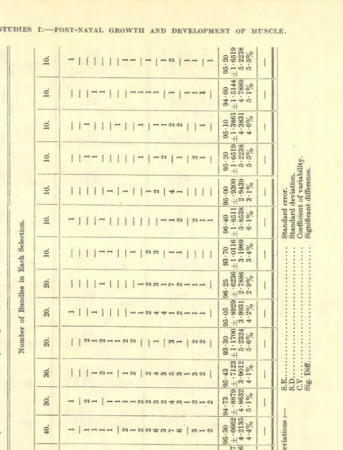
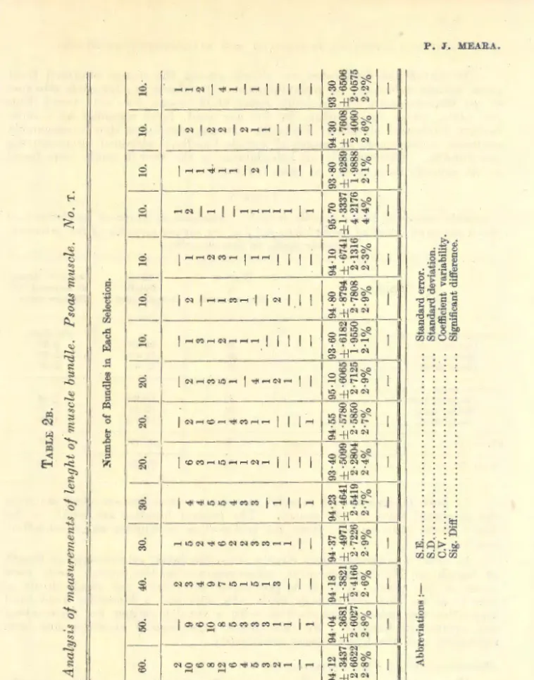
1. Length ............. : ....... ; ........................ : . . . 392 2. Number of fibres ... ·... . . 399 3. Cross-sectional area .... · ....................................... 399 (d) Muscle Fibre-
1. Length ........................................................... 408 2. Diameter. . . . . . 409
CHAPTER 5.
SUMMARY. . . 424
CHAPTER 6.
BIBLIOGRAPHY ........................................ ·. . . 426
CHAPTER 7.
APPENDIX. . . 433
CHAPTER 8.
(By D. van der Reyden, Section of Statistics, Onderstepoort).
RELATIVE GROWTH ........................................... ·. . . 455
CHAPTER I.-INTRODUCTION.
(a) INTRODUCTION.
HITHERTO the subject of meat production was approached mainly from the feeding side, and generally stopped at the digestion of the foodstuff and the body weight of the animal.
This traditional approach was broken by Hammond (1932). He made observations on the final product- meat -and worked backwards to deter- mine the conditions and factors which affect its formation. Macroscopic methods mainly, were applied in making a general survey of the scientific principles involved in the production of meat, from the physiological, anatomical and practical points of view.
Scattered throughout the literature are isolated references to micro- scopical meat studies, on small numbers of animals,"' of different species and varying uniformity. Due to the diversity of conditions under which the observations were often made, and in many cases too, due to a lack of accurate definition of procedure, these studies cannot easily be co-ordinated.
A wide field of investigation lies open to the worker who approaches the problem of meat quality from the histological point of view. Definition of the quantitative character of muscle, in terms of measurable biological entities such as muscle bundle and muscle fibre, constitutes a primary requisite for such an investigation. In addition, the qualitative changes ocGurring in meat must b~ considered in relation to the variations in the morphology of muscle.
Such a microscopic biological study will establish a basis for studying meat in the various domestic animals. It will facilitate evaluation of various factors which affect its formation. Furthermore, that elusive character, meat quality, may be brought a stage nearer to precise determi- nation when considered in terms of such study.
330
Accordingly, this work is devoted to a study of the morphological changes of muscle and its component units, during growth and develop- ment. The object is a general survey of the principles involved in muscle growth, particularly as it occurs within the individual muscle. It is hoped that these observations may suggest profitable lines of experimental work, dealing with development of muscle and meat quality.
A.lthough it would be of advantage to commence investigations on the domestic animals used for meat production, such observations would be costly and time-consuming. Preliminary observations on an animal species completing its life cycle in a short while yield information at less expense and in a shorter period of time. Such information may be of value in establishing various factors concerning growth of muscle and its develop- ment.
Small laboratory animals live under different conditions fromthe usual meat animals. Moreover, the general principles of growth may not be identical in small and large domestic animals. Nevertheless, the infor- mation obtained may serve a useful purpose, by making it easier to plan meat investigations.
Preliminary observations indicated that the rabbit was more suitable than the other laboratory animals for the purpose of this study.· Hence rabbit muscle was utilised for this work.
(b) OBJECT 01' WoRK.
The contractile properties of "voluntary muscle have been investigated almost exclusively in cold blooded animals such as the frog, mainly because such muscle may be isolated and kept alive for a considerable time. Study has largely been confined' to a few muscles such as Gastrocnemius, Soleus, and Sartorius. The physiology of warmblooded mammalian muscle is, to a large extent, interpreted in terms of the experimental behaviour of such frog muscle. This is not without difficulty. It is hardly surprising when the wide range of mammalian muscle, of varying architecture and function, is taken into consideration. Moreover, the work has been concerned with physiology (the nature of muscular contraction, its chemistry, and its efficiency), rather than morphology as such. Obviously there is still a wide field open for experimental investigation.
On the other hand, the long series of researches by Hammond (1932), and his co-workers PaJsson (1939-40), Verges (1939a, 1939b), and McMeekan (1940-41), represents , a return to the practice of the older days when animal physiology was not yet divorced from morphology ". These authors have dealt with the differential growth of constituent parts of the body in terms of muscle, fat, and bone, with the object of clarifying the biological problems involved in meat production. Muscle has been considered in terms of its proportional development in the different parts of the animal body.
As the economic value of meat depends primarily on the proportion of edible meat to inedible parts of the carcass, these workers have utilised weight as a basis for their investigations.
However, growth and development of muscle mass must ultimately depend on growth and development initiated in the micro-structure of the .muscle. Muscle is conceived as a network of connective tissue, binding together a mass of fibres which form the greater part of a muscle. Morpho- logical change of these muscle fibres must largely determine the change in morphology of the gross muscle, as well as its change in weight.
MEAT STUDIES I.-POST-NATAL GROWTfl AND DEVELOPMENT OF MUSCLE.
In the present work, the morphology of muscle has been studied, with the object of deciding how this changes during growth and develop- ment: This is necessarily the first step. The next step is to determine what growth and developmental changes occur in the individual muscle fibre, and whether they account for the coincident change in size and shape of the muscle mass. Special emphasis will be laid on the relation between the growth made during successive periods, with the object of deciding whether further research on these lines is likely to prove profitable.
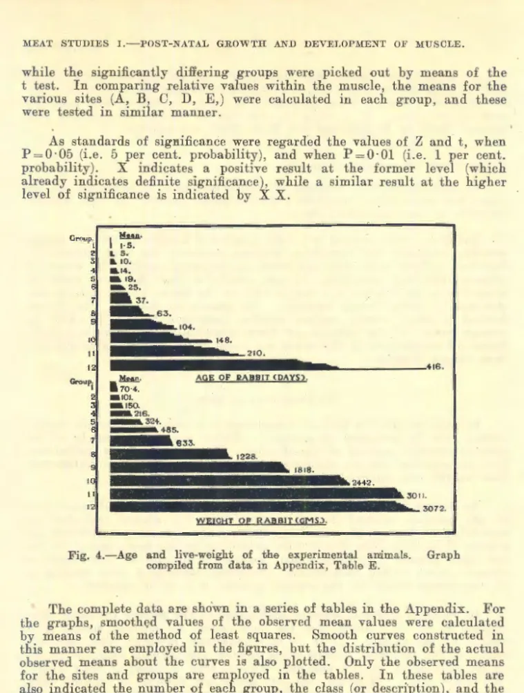
On account of the nature of the investigation, statistical treatment of the data collected is essential in order to obtain reliable quantitativ~ results.
However, the laborious character of the microscopic measurement in work of this nature places severe limitations on the extent of work which can be undertaken. This is likely to limit the value of the results by reason of the restricted scope of the investigation.
Hammond (1932) showed, that if muscles are arranged in different anatomical groups, growth follows well-defined gradients. However, individual muscles within the different groups " vary in their rate of growth and overlap in many places those of other groups ''. It follows that the behaviour of the muscle group as a whole cannot be accurately assessed from any single muscle within the group.
It can be inferred that similar difficulties are inherent in the present study, if attention is confined to a limited number of muscles. There is the danger fallacious properties may be attributed to musculature in general.
Furthermore, although a localised description of isolated muscles may show up minor variations it is not likely to affqrd any idea of the general laws
· of growth. Justification of the method lies in the fact that the present
investigation is only a preliminary step to decide whether morphological analysis of muscle growth is likely to prove a profitable avenue of meat research. Subsequently the study may be expanded to include muscular tissue throughout the animal body, in order to establish more closely the relationship between muscle growth and development, and muscle type and structure.
Morphological study o£ muscle growth may appear remote from the basic problem in mind, namely meat investigation. It is to be emphasised that know ledge regarding the structural composition of muscle affords a ready means of comparison of meat, not only from different muscles within the same carcass, but also from different carcasses of varying grade and quality.
(c) AcJ{NOWLEDGEMENTS.
It is a great pleasure to express my tha:p.ks to Dr. P. J. du Toit, Director of Veterinary Services, who provided the facilities for undertaking this study.
To Professor J. H. R. Bisschop, Section of Zootechny and Meat Research, who suggested an histological method of approach to the problem,
·thanks are due for his interest in the work and his willingness to assist
with helpful advice.
Both to Dr. G. B. Laurence and Mr. D. van der Reyden, Section of Statistics, I am particularly indebted for advice and guidance.
Dr. Laurence scrutinised the preliminary observations, and made valuable 332
suggestions. Without the guidance of Mr. van der Reyden, at the con- clusion of the experiment, it would have been extremely difficult to interpret the vast amount of data collected.
To Mr. G. Wilson-Jones, Technical Assistant in charge of the small animal section, I wish to express appreciation for his willing co-operation and assistance at all times.
My thanks are due to Mr. Theo. Meyer for the able manner in which he photographed the figures and plates. The quality of the photographic reproductions affords eloquent testimony of the efficient and conscientious manner in which Mr. Meyer applied himself to obtaining first-class results.
To Miss A. N. MacWhirter thanks are due for her assistance through- out the course of the experiment. She has been responsible for preparing all muscle specimens for measurement, as well as most of the graphs for this article.
Finally, I have great pleasure in expressing gratitude to my wife for her valuable services in the preparation of the manuscript. Besides typing a large portion of the work she also assisted in innumerable other ways.
CHAPTER 11.-REVIEW OF LITERATURE.
For convenience the literature will be discussed under headings corres- ponding with those employed in the treatment of the experimental data.
First, however, it .·is appropriate at this stage to consider briefly growth and relative growth.
By means of his autocatalytic theory of growth Robert~on (1923) holds that master reactions regulate growth cycles, repres@ted by peaks in the growth curve. However, Robb (1929) discredits this theory, as the cycles may be explained by natal and juvenile growth retardations, inevitably associated with the disturbance of birth, and of endocrine re-organisation at a later stage of life. Snell (1929) points out an inherent defect in the theory that growth rate is controlled by autocatalytic processes. Mac- Dowell, Gates, and MacDowell (19_30) also do not favour this interpretation.
Hammond (1932), and Walton and Hammond (1938) disagree with the autocatalytic theory as they show that the growth curve is· dependent on the food supply; thus, " these peaks at regular times in the life of an animal are due to the nutritive conditions usually existing at these times ''.
Recently analysis of growth has become increas{ngly directed towards the mathematical generalisation of experimental data. General formulae have been devised for describing the growth of the organism as a whole.
Although these theoretical expressions are useful for comparison and tabu- lation, there is danger in attempting to define the fundamental growth process itself from empirical formulae.
In order to understand the growth of an organism it is essential to analyse the changes in form of the organism. Huxley (1924, 1932), and Huxley and Teissier (1936), formulated a law of simple allometry which is applicable over long periods of the animal's life, after completion of the stage of histological differentiation. When the growth of a part is con- sidered in relation to the rest of the body, the relative rate o£ growth of thf' part and o£ the body remains constant. Huxley's equation expresses this law of relative growth by means of a formula y= bxa, where y equals ·the
333'
MEAT STUDIES I.-POST~:KATAL GROWTH A:KD DEVELOPllfE:KT OF MUSCLE.
part, a; the whole, b a constant representing the value ?f y when a: equals 1, and a the equili}rium constant of the part. When a 1s greater or less than unity, the part is growing more or less rapidly respectively than the whole, that is, positive or negative allometry.
For a wide variety <Jf data, this equation has been fitted to express the relation between a part and the whole, as the organism increases in size.
Cursory inspection of the literature discloses an amazing diversity of interests. [Pearsall (1927), Keys (1928), Robb (1929), Hersh (1931, 1934, 1938), Green and Fekete (1933), Needham (1932, 1934), Dawes and Huxley (1934), Lerner (1936), Rytand (1937-38), Gray and Newcombe (1938), Hamilton and Dewar (1938), Clark and Hersh (1939), Huggins (1940), Crozier (1940), Brody (1942), Kibler, Bergman and Turner (1943), Richards and Kavanagh (1943)]. The list is by no means complete, and it is intenti<lnally selective in order to emphasise the general manner in which Huxley's equation has been applied.
There is no ·doubt that the formula affords a useful method of com- paring curves of growth. Thus, instead of only being able to present a record of the differences of absolute size with age, the formula makes it possible to disclose more clearly the underlying morphological changes by showing alterations in the proportions of individual parts with increasing total size. By means of the equilibrium constant a, a measure is obtained of the relative increase <lr decrease of the part with increase in the absolute size of the organism.
On the other hand, doubts exist as to whether biologists have been sufficiently critical regarding the application of the formula, and whether the fit of their data to this equation is real or not. Discussion has taken place regarding1the implications of Huxley's formula, both from the view- point of possible shortcomings as well as its undoubted advantages [Robb (1929), Davenport (1934), Bernstein (1934), Wilson (1934), Feldstein and Hersh (1935), Richards (1936), Lumer (1936, 1939), Kavanagh and Richards (1942), Lumer, Andersen and Hersh (1942)].
Huxley makes it clear that the allometric law is limited to the growth occurring after the processes of histo-differentiation have been completed. Thus, the varying proportions of newly-born reciprocal crosses between the large Shire horse and the small Shetland pony are determined by genetic influences acting befme birth (Walton and Hammond, 1938). Although the proportions of the body subsequent to birth follow the law of allometry, nevertheless the constant _a is approximately the same for the different crosses. Similarly, Pontecorvo (1929, 1938) finds that the equilibrium con- stant a is very nearly the same for widely differing breeds of cattle. Nat- withstanding this similarity in their relative growth rate, the different breeds vary greatly in the adult conditi<ln. Apparently the initial absolute size <lf the parts and of the bod:v at birth must play a big role in determining body size and pr<lportions in later life.
Huxley also dem<lnstrates the presence of certain growth gradients where the values of the relative growth rate a, obtained for a series of parts arranged in order along the organism, change systematically from <lne end of the series to the other. Similarly, gr<lwth centres are found from which the growth intensity grades downwards, by comparing the parts of an organism on the assumption that the values of a are constant within each portion. Alth<lugh it is reasonable to assume that the. equilibrium constant
334
will vary in a progressive manner from point to point within the limits of a single part, the analysis does not take account of this continuous variation.
Such an analysis is, however, likely to yie1d a more accurate knowledge of the growth mechanism.
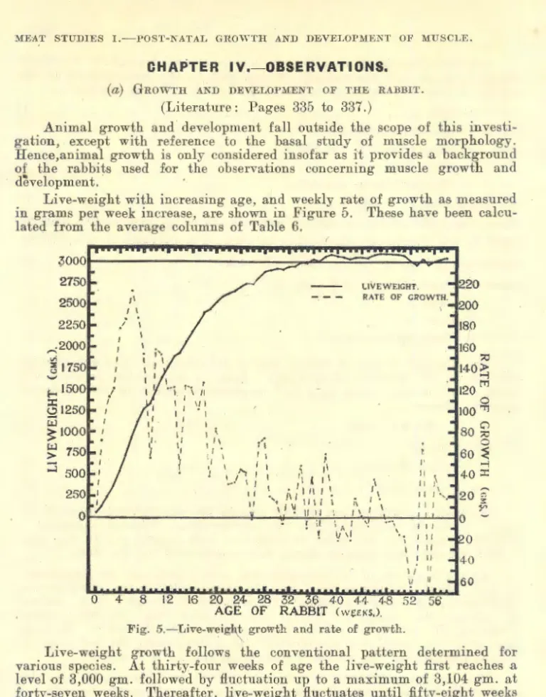
(a) GROWTH AND DEVELOPMENT OF THE RABBJT.
Post-n~tal growth has been extensively studied for most animal species.
Reference to Brody, Ragsdale and Elting (1926), or Hammond (1932, 1940a) gives ari idea of the extent of literature available.
Absolute weight increases slowly in the growing animal at first, then more rapidly. The live-weight curve for rabbits normally shows a steady rise gradually flattening with age as maturity is approached. About the period of sexual maturity it receives a temporary check. Later, an increase in weight is again noticeable in most cases and the animal subse- quently becomes somewhat heavier, largely due to deposition of fat after the cessation of growth (Punnett and Bailey, 1918; Castle, 1922; Pease, 1928).
While the animal is growing, body conformation and shape are under- going continuous change as a result of the different parts growing at different rates (Hammond, 1932; McMeekan, 1940-41). Jackson and Lowrey (1912-13) point out that, in the rat, the intensity of growth passes over the body like a wave, reaching the maximum first in the head and fore-limbs, and later passing backwards along the trunk to the abdominal portion and hind legs. Hammond (1940a) states: " In general, the wave of growth beginning at the head, spreads down the trunk, and secondary waves which start at the extremities gf the limbs pass upwards; these all meet at the · junction of the loin with the last rib, which area is the last part to develop.
Such growth gradients also exist between different tissues in the body which develop in the following order-brain, bone, muscle and fat."
Rate of growth varies in different breeds of rabbits. Large breeds usually mature more slowly than small breeds, hence, in general, the small rabbit will attain its mature weight earlier than a large rabbit (Dunlop and Hammond, 1937). Although heavy weight is closely associated with slow- ness of maturity, Pease (1928) 1.9oints out that many rabbits show conspicuous absence of thi~ association. Data have been presented showing a maximum rate of growth about 30 days after birth (Murray, 1921), sixty days (Wilson, 1930), whereas Dunlop and Hammond's (1937) large strains " E " and
" H " attained the maximum about 100 days compared with 40 days in their small strain " F ". Robb (1929) finds a distinct tendency for two peaks in the lifetime of the animal, one about 40 days after birth and the
o~her at about 100 days. These figures illustrate the differences inherent in different breeds.
It is hardly surprising that workers using different breeds in various parts of the world are not unanimous regarding the age at which the rabbit attains sexual maturity. Thus, Punnett and Bailey (1918) estimate puberty at 10 months in Polish and over 12 months in 'Flemish rabbits; Castle (1922) 6 to 7 months; Hammar (1932) 4 to 5 months; and Fangauf and Immenkamp
(1938) 5 to 7 months. ·
Earlier workers regarded mature weight as the maximum weight attained during the first year of life. However, Pease (1928) points out that this arbitrary measure has no relation to the rate of growth of the rabbit. He uses instead, the " turning point ", in his comparative growth
335
)fEAT STUDIES I.-I'OST-!'{.-\TAL GRO\YTH AKD DEVELOPMENT OF )fUSCLE.
studies. This is defined as the point where the live-weight curve for the individual rabbit slackens off at the oncome of puberty. Pease states the live-weight curve gradually rises again after the turning point, then declines, and finally rises once more to adult weight at about 400 to 500 days.
There seems to be little doubt that ao·e is not as important as weight in influencing· the normal body changes and proportions, as the magnitude of one body part tends to be a specific function of the total body mass (Robb, 1929; Huxley, 1932). In the albino rat, Outhouse and Mendel (1933) describe a close correlation between increase in weight and length. They found so little relationship to age, that body dimensions and proportions were identical in animals of the same weight irrespective of their age. Size of muscle, and organ too, is dependent on body size of animal, not age (Moment, 1933). Dunlop and Hammond (1937) show that changes in the body pron_ortion of the rabbit occur with weight rather than with age as such. For sheep too, weight classes rather than age classes at shows are suggested by Hirzel (1939), because the proportion of muscle within the sheep's body is influenced by increase in weight more than age. It follows, therefore, in planning comparative growth studies, live-weight rather than age must form the basis of comparison of normal animals. It is to be noted, however, where growth is suppressed by under-nourishment) the magnitude of an organ or system may vary markedly for any given body-weight according to the age of the animal and the general state of nutrition (Jackson, 1932).
In most species, the .male appears to be slightly heavier than the female at birth; in cattle (Hulce and N evens, 1917; Eckles, 1920); in sheep (Donald and McLean, 1935; Phillips and Dawson, 1937 ;,..Bonsma., 1939); in pigs (Carmichael and Rice 1920; Murray, 1934); in guinea-pigs, Haines (1931);
and in rats (King, 1935; Murray, 1941). As a rule the male continues to be heavier than the female, so that in most mammals the adult male is larger and heav:ier than the female. However, Kopec (1924) reports the weight of the two sexes is not essentially different in newly-born rabbits. Furthermore, Punnett and Bailey (1918) find the buck is in no case markedly heavier than the doe at maturity. Although the average weight is approximately equal in some cases, yet the doe is often markedly heavier than the buck. In the larger races of rabbits, the male has a bigger frame and is consistently larger in all bon.e measurements, nevertheless the female puts on more flesh and surpasses 'the male in weight (Castle, 1922). Castle cites breed standards for the various large breeds, in which rabbit breeders regularly specify a larger weight for females than for males~. MacDowell (1914) is of opinion that the growth subsequent to four months of age is greater in the doe than in the buck. Pease (1928) could find no difference in average weight of the two sexes, but bodyweight was nearly always more variable in does than in bucks. He suggests a heavier weight is prescribed for does by show stan- dards, because the female sex is more variable. This greater variability in live-weight of females is confirmed by Dudley and Wilson (1943). In addition, these authors find that the average live-weight of females after puberty is greater than that of males. Wilson (1930), and Wilson and Morris (1932), observed noticeable differences in the composition of male and female flesh. In general, the musculature of does at 11 months to 24 months of age contains 4 to 6 per cent. more fat than for the buck.
1t is not clear how much the growth of rabbits is affected by the seasons of the year. Under the conditions of Pease's (1928) experiment, growth rate is unaffected by the season of the year in which the rabbit is born, or in
336
which it reaches maturity. However, Wilson (1929) holds that Spring and early Summer are the most favourable periods for satisfactory growth. This is contradicted by Bertelli (1936), who shows rabbits born during Autumn obtain their complete development sooner than those born in Spring.
(b) GROWTH AND DEVELOPMENT oF MuscLE.
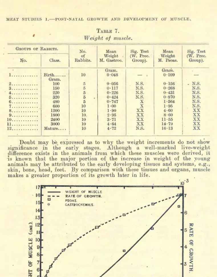
1. Weight.
Jackson and Lowrey (1912-13) cite numerous authors regarding the relative weight of skeletal musculature in widely varying species. Thus, in most adult mammals, between 40 per cent. and 50 per cent. of the body- weight is composed of muscle. Among mammals, the smallest relative weights are found in the large animals, while the largest percentage of muscle is recorded in comparatively small animals [rabbit, 49·7 to 57·2 per cent., Weiske (1895); 49·4 to 56·3 per cent., Levine et a,l (1941).]
Hammar (1932) presents evidence that the musculature of the rabbit grows most rapidly, and has its period of greatest growth about puberty.
He states muscle grows more than twice as much during the two months around puberty, as during the two months immediately preceding. Hence, the animal at puberty becomes muscularised to a striking degree.
Hammond (1932) and McMeekan (1940-41) show that, in the sheep and pig, bone makes its greatest growth in the early stages of life, followed by muscle at a later stage, while fat attains its maximum growth still later. In the rat, the skeletal musculature increases from a relative weight of 22·82 per cent. at one week old to 45·43 per c.ent. at one year of age, in sharp contrast to the skeleton\ which decreases from 18 · 4 7 per cent. to 10 · 91 per cent. (Jackson and Lowrey, 1912-13). In the fowl, the skeletal muscles increase from 21 OJ;' 22 per cent. at hatching to about 50 per cent. of the body weight in the adult, compared with a r·elative weight of the skeleton at hatching or slightly less than 16 per cent., afterwards decreasing to about 8 to 11 per cent. (Latimer, 1924). In the lmman, the relative weight of the skeleton remains practically unchanged from birth to maturity (17 · 69 and 17 · 60 per cent.), whereas voluntary muscle shows a marked increase from 24·80 per cent. in the newborn babe to 43·07 per cent. in the adult (Wilmer, 1940).
The increasing proportion of muscle with age is explained by Hammond (1932), by the greater rate of growth of muscle to bone in the different parts of the body, but also by the greater growth rate of the later maturing parts of the body which contain large proportions of muscle to bone (e.g. loin compared with head and limbs). Even after muscle has attained maximum development, there is an increas·e in inter- and intramuscular fat, which tends to increase the weight of muscle.
Hammond shows that weight of muscle, regarded as a measure, has a late period of maximum development, as it is an index of muscle and fat development. Length development is attained relatively early. Hence, because increasing muscle and fat development with age increase thickness of muscle,_ weight can also be considered as an indirect measure of muscle thickness.·
Hammond's studies make it clear that muscle groups develop serially in a definite manner, corresponding to the differential growth gradients existing between the different parts of the body. Growth waves pass Irom lower to upper limb, from the cranium backward, and from the tail forward,
337
llfEAT STUDIES I.-POST-NATAL GRO\YTH AKD DEVELOP?I!EKT OF 2\fUSCLE.
to meet in the lumbar vertebrae, so that muscle in the loin and pelvis makes the most growth post-natally. However, within each group of muscles the rate of growth of individual muscles varies greatly and overlapping occurs between groups. Hammond also shows how these normal age changes are emphasised by sex, breed, domestication, and fattening.
Hammond clearly indicates that if an individual muscle is to be taken as a sample of a carcass, it is advisable to select a muscle with a late rate of post-natal development. M. Psoas major has been studied as a physio- logical unit 'of musculature with a view to obtaining information on the musculature in general (Callow, 1935, 1936, 1937, 1938; Woodman, Evans, Callow and Wishart, 1936). 'l'his muscle has the advantage of relatively late development; moreover it can readily be removed without cutting the carcass. In the pig, McMeekan (1940-41) finds that the weight o£ the Psoas muscle has a significant correlation with the total weight of muscle in the carcass.
(2) Length.
By the time adult life is readied the long bones have achieved their maximum growth in length, the other tissues growing in proportion with the growth of the bones in length. Haines (1932) explains the growth in length of a muscle as following on the lengthening of the bones to which it is attached, in response to the traction set up within the muscle by the bone growth. This stretch, which the growing skeletal system places on the muscle, probably influences considerably the increasing strength of skeletal muscles in growing animals, as the period of greatest increase in strength coincides with the period of rapid increase in the length of the long bones (Knowlton and Hines, 1939). As muscle growth follows and is so closely dependent on bone growth, it is not out of place to digress for a moment to
consider the growth of bone. "
Hammond (1932) and McMeekan (1940-41) show that bone length reaches a maximum relatively early in life, before muscle development takes place. Hence, lengthening of-:any individual muscle must reach a maximum earlier than its growth in width and depth, which depend on the development of both muscular and fatty tissues.
With regard to the relative change in length of different muscles as the animal grows, the lengthening of muscle units in various parts of the body must be influenced by the well-defined differential growth relationship for individual bones, demonstrated by Hammond and McMeekan. In the heifer, Eckles and Swett (1918) report a greater degree of lengthening for the vete- bral column than for the hind limb (117·3 to 66·3 per cent.). The same is true of the sheep. Hammond (1932) gives measurements for Suffolk rams, from which the following table has been calculated.
Relative length .growth with age.
·Three Five Four
Months. Months. Years.
Birth.
Lumbar vertebrae....................... 100 305·6 330·0 427·8 Femur............... 100 214·8 247·9 309·9 Tibia..................... 100 197·1 224·1 278·7 Cannon........................ 100 176·5 187·9 213·6
Whereas the ruminant is horn in a relatively mature condition with long legs to follow its dam, the rabbit is comparatively immature at birth. Hence, caution must be exercised in drawing an analogy between diff.erent species.
However, for the pig, where the limb bones are not so well developed at birth compared with the sheep, the same tendency is shown by ~Ic:Me.ekan
(1940-41). The following table, compiled from his scale photographs of the lumbar vertebrae and femur, shows clearlv that the lumbar vertebrae lengthen to a relatively greater degree than the femur.
Relative length gro'wth with age.
Birth.
I
FourI
EightI
TwelveI
SixteenI
TwentyI Trenty- ~ T'~e~~y
Weeks. Weeks. Weeks.
1
Weeks. Weeks. W~~~s. ~'!ks.
It can be inferred that M. Psoas, which is closely adherent to the ver- tebral column, will show a similar difference in length g-rowth, as compar·ed with a muscle from the upper limb.
(3 and 4). Width and depth.
In general, width and thickness are late maturing body measurements (Bonsma, 1939). Latimer (1927, 1928) shows that, after puberty in the foal, there is no increase in the length o£ bone, but the bones become stouter and increase in weight. Similarly, in the rat, the adult bones ar·e wider and thicker than at an earlier stage of development (Hammett, 1924). Also in the pig, thickness growth of bone is a late developing character compared with length of bone (McMeekan, 1940-41). It is shown by Hammond (1932), that in the sheep, growth in circumference of bone, i.e., thickness, persists after bone has ceased growing in length. This author demonstrates how the growth changes in muscle groups copy, in an exaggerated form, the coin- cident changes in the bones they surround. By analogy, it can be inferred muscle width and depth increase after length has become stabilised. As muscle width and thickness are an indirect measure of muscle and fat development, i.e., weight, which is a later maturing factor than length, this is to be expected.
Hammond (1936) observed the' changes in shape of the Longissimus dorsi muscle, with increasing age of various .species of domestic animals. He shows the medio-lateral axis (width) reaches maximum development earlier than the dorso-ventral axis (depth), so that depth of muscle becomes rela- tively greater in proportion to width of muscle, as an animal becomes older.
McMeekan (1940-41) too, states that as the animal ages muscle width achieves stability, whereas depth increases at a still greater rate. A picture is presented of the muscle increasing equally in both width and thickness in the initial stages, later only by thickness growth in increasing amounts.
(c) GROWTH AND DEVELOPMENT oF MuscLE BuNDLE.
1. Technique of measurement.
Satisfactory demarcation of the bundle unit is an immediate difficulty in the morphological study of muscle.
339
MEAT STUDIES I.-POST-SATAL GRO\YTH AXD DEYELOPMENT OF :HUSCLE.
Many workers have utilised a variety of methods to measure length of muscle bundle. This will be considered in connection with the muscle fibre (pages 341-342). Accordingly, they are not mentioned at this stage.
Although bundle length may be measured fairly easily, thickness o£
bundle is not capable of rigid definition. The smallest units, the primary bundles, are formed by a number of closely adjoining parallel muscle fibres held together by interstitial connective tissue. Several primary bundles combine to form secondary bundles, secondary bundles combine to fqrm tertiary bundles, etc. A vast network of connective tissues binds together these bundles to constitute the individual muscle.
Hammond and Appleton (1932) judged bundle thickness by eye, because of the technical difficulties and labour involved in actual measurement.
Sections were cut across the grain, from samples taken £rom the middle of the muscle. These sections were then graded, according to the coarseness o£ the component bundles. Hammond and Appleton point out that sectioning may introduce artefacts, as the bundles tend to £all apart more easily in some muscles than in others. Apart from this fact, in some muscles there are large bundles which are sub-divided into a number of smaller bundles, whereas in other muscles the bundles are all small. These authors confirm Pierr:;ol's (1920) finding that in muscles of coa;rse texture each bundle includes a number of sub-bundles, whereas in muscles with fine texture the seeoudary bundles correspond with the fasciculi.
Brady (1937), and Satorius and Child (1938) obtained a measure o£
hundle thickness, by counting the number of fibres in 50 bundles from each muscle, 'and by measuring the diameter of 50 muscle fibres from each muscle.
Mc:Meekan (1940-41) counted the fibres in 20 bundles selected at random, as well as measuring 100 fibres in each muscle. ·
2. Thickness of muscle bundle (texture, " grain ").
Texture is important mainly because coarse texture is associated with tough stringy meat [Hammond 1940(a), 1940(b), 1942].. However, Beard (1924) finds that " the inherent properties of the endomysium contribute to the toughness of meat more than does the size o£ the fibre ". Although there is also a broad correlation between toughness of meat and its connective tissue content (Mitchell and Hamilton, 1927-28; Moran and Smith, 1929;
Mackintosh et al, 1936; Bate-Smith, 1942), observations by Hammond (Moran and Smith, 1929, page 42) show that the proportion of connective
ti~sue to muscle substance is considerably higher in the tender meat of foetal lamb than in the tougher meat of an adult sheep. This finding is corro- borated for the rat by Hines and Knowlton (1939). These authors calculate that the connective tissue decreases from 40 per cent. of the total muscle mass at 15 days to 15 pe:t cent. at 90 days of age. Hirzel (1939) comes to the conclusion that " evidence on tex~ure and connective tissue, their inter- relation and the effect on toughness of meat is still scarce and inconclusive ".
Muscle texture is dependent on the size of the muscle bundles, which again depends on the number and size of the fibres comprising the bundle.
Hammond and Appleton (1932) cite many authorities regarding texture of meat. Different muscles vary in texture; for example, Moran and Smith (1929) arrange beef muscles in order of increasing coarseness and toughness, from the M. Psoas (fillet), to Longissimus dorsi (rib), Biceps femoris (top- side) and lastly Semimembranosus (silverside). Muscles are fine-grained at
birth, but corresponding with the degree of enlargement of the muscle fibres, so does texture become coarser as the animal becomes older. Hammond and Appleton (1932) are of opinion that where the fibres are small, texture does not coarsen with age as much as in large-fibred muscles. Apart from differences within the animal, species differences are also evident. ln general, a large species (ox) has muscles with coars·er texture than a small species (sheep). Hammond and his co-worker show that, within a species, similar differences are present between large breeds and the smaller breeds.
The niceties of gradation of texture largely remain to be worked out.
The extremes ar·e probably represented by bundles with small numbers of fine fibres, as opposed to bundles with large numbers of thick fibres. 'fheo- retically, there is possible an enormous range of intermediate gradations and combinations-small numbers of thick fibres, large numbers of fine fibres, etc. Possibly, size of bundle as such, is less important than the coincident association of thick bands of connective tissue in coarsely grained muscle, such as has been observed by Hammond and Appleton (1932).
(d) GRoWTH AND DEvELOPMENT OF MuscLE :FrBRE.
Cobb (1925), Needham (19"2!:i), Hines (1927), Denny-J3rown (1929), and Hammond and Appleton (1932), have reviewed the literature dealing with the histology of muscle. Most of the original articles are not obtainable in this country. This is understandable, as Needham remarks on the fact that the field of muscle histology has been almost deserted since 1909, when attention became focussed on the chemistry of muscle.
1. Length of fibre.
Maximow and Bloom (1930) state that muscle fibres are entirely inde- pendent structures, of cylindrical or prismatic shape, gradually constrict- ing towards the ends and terminating in fine points. Particularly at the union of muscle with tendon, the end of the fibre may appear rounded, notched, or provided with teeth-like projections. These authors estimate the length of striated muscle fibres may vary from 1 to 41 mm. In short muscles, the fibres may continue through the entire muscle. In the larger muscles, the fibres are usually shorter than the muscle itself, and one or both ends may lie free within the muscle. ·
Huber (1916-17), working with adult rabbit muscle, dissociated single fasciculi into their component fibres. He found, in muscles with relatively short fasciculi (not longer than 2 · 5 em.), the fibres extend from tendon to tendon. In semi-pinnate, pinnate, or compound pinnate muscles, also where the distal and proximal tendons overlap, the respective fasciculi are much shorter than the muscle itself. No fibres longer than 2·5 em. were seen in the longest fasciculi teased out. In other words Huber found no fibres reaching from end to end of any fasciculi longer than 2 · 5 em. In longer fasciculi, the fibres had either one blunt tendon end and one filamentous intra-fascicular termination, or the fibres were spindle-shaped ending in hair-like processes within the fasciculus. It is noteworthy, in only two fasciculi of a number teased from the Gastrocnemius muscle, one single fibre was found which did not extend from tendon end to tendon end.
Lindhard (1929) measured Gastrocnemius fibres in the frog. He reports the fibre runs from one terminal tendon of the fasciculus to the other ter- minal tendon. It is interesting to observe differences in two species of frogs
MEAT STUDIES I.-POST-NATAL GROWTH AND DEVELOP:J.lENT OF MUSCLE.
examined. In R. esculenta fibres are bluntly conical, whereas in R. tem- poraria the fibres are irregularly cylindrical, arranged in pairs, a thick and a thin fibre alongside each other.
Denny-Brown (1929) says of the Gastrocnemius medialis muscle of the cat: " Careful dissection of the fresh muscle with a wet knife shows every fasciculus runs from aponeurosis to aponeurosis. It was further found . . . that in any particular fasciculus the fibres run from end to end of the fasci- culus. . . . No fibre was found which did not reach from aponeurosis to aponeurosis. All fibres, thick and thin alike, found their way from end to end of th_e fasciculus."
Buchthal and Lindhard (1939) give an excellent review of work dealing with the anatomy of the striated muscle fibre. They establish certain general types of fibre. Thus, cylindrical or bluntly conical fibres are com- paratively short. Long muscle fibres are flagelliform, or lanceolate, con- nected to the terminal tendons by the thick rounded end, while the tapering end is lost in the endomysium. On the average, fibres shorter than the bundle are more than hal£ the length of the bundles. 'fhin fibre-ends over- lap at varying points within the bundle. Varying numbers of elongated spindle-shaped fibres, with both ends terminating in the endomysium, furnish additional mechanical support.
Hammond and Appleton (1932) measured only thickness of fibre. They point out, however, the size of the muscle is determined mainly by the number or length of the fibres, rather than by their thickness.
As muscle fibres are often of considerable length it is difficult to measure their length under the microscope. Moreover, the fibres are intimately inter- woven and overlapped by other fibres, so that it is almost impossible to measure their length without completely isolating the individual fibres. This process is so laborious, it is incapable of routine application. Length of fasciculus, however, is more easily determined. Where fibres pass from end to end of the fasciculus, this measurement affords an idea of the fibre length. From the evidence cited, it appears that the Gastrocnemius muscle falls within this category.
2. Diameter nf fibre.
(i) Technique of. measurement.
Various methods of isolating muscle fibres for measurement of the shape and the dimensions are reviewed by Buchthal and Lindhard (1939). These authors stress the difficulty in evaluating the comprehensive histological literature concerning the muscle fibre, becau e most observations have been made on fixed and stained fibres. Diffetent methods have been applied for measuring muscle fibre diameter by various workers, and only in very few cases have attempts been made to examine living fibres.
Lindhard (1926) boiled the muscle in situ for two hours in water. After isolating individual fibres under the low-power binocular microscope, he measured uninjured fibres with the aid of an ocular micrometer.
_ Paff (1930) made camera lucida drawings, on squared graph paper, of transverse paraffin sections of skeletal muscles of the rat, guinea-pig, and cat. He computed the average area of muscle fibres, by counting the square millimetres enclosed by the drawn outlines of six hundred different fibres, making seventy-five measurements for each muscle.
Clark (1931) used stained celloidin cross-sections in order to oount the total number of fibres in skeletal muscles of the cat. The sections were pro- jected on bromide paper at a magnific.ation of seventy-five to a hundred diameters, and the fibres in each photograph were counted.
Hammond and Appleton (1932) cut free-hand shavings from a formalin- fixed strip from the middle of each muscle, and teased out the shavings on a slide in a drop of dilute glycerin. Average diameter was calculated by measuring the cross-diameter of fifty fibres by means of an eye-piece micro- · meter. The diameters of fibres in the middle and at the end of the muscle, in ten different muscles, from four animals, averaged 40 ·10,u. for the middles, and 42·37,u. for the ends. In six out of the ten muscles the ends had the slightly thicker fibres. Hammond and Appleton conclude there may be slightly more small fibres than usual, found in measurements of cross- diameters of fibres taken from the middle of the muscle.
McMeekan (1940-41) employed essentially the same method. He stained the shavings with picrie acid, and mounted them in li'arrant's solution.
Robertson and Baker (1933) macerated slender strips of fresh muscle in twenty per cent. nitric acid for two to four days. The macerated muscle fibres were washed with distilled water, and mounted in glycerin. Average diameter was calculated from the measurement of two hundred fibres. In addition, fibre size was indirectly estimated, in transversely cut sections, by counting the number of muscle fibres in an area 0·207 sq. rum. Twenty-five crorss-section areas of the muscle were counted in order to calculate the average number of fibres in a square.
\Toss (1935) utilised two methods to measure size of fibre. In a study of the leg muscles of the frog, he used a planimeter to record the area of cross-section of the fibres at a magnification of six hundred diameters. In an extensive tabulation of muscles from' the human, dog, sheep, and hedgehog, a different method was employed. Here Voss counted the number of fibres in a square millimeter of cross-section to obtain . an estimate of fibre diameter.
Brady (1937) i~olated fibres by micro-dissection. A filar micrometer was used to measure the diameter of fifty fibres. Satorius and Child (1938) followed Brady's technique. It is worthy of mention that fresh unfixed
. tissues can be examined in this way.
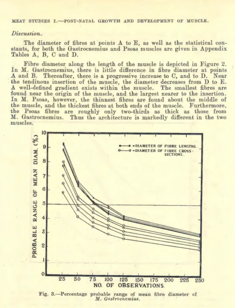
(ii) F1:bre diameter.
Perusal of the literature makes it readily apparent that the causes of differences in size of muscle fibres are a matter for speculation rather than assertion.
Hammond and Appleton (1932) find that the size of dark and of clear fibres, occurring side by side in the same muscle, varies independently of the colour of the fibres. Average size of fibre varies from muscle to muscle, but there is a more or less constant difference between the relative size of fibres in the different muscles. Denny-Brown (1929) maintains that there is no histological criterion of the speed of contraction of a muscle fibre. Although redness is generally associated with slowness of contraction, this is only a chance association with many ·exceptions. There is no relation between histological features, such as thickness or thinness of individual fibres, red- ness or paleness, and the rapidity or slowness of contraction. Voss (1935)
536~9 343
MEAT STUDIES I.-POST-NATAL GROWTH AND DEVELOPMENT OF MUSCLE.
tested the correlation between fibre size and delicacy of movement of the muscle. He believes the more delicate the motion of which a muscle is capable, the finer is the degree. of sub-division of the contractile mass.
Hammond and Appleton (1932) are of opinion that the differences in growth and development of muscles are determined by the interplay between complex factors such as evolution, function, and rate and degree of post- natal growth. Muscles used mainly for movement have on the whole smaller fibres than those us.ed for maintaining posture probably because the small size of the fibre facilitates quick respiratory exchange (small pale fibre).
Generally speaking, in the evolution of a muscle increasing in the history of a species, there is an increase in the number of fibres rather than their size, in order to increase activity and function of the muscle. With r·egard to rate and degree of post-natal growth, the earlier differentiating muscles tend to increase in size of fibre alone, whereas the latter differentiating muscles tend to be developed in number of fibres as well as size of fibre. On the other hand, in the individual after birth increase in number of fibres is not possible. Consequently, with increase of muscle function, an hyper- trophy of the fibres occurs, together with an extra supply o£ myoglobin to facilitate respiratory exchange (large red fibre).
Donaldson (1915) cites Morpurgo's (1898) data regarding the number of muscle fibres in M. Radialis of the albino rat, from which it would appear that the fibres have increased by twenty-three per cent. at fifteen days, as compared with the new-born animal. Thereafter, until 420 days of age, cell multiplication is insignificant. Schultz (1934) reports that the musole fibres of the frog increase in number with increasing age. She finds this post-natal increase proceeds more rapidly in younger than in older frogs, and continues until the number of muscle fibres is doubled. However, Hammond and Appleton (1932) state muscle growth after birth is mainly due to increase in size of the muscle cell~ although they were unable to deter- mine precisely at which stage muscle cell 'formation ceases in the sheep .
. McMeekan (1940-41) is unable to detect any increase in the number of fibres
per bundle, in pig muscles, from birth to twenty-four weeks of age. Eliot, Wiggington and Corbin (1943) find that the number of muscle fibres in M.
Soleus of the rat is not influenced by age of the animals. Concensus of opinion seems to favour this point of view, that growth of muscle occurs by hyperplasia in pre-natal life, and by hypertrophy in post-natal life (Mac- Callum, 1898; Schiefferdecker, 1919). Hence, it is to be expected that fibre diameter increases as the animal becomes older.
Apart from this thickening with age, good nutrition also increases the size of the muscle fibre. · Conversely, defective nutrition reduces fibre diameter. Thus, Robertson and Baker (1933) find that muscle fibres of full- fed yearling steers are greatest in diameter and rough-fed smallest, while fibres from half-fed steers are intermediate in size.. Black et al (1931) show that muscle fibres from steers fed a supplementary ration are slightly larger than those from steers on grass alone. Primitive breeds of sheep kept under poor nutritive conditions--semi-:.wild Shetland 45·5,u- have smaller muscle fibres than a highly improved breed reared on high nutrition-Suffolk 49·2,u (Hammond and Appleton, 1932). Similarly, McMeekan (1940-41) observes that pigs reared on a high plane of nutrition until sixteen weeks, have fibres roughly fifty per cent larger than individuals of the same breed reared on low nutritive conditions (12·08,u-8·52,u). Moreover, this difference in fibre diameter is closely related to differences in the weights o£ both pig and muscle.
344
Kremer (1930) indicates that the musculature acts as a food reservoir in the hibernating frog. In consequence, the striated muscle is altered as this reserve is used. Voss (1937) maintains that starvation decreases fibre thickness in the muscles of the frog. Greene (1912), in an extremely interesting study, obs'erve's that the king salmon stores large quantities of fat in the muscular tissues, during its life in the ocean. It ceases to take food when it enters the fresh waters of the rivers in the journey to the spawning ground. Fat is gradually removed from the muscle during the migration period, so that it has almost disappeared when the fish has reached the spawning stage. Verne (1938) reports a marked decrease in the lipids in muscle fibre during fasting. Bell (1909) and Bullard (1916) describe lipoidal granules in muscle fibres, which are increased by feeding and reduced by starvation. Denny-Brown (1929) shows an increased granu- lation in the muscle of fattened cats, whereas the granulation seems to
vanish in emaciated muscle. ·
As regards sex differences in size of muscle fibre, Eliot, Wiggington and Corbin (1943) observed no difference in size of fibre in M:. Soleus of male and female rats. However, Hammond and Appleton (1932) report that the ram has larger fibres than the ewe, and wethers have fibres inter- mediate in size. M:ehner (1938) states the muscle fibres·, from M. Gracilis and M. Sartorius of chickens, are larger in the male. On the other hand, Brady (1937) and Satoril!s and Child (1938) find that cows have significantly larger fibres than steers. It must be remarked that their experimental material comprised six Hereford-Shorthorn yearling steers and seven
~ature Holstein cows. As both age and breed are known to influence fibre diameter, it is unfair to attribute this difference to sex alone.
The effect of breed differences have been studied by Hammond and Appleton (1932). These authors show that +.he muscle fibrP.R are larger in an improved breed of sheep than those of an unimproved breed. They believe there has also been an increase in the number of fibres in each muscle in the improved breed. They cite M:alshurg (1911) to the effect 'that the heavier breeds of farm animals have larger fibrE's than the lighter breeds. Mehner (1938) confirms this finding. ·In a study of twelve races and crosses of ninety chickens, he finds the distinct racial differences in diameter of muscle fibre are almost parallel to the racial differences in body size. M:ehner is of opinion the variations in size of the muscle fibres are almost enough by themselves to account for the differences in body size.
The comprehensive literature dealing with the effect of exercise on muscle has been· reviewed by Steinhaus (1933). Various aspects of the problem have been investigated by Eliot, Wiggington and Corbin (1943), Fischer (1940), Bruman and Jenny (1936), Petren (1936), P.etren et a:l (1936), Frey (1936), Rein et al (1935), Donaldson et al (1932, 1933), Donaldson f1935 (a), 1935(b)l, Vannotti and Mageday (1934), Thorner (1930, 1934) Vannotti and Pfister (1933), and Regnault (1927). Consensus of opinion indicates that increased exercise produces increased vascularisation and hypertrophy of muscle. Steinhaus cites Siebert (1928), who states exercises of speed, strength, effort, induce hypertrophy of skeletal muscle, whereas exercises of endurance leave the body muscles unchanged ~n :"ize. Morpurgo (1897), attributes hypertrophy to true enlargement of ex1stmg fibres solely due to formation of an increased amount of sarcoplasm. Thus, there is
"no change in fibre length nor in the number of nuclei, nor the number or size of the fibrilli in the muscle cell."
MEAT S1.TDIES I.-POST-XA'IAL GUO""Tll .-L\D DEVELOl'~lEJ'\'1' OF .HUSCLE.
(iii) Colour OJ' structwre of 111/Uscle fibre.
Colour of meat in relation to breed, condition, age, sex, feedmg, management, exercise, and storage, is discussed by Hirzel (1939). In the higher mammals all muscles with few exceptions are red, but differences exiRt in the degree of redness between ~Efferent muscles, also under different environmental conditions. For example, it has been found that the thigh muscles of the sheep are paler than the leg muscles. Moreover, the redness of these muscles increases with age and activity (Hammond and Appleton, 1932; Griffiths, Vickery; and Holmes, 1932). This redness is due to the haemoglobin content (myoglobin) of the muscle (Kuhne, 1865; Whipple,
1926). .
Millikan (1939) states: " Muscle haemoglobin is generally found in large quantities in those muscles requiring slow repetitive activity of con- siderable force.'" Hammond (1942) deduces the fatigue-resisting function of myoglobin, from the dark red colour of muscles of game animals livmg an active life, such as the hare, grouse, deer. These animals have darker coloured muscles than the domesticated rabbit, fowl and sheep. Within the same animal, colour differences may be explained on a like basis. For example, in the leg of the sheep, the muscle Extensor pedis which functions continually in maintaining posture is dark red in colour (Hammond and Appleton, 1932 (p. 497). In the rabbit, Roberis (1916) believes the red muscles play a prominent part in maintaining posture and fixing joints (M. Soleus, M. Crureus, deep head of M. Triceps).
Mention has been made of the extensive use of frog muscle f()r physio- logical investigation of muscle contractility. Although this muscle is pale ::tnd unpigmented, histological study reveals the presence of granular (sar- coplasmic, protoplasm-rich) and clear fibres (aplasmic, protoplasm-poor).
Earlier workers attempted to homologise these two types of muscle fibre with the red and white muscles of birds and mammals.
Early workers studied especially the red and ''"hite muscles in the rabbit. They were inclined to homologise these two types \Yith the histo- logirally different dark and clear muscle .fibres. Morphological differen- tiation was based rhiefly on the relatively greater amount of sarcoplasm in the dark fibre, also the fact that the nuclei are not always found immediately beneath the sarcolemma as in the clear fibre. Hines (1927) c-ites Schaffer (1893), who reported also that -the clear fibres in man contain sma l1 myofibrils arranged rat her regularly, where as in the g-ranular fibres the :fibrils are large and the arrangement without order.
Although striated muscle of higher vertebrates is 1.-ed in coiour, both
<lark and clear fibres are present so that few muscles are exclusively " red "
or " white " in their make-up. Hammond and Appleton (1932), in a macroscopic examination of the leg muscles of the sheep, find all shades of colour between red and white linking up the extremes. Microscopical
~xaminat_ion of these muscles showed that the proportion of <lark, clear, and mte~med1ate :fibres varies accor<ling to the colour of the muscle, but inter- mediate :fibres are numerous in practically all muscles.
Contradictory evidence is presented reo·ardino· aa-e chano·es i11 the colour of muscle :fibres. Denny-Brown (1929) states that tbe fibres "'of the pale muscle of the new-born kitten appear dark in cross-&l"ction due to the presence of numerc;ms granules of some complex lipoidal substance. At fourteen days
~~ proportwn of fibres are clear and' by a ·continuation of" the p'rocess the