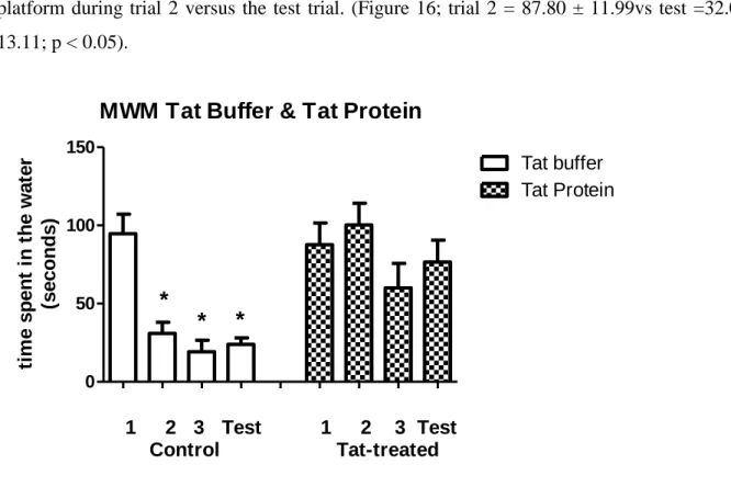
16 This graph shows the time spent by animals treated with tat protein plus picolinic acid during a three-day trial session/next day test session in a Morris water maze. Our gene expression data showed a significant increase in the regulation of the caspase 3 gene in the tat protein-treated group, compared to controls.
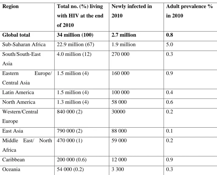
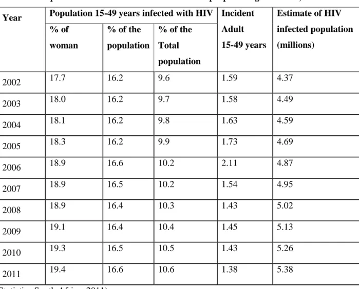
Scope of the HIV problem
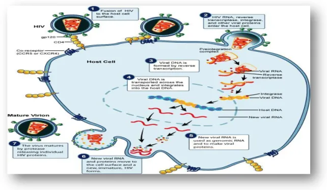
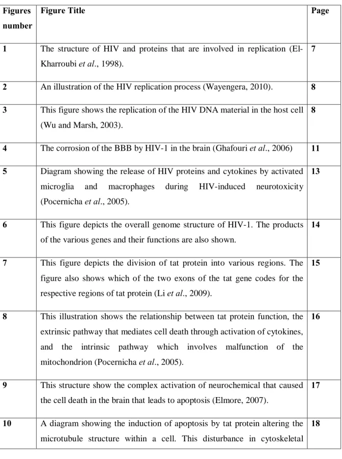
7 Figure 1: The structure of HIV and proteins involved in replication (El-Kharroubi et al., 1998). Memory loss, poor attention and visuospatial functioning have been observed in patients infected with HIV (Cohen et al., 2010).
Treatment of HIV associated dementia (HAD)
They are nimodipine (an L-type calcium channel antagonist) and memantine and nitroglycerin (both NMDA receptor antagonists) (Lipton and Gendelman, 1995; Parsons et al., 2007). Activation of the kynurenine pathway in the brain has been associated with inflammatory neurological diseases (Heyes et al., 1992).
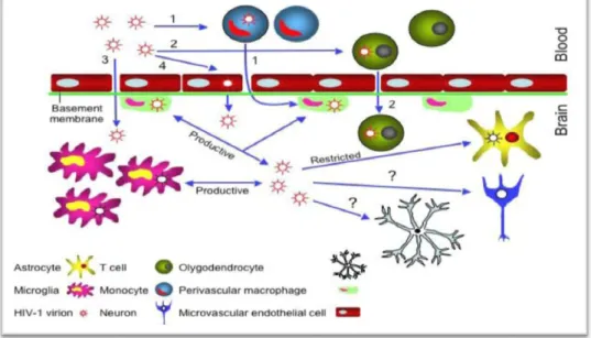
HIV-1 neurophathogenesis in the CNS
Picolinic acid is another pyridine compound that is formed along with quinolinic acid during the catabolism of L-tryptophan (Grant et al., 2009). In the brain, macrophages and microglia serve as scavengers and control cells that non-specifically remove foreign matter and secrete trophic factors that contribute to the maintenance of homeostasis in the CNS (Kaul et al., 2005). However, the sustained release of neurotrophins itself can have deleterious consequences in the brain (Rappaport et al., 1999; Kaul et al., 2005).
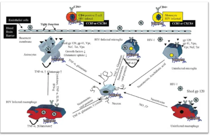
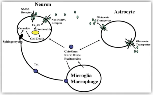
In general, the deleterious effects of these trophic factors are mediated by the activation of cytokines such as TNF-β and TNF-α (Rappaport et al., 1999). It has been proposed that this mechanism may be particularly relevant to the development of HAD (Figure 5; Kaul et al., 2001). However, astrocytes also express other HIV-1 co-receptors, such as CXCR4 and CCR5, which can facilitate virus entry ( Kaul et al., 2001 ; Ghafouri et al., 2006 ).
These neurochemical changes can result in glutamate toxicity, and the eventual death of neurons (Figure 5; Kaul et al., 2001).
The effects of HIV-1 proteins in the brain
Tat is a very basic, non-specific protein and thus has the ability to bind to many different RNAs (Ruben et al., 1989; Li et al., 2009). The Tat protein is produced early in HIV-1 infection, implying its important role in regulating the transcription of the viral genome especially through the transactivation of the TAR (Tat-associated region) (Campbell et al., 2005;. The first exon codes of the tat gene for 5 functional regions, the first region has 1-21 amino acids and is responsible for the biological activity of the tat protein.
Jeang et al., 1999; Campbell et al., 2005), while the cysteine residue at position 31 is responsible for binding to the N-methyl-D-aspartic acid (NMDA) receptor on neurons. The third and fourth regions are jointly responsible for binding and nuclear localization, and uptake of tat protein by cells (Campbell et al., 2005; Li et al., 2009). This part of the tat protein is important for transactivation, transrepression of transcription factors such as AP-1 and NF-kB, and virus replication (Li et al.
The figure also shows which of the two exons of the tat gene encodes the respective regions of tat protein (Li et al., 2009).
Apoptosis
16 Figure 8: This illustration shows the relationship between tat protein function, an extrinsic pathway that mediates cell death through cytokine activation, and an intrinsic pathway that involves mitochondrial damage (Pocernicha et al., 2005). The function of caspase-8 is to cleave procaspase-3, resulting in the production of caspase-3, one of the main enzymes responsible for the induction of apoptosis (Li et al., 2005; Pocernicha et al., 2005; Romani et al., 2010). . Caspase-3 is responsible for the activation of a nuclease known as caspase-activated DNase (CAD) or endonuclease (ENase), whose function is to cleave DNA into fragments of 180–200 base pairs, which is one of the main signs of apoptosis (Li et. al. , 2005; Elmore, 2007; Kanwar et al., 2011).
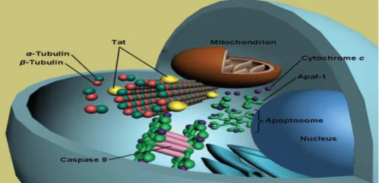
It has been proposed that tat protein can promote apoptosis by upregulating tumor necrosis factor-related apoptosis-inducing ligand (TRAIL) and eventually activate caspase-3 (Huang et al., 2005). Studies showing the tat protein to upregulate Fas, Fas ligand (FasL/CD95L), and TNF-α ( Song et al., 2006 ) supported this proposal. 18 microtubules (α and β-tubulin dimers), as well as polymerized microtubules, and this interaction can lead to mitochondrial induction of apoptosis (Figure 10; Romani et al., 2010).
This disturbance in cytoskeleton stability leads to the activation of the intrinsic apoptosis pathway (Romani et al., 2010).
Other mechanisms of tat-mediated cell death
Alternatively, tat-mediated glutamate excitotoxicity may originate from the interaction of tat protein with glutamate transporters located on glial cells. To prevent excitotoxicity, the proper function of Na+-dependent glutamate transporters or extracellular excitatory amino acid transporters (EAAT-1 and EAAT-2) on astrocytes and EAAT-3 on neurons is important to maintain a low concentration of glutamate in the brain (Figure 11; Chrétien et al., 2002; Cross et al., 2011). Tat protein has been shown to specifically derail the normal function of the cystine/glutamate antiporter through activation of stress-related signaling pathways such as p38 and p42/44 (ERK) (Gupta et al., 2010).
During HIV replication, the virus also consumes significant amounts of cysteine, which increases the activity of the antiporter and subsequently increases glutamate release into the extracellular space (Cross et al., 2011). It is interesting to note that the presence of oxidative stress has been observed in the brains and cerebrospinal fluid of HAD patients (Bottiggi et al., 2007; Romani et al., 2010). 20 Figure 11: A diagram illustrating the involvement of glutamate transporters in the regulation of the extracellular concentration of glutamate in the brain.
The diagram also indicates potential areas of interference in glutamate homeostasis during HIV replication (Cross et al., 2011).
Models to study cognitive function in rodents
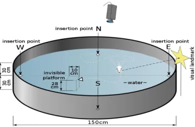
Also, the water temperature is maintained between 22-25 °C, so that the animal is not exposed to hypothermic stress when it is placed in the water. If it does not find the platform, the animal is guided to it and allowed to remain on the platform for at least 10 seconds to familiarize itself with the position of the platform in the maze. For each subsequent training session, the animal is placed in a different starting quadrant.
To perform the test, the rodent is placed in any quadrant other than where the hidden platform is located, with the animal facing the wall of the water maze. Control animals usually learn the position of the hidden platform quickly, and therefore the time period required to find the hidden platform gradually decreases with each training session. On the other hand, cognitively impaired animals struggle to learn the position of the hidden platform and, as a result, these animals require significantly longer time to locate it.
This arrangement increased the difficulty of the new path for the animal to find its way into the dark room.
Experimental design
Materials 1. Animals
These were at the following coordinates: Anterior-posterior (AP) = −3.7 mm posterior to bregma, Medio-lateral (ML) = ±2.6 mm left or right of the midline, and Dorsal-ventral (DV) = - 3.2 mm from the surface of the skull. After dura puncture, a Hamilton syringe was used to infuse tat protein into the CA1-2 region of the dorsal hippocampus. This additional time period was allowed to facilitate maximal diffusion of Tat protein into the hippocampal tissue.
Abnormalities in these aspects of memory in the rat have been suggested to reflect some of the neurocognitive symptoms of the HIV patient, such as poor concentration, slow thinking, difficulty in. The purity of the RNA was determined using a Nano-drop® ND-100 spectrophotometer (Thermo Scientific, SA) with the purity of RNA taken at the absorbance ratio A260/A280. The samples were run on the SDS-PAGE gel at 200V (400 mA) for approx. 45 to 50 minutes or until the lower marker band reached the bottom of the gel.
The Medicon tube was then placed in the Medi machine (Becton Dickinson, SA) and the tissue was homogenized for 30 seconds.
Statistics
Frozen hippocampal tissue was cut into 2 mm pieces, placed on the mesh portion of a Medicon tube, and 1 ml of PBS was added to the tissue. The homogenate was filtered through Falcon filters into Falcon tubes and centrifuged for 10 min at 4500 r.p.m. This was necessary to ensure that at least 1x10⁶ cells were present in each sample tube for analysis.
The pellet was now resuspended in 0.5 ml cytofix/cytoperm solution (Becton Dickinson, SA) and incubated on ice for 20 minutes. After that, the resuspended cells were washed with 0.5 ml of Perm/wash (Becton Dickinson, SA). Cells were then incubated with rabbit polyclonal anti-active caspase-3 antibody (Caspase 3 Kit, The Scientific Group, SA) in the dark for 30–50 min at room temperature.
The cell suspension was then centrifuged (10 min at 4500 rpm), washed with 1 ml BD Perm/wash and finally resuspended in 0.5 ml BD Perm/wash for analysis on a FACS Calibur flow cytometry machine (Becton Dickinson, SA) .
Results
31 Figure 14: This graph shows the time spent by tat buffer (control) and tat protein treated animals moving from the light compartment to the dark compartment of a light/dark box. In contrast, rats treated with tat protein showed poor learning and memory abilities. Tat protein-treated rats that were also given picolinic acid showed partial recovery in their learning and memory abilities when tested in the Morris water maze (Figure 16).
33 Figure 16: This graph represents the time spent by animals treated with tat protein plus picolinic acid during a 3-day trial/next day test session in a Morris water maze. Real-time PCR was used to determine the effects of tat protein on the transcription levels of the caspase 3 gene in the hippocampus. Our data showed that injection of tat protein led to a significant increase in caspase 3 mRNA levels compared to the tat-buffered control group (Figure 17).
This increase was not observed in the tat protein group treated with picolinic acid, which showed caspase 3 mRNA levels similar to control animals (Figure 17).
In our study, we then focused on the activity and expression of caspase 3, as this is one of the executioner enzymes that is central to apoptosis.
GENE
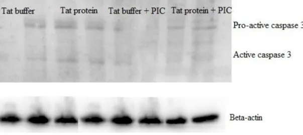
Figure 18: Representative Western blot of 3 experiments showing pro-caspase 3 and active caspase 3 staining in animals treated with tat buffer, tat protein, tat buffer plus picolinic acid, and tat protein plus picolinic acid. Flow cytometry techniques were used to assess the intracellular expression of caspase 3 in hippocampal cells. However, statistical analysis of the averages of the results from the 3 animals showed that these increases were not significantly different (Figure 21). a) b).
Tat protein-treated group, c) Tat buffer plus picolinic acid, and d) Tat protein plus picolinic acid.
Activity
WHO/UNAIDS/UNICEF (2011)