Page | 1
Chapter 1 INTRODUCTION
Aquatic environments have been grossly polluted by heavy metals in recent times.
Thus heavy metal contamination of aquatic environments has become a problem of great concern to man. This situation has arisen as a result of the rapid growth of population, increased urbanization and expansion of industrial activities, exploration and exploitation of natural resources, extension of irrigation and other modern agricultural practices as well as the lack of environmental regulation (Idowu, 2004).Environmental pollution is the universal problem and most important pollutants are the heavy metals (HMs) in aquatic network because of their toxicity, accumulation and bio-magnification by aquatic organisms. Among the pollutants, HMs consist a major portion. The HM pollutions have dreadful effects on the environmental equilibrium and a variety of aquatic entities (Akinmoladun AC, Ibukun E, Afor E, Obuotor E, Farombi E ,2007; Vosylien AMZ, Jankait AA ,2006). The pollutants like HMs gradually accumulate in food chain and cause the antagonistic effects, even death. Generally, accumulation relies on metal concentration, time of exposure, way of metal uptake, environmental conditions (water temperature, pH, hardness, salinity) and intrinsic factors (fish age, feeding habits)(Jezierska B, Witeska M ,2006).Commonly encountered HMs is chromium, cobalt, nickel, copper, zinc, arsenic, selenium, silver, cadmium, antimony, mercury, thallium and lead (Abdul rehman FI, Akan JC, Chellube ZM, Waziri M ,2008).
Heavy metals from natural sources and anthropogenic activities are continually released into aquatic systems, causing serious threat because of their toxicity, bioaccumulation, long persistence and bio-magnification in the food chain (Eisler, 1988). Fish are considered as one of the most indicative factors, in freshwater ecosystems, for the estimation of trace metals pollution (Rashed, 2001).Fish are at the higher trophic level of the food web and may absorb large amounts of heavy metals from the water and often in concentrations several times higher than in the ambient water. Heavy metals are taken up through different organs of the fish because of the interconnection between them. In this process, many of these heavy metals are concentrated at different levels in different organs of the fish body (Rao and Padmaja, 2000; Bervoets et al., 2001). Heavy metals like copper, iron and zinc are vital for fish
Page | 2 metabolism, while some others such as mercury, cadmium and lead have no known function in biological systems. For normal metabolism the essential metals must be taken up from water or food, but excessive intake of the essential metals can produce toxic effects (Yousafzai, 2004). Studies from the field and the laboratory experiments reveal that accumulation of heavy metals in fish is mainly dependent upon metals concentration in ambient water and exposure period, although some other factors such as water salinity, pH, hardness and temperature, ecological needs, size and age, life cycle, capture season and feeding habits of fish also play significant role in metal accumulation (Canli and Atli, 2003). The pollution of aquatic resources with a wide range of contaminants has become a matter of concern over the last few decades
Natural aquatic environments are heavily polluted with heavy metals emitted from domestic, industrial and other anthropogenic activities. Heavy metal contamination may have harmful effects on the ecological balance of the recipient environment and a diversity of aquatic species. Fish are widely used to estimate the status of aquatic ecosystems because pollutants build up in the food chain and are responsible for adverse effects and death in the aquatic systems. Studies carried out on various fishes have shown that heavy metals alter the physiological activities and biochemical parameters both in tissues and in blood (Basa and Usha Rani, 2003; Canli, 1995).
Bangladesh is one of the world's leading fish producing countries with a total fish production of 42.77 lakh MT in FY 2017-18, where aquaculture production contributes 56.24 percent of the total fish production. Aquaculture shows a sturdy and consistent growth average growth rate is nearly 10 percent during the same timeframe. It is assumed that if the increasing trend of fish production continues, it will be possible to attain the estimated production target of 45.52 lakh MT by 2021 in conformity with the targets of Vision-2021of the current Government. After 48 years of independence, Bangladesh becomes a self-reliant country in fish production, with a per capita fish consumption of 62.58g/day against set target of 60 g/day. According to FAO report The State of World Fisheries and Aquaculture 2018, Bangladesh categorized 3rd in inland open water capture production and 5th in world aquaculture production. Recently Bangladesh ranks 4th in tilapia production in the world and 3rd in Asia. Hilsa which is known as national fish has been making the highest contribution (around 12 percent) to the country’s total fish production as a single
Page | 3 species. Geographical Indication Registration Certificate has been achieved for our national fish Hilsa.
It is known to all that ponds are very important water bodies in the country. The total pond fisheries production is 797851mt (DoF, 2018) where in Rajshahi district the total fish production is 67093 mt (DoF 2018). The roles of these ponds are very functional for freshwater aquaculture. Most of the research works have been focused on the determination of water quality like physic-chemical or biological parameters.
But very little works have been done to generate data on the others components such as heavy metal in fish ponds. Some related works in this point were done by Jop (1980), Manga (1983), Gaigher and Krause (1989), Tariq et al. (1996), Ehshan et al.
(1997), Kabir et al. (1997), Hossain (1998), Saha and Hossain (2002) and Javed (2003). So, it is necessary to evaluate water pollution status and its effect on fisheries resources of pond considering the adverse effects of municipal sewage, industrial effluents on water quality.
During the past several decades, the increasing usage of heavy metals in industry has led to severe environmental pollution through effluents and emanations. Heavy metals are of particular concern because of their toxicity and tendency to be bio accumulated in aquatic ecosystems, as well as persistence in the natural environment. Among the different metals analyzed cadmium (Cd), lead (Pb), mercury (Hg), chromium (Cr) and arsenic (As) are categorized as chemical hazards and maximum residual levels have been prescribed for humans. Fish are one of the most significant and the largest groups of vertebrates in the aquatic system. Trace metals can be accumulated via both food chain and water in fish. Fish have been considered good markers for heavy metal contamination in aquatic systems because they live in different trophic levels with different sizes and ages .In the meantime, fish are widely consumed in many parts of the world by humans and polluted fish may endanger human health.
ATPase are a group of enzymes that catalyze the decomposition of ATP into ADP and a free phosphate ion or the inverse reaction. ATPase found in the plasma membrane of all animal cells. It carry out several functions in cell physiology.
ATPase are the membrane-bound enzymes responsible for the transport of ions through biological membranes and thus regulate, e.g., cellular volume, osmotic
Page | 4 pressure, and membrane permeability (Sancho et al. 2003). Alkaline phosphatase (ALP) catalyzes the hydrolysis of different physiologic and non-physiologic phosphoric acid esters in alkaline medium (pH optimum 10). ALP is used to assess the integrity of plasma membrane and endoplasmic reticulum. Liver and biliary tracts are the sources of alkaline phosphatase. Phosphatase is a hydrolytic enzyme that causes ortho-phosphate to be released from the phosphorus compound. Any change in acid and alkaline phosphatase activities can affect the metabolism of the fish. In fisheries sciences, changes in phosphatase activities have been regarded as indices of growth, illness and spawning of fish (Goldemberg et al., 1987; Matusiewicz, M., Dabrowski, K. 1996).
Although 260 freshwater indigenous fish species were documented in Bangladesh only a few species are cultured. Traditionally, the culture of both indigenous and exotic fish species has been practiced in Bangladesh. Tilapia and Thai Pangus has become popular in Bangladesh over the last decade. The culture of tilapia and Thai Pangus has been growing well and those are commonly referred to as high value fish species in Bangladesh. Tilapia provides us 381215MT production and Thai pangus provides 453383MT production on this year. Tilapia has great role in terms of food security and nutritional benefit. Nowadays tilapia is being popular among consumers because of taste and less bones making it easier to eat. Tilapia are highly preferred by all religious, social and economic groups in Bangladesh. The Pangus aquaculture in Bangladesh has been emerged with an exotic species P. hypophthalmus also known as
‘Pangus’ or ‘Thai Pangas’.Pangushas been found cheaper protein source than other fish, meat and dal (lentil soup). This indicates protein rich food are being easily available by poor people that produced within their community. In order to conduct this research successfully, these two commercially important fish species are selected.
Considering this issue, two commercially valuable fish species were collected from four well known farms located in the Chattogram district. Heavy metals taken up by an organism are dispersed to different organs of the fish, because of the chemical assemblage between them. The research was conducted to know about the accumulation of heavy metal in different organs of fishes and their affinity to accumulate on different organs. Heavy metals when undergo metabolic activation provokes a cellular alteration in the affected fish. A simulation study was designed to
Page | 5 understand the enzymatic activities (ATPase and ALP activity) in the investigated organs of cultured Tilapia and Thai Pangus. Therefore, objectives of the current research can be narrated as:
To develop a comparison of heavy metal accumulation in different organs (kidneys, livers, gills and muscles) of examined fishes.
To compare the levels of heavy metal accumulation among the two investigated fishes.
To evaluate the ALP and ATPase activities among those investigated organs of these two fishes.
Page | 6
Chapter 2
REVIEW OF LITERATURE
Before conducting an experimental research procedures, it is important to have a look on the previously conducted research activities on the related topics. So a review of the literature relevant to the present research work has been given below:
2.1 Heavy metals
Heavy metals are the metallic chemical component with relatively high density but toxic or poisonous at low level. Mercury (Hg), thallium (Tl), arsenic (As), cadmium (Cd), chromium (Cr), and lead (Pb) are some heavy metals. Heavy metals are natural constituents of the earth's crust. These metals do not get degraded or destroyed. Heavy metals are hazardous because they accumulate bio in organisms. Heavy metals deposit in living things by taking up as food and accumulated faster than they are metabolized or excreted.
2.2 Toxic heavy metals
Toxic heavy metal is any comparatively dense metallic compound that’s noted for its potential toxicity, mostly in environmental contexts. The term has specific application to cadmium, lead, mercury, and arsenic all of them seem within the World Health Organization's list of ten chemicals of major public concern. Heavy metals become poisonous when they are not dissolved by the body and accumulated within the soft tissues. Heavy metals may enter the human body through food, water, air, or absorption through the skin when they come in contact with humans in agriculture and in manufacturing, pharmaceutical, industrial, or residential settings.
2.3 Fish as a bio-indicator of heavy metal pollution
Bio indicator that says that is an organism or part of an organism and a community of organisms containing a series of information on the quality of the environment or its parts. Fish are considered the key indicators of heavy metal enrichment in aquatic environment. Metals are shifted to the fish through food chain that could eventually impact on the health of people who are consuming this fish. They are known for their persistent toxicity and affinity to bio-accumulate in aquatic food chain.
Page | 7 2.4 Route of exposure, bio-uptake and bioaccumulation of heavy metals
Human beings can come into direct contact with heavy metals by eating contaminated foodstuffs. The contamination chain of significant metals typically follows cyclic order: from manufacturing to the atmosphere, soil, water and foods then human.
There are several roads that can take up these heavy metals. Some heavy metals such as lead, manganese, cadmium and arsenic can enter the body through the gastrointestinal route; that is, through the mouth when eating food, fruits, vegetables or drinking water or other beverages. Most significant metals are distributed within the body through blood to tissues. Lead is carried by red blood cells to the liver and kidney and subsequently decentralized to the teeth, bone and hair mostly as phosphate salt. Arsenic is spread throughout in blood and accumulates in liver, kidney, lung, brain, muscle and neural tissues as well as in the skin, nails and hair.
2.5 Health effects of heavy metal toxicity in humans
The toxicity of heavy metals have several health effects in the body. Heavy metals can affect or change the functioning of organs like the brain, kidney, lungs, liver, and blood. Toxicity of heavy metal can either be acute or chronic effects.
Arsenic
Exposure to arsenic can result in either acute or chronic toxicity. Acute arsenic poisoning can damage blood vessels, gastrointestinal tissue and can also affect the heart and brain. Chronic arsenic toxicity known asarsenicosis, usually focuses on skin manifestations including pigmentation and keratosis. Long-term exposure can lead to the formation of pulmonary disease, skin lesions, neurological problems, cardiovascular disease, peripheral vascular disease, diabetes mellitus, and hypertension
Lead
Lead poisoning known as toxicity due to lead exposure. Lead poisoning in children and adults is mostly concerned with the gastrointestinal tract and central nervous system. Lead poisoning may be either acute or chronic. Acute exposure of lead can cause loss of appetite, headache, abdominal pain, sleeplessness, hallucinations, vertigo, renal dysfunction, hypertension, fatigue, and arthritis while chronic exposure can result in mental retardation, birth defects, autism, brain damage, coma and may even cause death.
Page | 8 Chromium
Chromium is the most toxic element in its hexavalent form. Chromium (III) compounds are much less toxic and cause little or no health problems. Chromium (VI) appears to be corrosive and also to cause allergic reactions to the body. Exposure of tremendously high doses of chromium (VI) compounds may lead to severe cardiovascular, hematological, renal, hepatic, gastrointestinal, respiratory, and neurological effects and possibly death.
2.6 Environmental impacts of heavy metals
The existence of heavy metals in the environment leads to a number of adverse impacts. Such impacts affect all spheres of the environment, that is, hydrosphere, lithosphere, atmosphere and biosphere.
Effects on soil
Discharges from activities and sources such as agricultural operation, industrial activities, leaded gasoline and paints, mine tailings, disposal of high metal wastes, sewage sludge, pesticides, land application of fertilizers, animal manures, waste-water irrigation, coal combustion residues and spillage of petrochemicals result in soil contamination by heavy metals. Most heavy metals are not subject to microbial or chemical break down because they are non-degradable, and thus their total concentrations last for a long time after being released to the environment.
Effects on water
Heavy metals can be found in traces in water sources and are still highly toxic and possess serious health problems to humans and other ecosystems. The toxicity level of a metal depends on factors such as the organisms that are exposed to it, its existence, its biological function and the duration of exposure of the organisms to the metal.
Food chains and food webs represent the interactions amongst organisms. Therefore, the pollution of water by heavy metals actually affects all organisms.
Effects on air
Industrialization and urbanization which occurs due to rapid world population growth, have recently made air pollution as a major environmental concern around the world.
Natural processes that release particulate matters into atmosphere include soil erosion, volcanic eruptions and rock weathering, while anthropogenic activities are more industrial and transportation related.
Page | 9 2.7 Related work done
Over the last few decades, there has been growing interest in determining heavy metal levels in the aquatic environment and attention was drawn to the measurement of contamination levels in public food supplies, particularly in fishes (Kalay et al. 1999).
The levels of heavy metals in fish have been extensively studied in different parts of the world over the few past decades. Most of these studies focused on the heavy metals in the edible part (fish muscles). However, other studies reported the distribution of metals in different organs like the kidneys, liver, heart, gonads, bone, digestive tract and brain.
Afshan et al (2014), conducted a study aimed at testing the accumulation and concentration of heavy metals in various organs of cultured fishes that come into contact with the water polluted with the heavy metals. The subjected fish were exposed to cadmium (Cd), zinc (Zn), chromium (Cr) and lead (Pb) at sub lethal levels.
Heavy metals entered in bodies of fishes by three potential ways: by gills, by body surface and digestive track. The calculated deposition of heavy metals in the liver and gills were in order of Pb> Cd > Ni > Cr and Cd >Pb> Ni > Cr. Similarly, the order in case of kidneys and flesh tissues was and Pb> Cd > Cr > Ni and Pb>Cr > Cd > Ni. In Cyprinus carpio(Common carp), the level of cadmium and lead was intentionally raised in tissues as compared to the other heavy metals.
Shahid M et al. (2016), evaluate the effect of heavy metals on a substantial tissue of two fish species Cyprinus carpio and Wallago attu, collected from the Indus river, Mianwali District, Pakistan. The concentration of selected heavy metals Fe, Cr, Cu, and in gills, muscles, liver and kidney was compared with an International standard of food fish. In W. attu the overall accumulation of these metals were, in order of Fe >
Cu > Cr >Pb. The overall metal concentrations among different weight categories in C. carpio were in the order of Fe > Cu > Cr>. The order of accumulation of metals in gills and muscle of C. carpio was Fe > Cr >Pb> Cu; kidney and muscles of W. attu was Fe > Cr > Cu >Pb; liver Fe > Cu > Cr >Pb. An increasing trend of concentration of copper, iron, chromium and lead occurred with an increase in weight of C. carpio and W. attu. There was a significant difference (p<0.01) in the accumulation of heavy metals in different organs of both species. All studied heavy metals excluding Cr were
Page | 10 within permissible limits described by various international agencies like WHO, FAO and FEPA in edible tissues of C. carpio and W. attu.
Hossain et al. (2016), determine the concentration of heavy metals in some commonly used marketable fish feeds and to observe the bioaccumulation of Cu, Cr, Cd and Ni in muscle, liver and gills of tilapia Oreochromis niloticus after culturing them for 60 days by feeding those commercial feeds. The study revealed that the concentration of Cu was the highest (65.08 mg/kg)) among four heavy metals in handmade feed (B1).Cr concentrations in collected feeds were 1.75 to 3.04 mg/kg, which exceeds the permissible limit set by FAO. For cultivated tilapia the concentrations of studied heavy metals were found higher than initial concentration in fish feeds and in tilapia fingerlings. Metal levels in cultured fish followed the order of Cu>Cr>Ni>Cd and ranking in individual organs was liver>gill>muscle. The maximum concentration of Cu (72.86 mg/kg) was found in liver receiving S feed and the minimum concentration (0.67 mg/kg) was in muscle given Q feed. Bioaccumulation of Cr was the highest (23.95 mg/kg) in liver taken B1feed and the lowest (9.29 mg/kg) in muscle of tilapia cultivated with C feed. Level of Cu exceeded the tolerable limit in fishes reared with S and C feeds. But Cr concentration exceeded the allowable limit in every feed studied. The concentrations of Ni and Cd were below the acceptable range approved by FAO. Present study shown that tilapia cultured with these experimental feeds is unsafe for human consumption.
Ahmed et al.(2009),studied heavy metal(As, Pb, Cr, Ni, Hg and Cd) concentrations in various organs(intestine, liver, gill, scale and muscle from different portions of body along with whole body) of three fish species (Channa striatus, Glossogobius giuris and Clupisoma garua) of Fishes of the River Meghna, Bangladesh. Among all the measured heavy metals Pb was highly concentrated in different organs of fishes.
Cumulative mean concentrations of heavy metals in different organs of the analyzed fishes were observed in the order: liver > intestine >gill > scale > muscle. Two age groups of G. giuris were also studied to find out the variation of heavy metal concentrations within age group (3-to-4 and 7-to-8 months’ old); except for Hg, the pollution level was higher in the tissues of the younger age group compared to the older group.
Page | 11 Yousafzai A M et al. (2010), examined Ni, Zn, Cu, Cr, Cd and Pb in the intestine, skin, gills, liver and muscle of two fresh water fishes, Wallago attu and Labeo dyocheilus. The goal of the study was to apprehend the metal accumulation pattern of two species occupying different feeding areas in the same habitat. Metals amassed in the order Zn>Cr>Cu>Pb>Ni>Cd in the body of Wallago attu. Metal exorbitance in different organs of this fish was skin>gills>muscle>intestine>liver. Correspondingly, the sequence of metal accumulation in the body of Labeo dyocheilus was Zn>Cr>Cu>Pb>Ni>Cd, while metal abundance in various organs of this fish was in the series liver>muscle>skin>intestine>gills. The sequence of metals bioaccumulation in both the species was different, but in both species Zn was the highest and Cd was the least accumulated metal. Generally, Labeo dyocheilus accumulated 65.2% more heavy metals burden as compared to Wallago attu. Our results suggest that omnivorous fish may bio accumulate extra heavy metals than the carnivorous fish in natural habitats.
Udotong J I R (2015), studied diagnostic enzymes such as aspartate aminotransferase (AST), alanine aminotransferase (ALT) and alkaline phosphatase (ALP) in Tilapia guinensis as indices of heavy metal contamination. For the study, three different sets of fishes treated with lead (Pb), iron (Fe) and copper (Cu) were used while a fourth group with no heavy metal served as a control. Fishes in each of the groups were exposed to 2.65mg/l of Pb, 0.85mg/l of Fe and 0.35 mg/l of Cu in aerated aquarium for 96 hours. Liver tissue fractionation was performed and the three diagnostic enzymes (AST, ALT and ALP) were determined. Serum levels of the same diagnostic enzymes were also analyzed. The mean serum enzyme activity values for ALP in each experimental group were 19.5±1.62, 29.67±2.17 and 1.15±0.27 IU/L for Pb, Fe and Cu groups compared to 9.99±1.34 IU/L enzyme activity in the control. This outcome showed that Pb and Fe caused increased release of the enzyme into the blood circulation indicating augmented tissue damage while Cu caused a decrease in the serum level as compared with the level in the control group. The mean values of enzyme activity found in the liver were 102.14±6.12, 140.17±2.06 and 168.23±3.52 IU/L for Pb, Fe and Cu groups, correspondingly compared with 91.20±9.42 IU/L enzyme activity for the control group. The serum and liver AST and ALT activities obtained in Pb, Fe, Cu and control groups are identified. It was commonly found that
Page | 12 the presence of the heavy metal caused damage to liver tissues and thus increased level of the diagnostic enzymes in the serum.
Atli G, Canli M (2007), performed a study on enzymatic responses to metal exposures in a freshwater fish Oreochromis niloticus. Freshwater fish Oreochromis niloticus were treated with 5, 10 and 20 μm concentrations of Cu, Zn, Cd, and Pb for 14 days and responses of several enzymes were estimated subsequently. Liver catalase (CAT) activity was influenced by Cd and Pb exposures while Zn exposure was inhibited.
Copper did not cause significant changes in CAT activity. The activity of liver alkaline phosphatase (AP) was initially stimulated at lowest (5 μm) exposure level, whereas there were significant inhibitions at higher (10 μm) exposure concentration.
AP activity at the highest (20 μm) exposure concentration was compensated coming to the control level, except Pb exposure. AP activities of intestine and serum were stimulated by all Zn exposures and 10 μm Cu exposure, while other exposures did not cause substantial changes on ALP activity.Na2+,K+ATPase activity in the gill and intestine was obstructed by all the metal exposures, except 20 μm, Pb exposure that resulted in an increase in the activity in the gill. Likewise, muscle Ca2+ ATPase activity was clogged by all the metal exposures, except Cu exposures. This study demonstrated that enzymatic activity may be used as a sensitive bio-indicator of metal contamination in aquatic environment.
Ahmmed M K et al. (2017), determine biochemical changes a few important physiological parameters were observed after 90 days of exposure. Level of glycogen in liver and muscle in the fish cultivated at 9 ppt salinity reduced significantly (P <
0.05) as compared to the control. Glucose level in blood and liver was also found to be raised in fish with raised in salinity. The activity of ALP and ATPase were reduced significantly in both muscle and liver tissues at higher salinity, indicating the stress mitigation impact. However, all the biochemical factors were found in normal condition up to 6 ppt compared to control. This evidence indicates that H. fossilis can withstand and grow well below 6 ppt and can be a potential candidate for coastal cultivation after heavy downpour when the salinities level drops to 6 ppt or lower.
Page | 13
Chapter 3
MATERIALS AND METHODS 3.1 Sample collection
Four fish samples for each species were netted with the help of the local fishermen from the pond of different region of Chattogram district. Fish samples were shifted to ice box and were transported to the laboratory of Applied Chemistry and Chemical Technology Department where fish were washed with distilled water and stored in plastic bags at -20º C until dissection and will be marked accordingly.
.
Plate 1: Sampling area
Page | 14 Plate 2: Sample storage Plate 3: Sample weighing
Plate 4: Measuring length Plate 5: Labelling
Page | 15 3.2 Dissecting and preserving of fish sample
Fish were dissected on a clean working glass surface for taking out the desired tissues.
A weighed portion of each of muscle, gills, kidney and liver were separated and rinsed with distilled water. Then 32 organ of 8 samples were stored in 10% formalin and will be marked carefully.
Plate 6: Fish dissecting Plate 7: Dissect fish organs
Plate 8: Adding formalin Plate 9: Preserving fish
Page | 16 Plate10: Sample weighing for drying Plate 11: Hot air oven dry
3.3 Digestion and sample analysis
Sample preparation and analysis was carried out according to the procedure described by UNEP Reference Methods (1984). The tissues was digested with concentrated nitric acid and per chloric acid (2:1 v/v) at 60 ºC for 3 days and all samples were diluted with double distilled water. Following acid digestion, all samples were analyzed for three elements by atomic absorption spectrometry (Phillips AAS with double beam and deuterium background corrector).
Plate 12: Acid mixing for digestion Plate 13: Digestion tube setting
Page | 17 3.4 Heavy Metal Analysis
As, Pb and Cr was analyzed in a graphite furnace (GBCGF 3000 with Zeeman background corrector) with an auto sampler. All digested samples were analyzed three times for each metal. The standard addition method was used to correct for matrix effects. The instrument was calibrated with standard solutions prepared from commercial materials. Analytical blanks were run in the same way as the samples and determined using standard solutions prepared in the same acid matrix.
3.5 Enzyme analysis
ALP (alkaline phosphatase) activity of liver and muscle was assayed using the standard method described by Garen and Levinthal (1960). The solution was prepared with 0.1 mL of 0.1 M magnesium chloride, 0.1 mL of 0.1M para-nitro-phenyl phosphate, 0.05 ml tissue homogenate, 0.2 mL of HCO3 buffer, and 0.5 mL of distilled water. Water bath is used for incubating the mixture at 37°C for 20 min.
Optical density was recorded at 410 nm using UV–visible spectrophotometer (LT- 2900, Germany). Total ATPase enzyme activity was determined following the standard method described by Post and Sen (1967). A mixture of 100 mm Nacl, 3 mm magnesium chloride, 20mM potassium chloride, 0.1 M HCl buffer, 0.1 mL tissue homogenate and 5 mm ATP was used. The reaction mixture was incubated for 15
Plate 14: Sample ready for heavy metal analysis
Plate 15: Atomic Absorption Spectrometry for metal analysis
Page | 18 min, and the reaction was stopped using 10% TCA. Optical density was maintained at 660 nm (Fiske &Subbarow 1925).
3.6 Data Analysis
Statistical analysis of data was carried out using SPSS statistical package program.
One-way analysis of variance (ANOVA) and Duncan multiple range test was used to assess whether metal concentrations varied significantly among tissues & seasonally.
As the distribution of metals data was markedly skewed, logarithmic transformations of the data were applied. A 5% level of significance was used (P<0.05).
Page | 19
Chapter-4 RESULTS
Concentration of As, Pb and Cr in gills, livers, kidneys and muscles of cultured Pangus:
4.1 Different heavy metals concentration in gills of cultured pangus:
The values of heavy metal concentration (As, Pb and Cr) in gills of cultured pangus are shown in (Fig.1). Among these heavy metals, Arsenic concentration was the highest (0.047ppm) where Chromium concentration was the lowest (0.018 ppm) in gills of cultured pangus. The obtained data varies statistically in terms of heavy metal concentration in gills of examined fishes.
Figure 1: Different heavy metals concentration in gill of cultured Pangus
4.2 Different heavy metals concentration in liver of cultured pangus:
The recorded values are shown below. The concentration of Arsenic was found to be the highest (0.056ppm) followed by Pb (0.021ppm) and Cr (0.006ppm) respectively.
The obtained data values are statistically significantly varied among different heavy metals.
0 0.01 0.02 0.03 0.04 0.05 0.06 0.07
As Pb Cr
Concentration inppm
Heavy metals
gill
a
b
c
Page | 20 Figure 2: Different heavy metals concentration in liver of cultured Pangus
4.3 Different heavy metals concentration in kidney of cultured pangus:
The following graph shows that the concentration of Arsenic was the highest (0.062ppm) with a significantly lower values of lead (0.021ppm) and chromium (0.002ppm).
Figure 3: Different heavy metals concentration in kidney of cultured Pangus
4.4 Different heavy metals concentration in muscles of cultured pangus:
The values of Arsenic were found to be the highest in muscle (0.036 ppm) followed by lead (0.012ppm) and Chromium (0.001 ppm) respectively. The values of lead and chromium (0.012 ppm and 0.001ppm) are significantly different compared to Arsenic in muscles of cultured pangus.
a
b
c 0
0.01 0.02 0.03 0.04 0.05 0.06 0.07 0.08
As Pb Cr
Concentration in ppm
Heavy metals
liver
0 0.01 0.02 0.03 0.04 0.05 0.06 0.07 0.08 0.09
As Pb Cr
Concentration in ppm
Heavy metals
kidney
a
b
c
Page | 21
Figure 4: Different heavy metals concentration in muscle of cultured Pangus
Concentration of As, Pb and Cr in gills, livers, kidneys and muscles of cultured Tilapia:
4.5 Different heavy metals concentration in gills of cultured Tilapia:
The Lead concentration was recorded as the highest (0.038ppm) in gills of tilapia followed by Arsenic and Chromium (0.033ppm and 0.002ppm). The values of Arsenic and Lead (0.033ppm and 0.002ppm) are significantly different in comparison with Chromium in gills of cultured tilapia.
Figure 5: Different heavy metals concentration in gill of cultured Tilapia
0 0.005 0.01 0.015 0.02 0.025 0.03 0.035 0.04 0.045 0.05
As Pb Cr
Concentration in ppm
Heavy metals
muscle
aa
b
b
0 0.01 0.02 0.03 0.04 0.05 0.06 0.07
As Pb Cr
Concentration in ppm
Heavy metals
gill
a
a
b
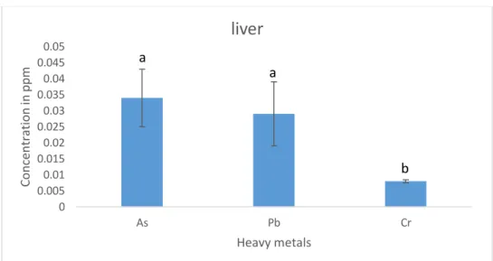
Page | 22 4.6 Different heavy metals concentration in livers of cultured tilapia:
The values of Arsenic were found to be the highest in liver of tilapia (0.034 ppm) followed by lead (0.029ppm) and Chromium (0.008 ppm) respectively. The values of Chromium varies from other two heavy metals.
Figure 6: Different heavy metals concentration in Liver of cultured Tilapia 4.7 Different heavy metals concentration in kidneys of cultured tilapia:
The concentration of lead (0.031 ppm) and chromium (0.007ppm) in kidneys of tilapia fishes were found to be comparatively lower than the values obtained for Arsenic (0.071ppm).The obtained values are significantly varied among each other statistically.
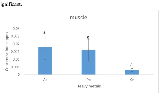
Figure 7: Different heavy metals concentration in kidney of cultured Tilapia 4.8 Different heavy metals concentration in muscles of cultured Tilapia:
0 0.005 0.01 0.015 0.02 0.025 0.03 0.035 0.04 0.045 0.05
As Pb Cr
Concentration in ppm
Heavy metals
liver
a
a
b
0 0.01 0.02 0.03 0.04 0.05 0.06 0.07 0.08 0.09 0.1
As Pb Cr
Concentration in ppm
Heavy metals
kidney
c a
b
Page | 23 Among the investigated heavy metals (As, Pb, Cr), Arsenic concentration (0.018ppm) was the highest in muscles of tilapia followed by the concentration of lead (0.016ppm) and chromium(0.003ppm) though the values are not statistically significant.
Figure 8: Different heavy metals concentration in muscle of cultured Tilapia
4.9 ATPase activity in different investigated organs of cultured pangus:
The graph illustrates the ATPase activities of different examined organs in pangus.
From the obtained values, it is observed that the ATPase enzymatic activities was found to be the highest in kidneys with insignificant difference for the values obtained in livers. The figure also suggests that the lowest ATPase activity was recorded in muscle which was statistically significant varied with the above two organs along with the values obtained for gills.
0 0.005 0.01 0.015 0.02 0.025 0.03
As Pb Cr
Concentration in ppm
Heavy metals
muscle
a a
a
0 0.5 1 1.5 2 2.5 3 3.5 4 4.5
gill liver kidney musscle
ATP ase activity
Organ wise
Pangus
a
b b
c
Page | 24 Figure 9: ATPase activity as µg of phosphorus mg protein-1 (37°C) in different organs of cultured Pangus
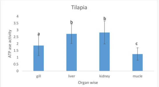
4.10 ATPase activity in different organs of cultured Tilapia:
The recorded ATPase activities in different organs of cultured Tilapia are in a line of match with the results obtained for pangus depicting the highest in kidneys followed by livers, gills and muscles respectively. The values here for kidney and livers are significantly different from those for gills and muscles.
Figure 10: ATPase activity as µg of phosphorus mg protein-1 (37°C) in different organs of cultured Tilapia
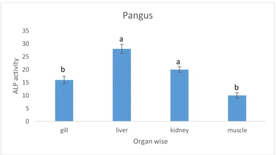
4.11 ALP activity in different organs of cultured pangus:
The concentration of ALP activity was found highest in livers (28ppm) with insignificant statistical difference for that of kidneys (20 ppm) in tilapia. The lowest value observed in muscles (10 ppm) with insignificant difference for the recorded values in gills (16ppm).
0 0.5 1 1.5 2 2.5 3 3.5 4
gill liver kidney mucle
ATP ase activity
Organ wise
Tilapia
a
b
c b
Page | 25 Figure 11: ALP activity as n moles of para-nitrophenol mg protein-1 (37°C) in different organs of cultured Pangus
4.12 ALP activity in different organs of cultured Tilapia:
The concentration of ALP activity was found highest in kidney (26 ppm) with insignificant statistical difference for that of liver (22 ppm) in tilapia. The lowest value was observed in muscles (08 ppm) with significant difference from above mentioned three organs.
Figure 12: ALP activity as n moles of para-nitrophenol mg protein-1 (37°C) in different organs of cultured Tilapia
4.13 Average concentration of different heavy metals in examined organs of cultured Pangus and Tilapia:
0 5 10 15 20 25 30 35
gill liver kidney muscle
ALP activity
Organ wise
Pangus
b
a
a
b
0 5 10 15 20 25 30
gill liver kidney muscle
ALP activity
Organ wise
Tilapia
b a
a
a
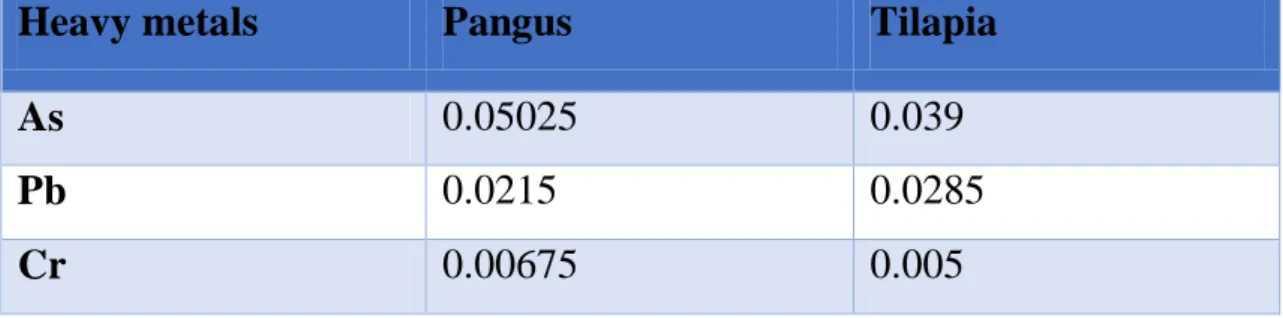
Page | 26 The recorded values in pangus demonstrated that the higher concentration is observed in case of Arsenic (As) with a mean value of 0.05025 ppm which is higher than the recommended value of 0.01 ppm (WHO/FAO, 2005). The mean average value of Chromium was the least among the three investigated heavy metals (0.00675ppm) which is far lower than the standard value (0.1 ppm) of Chromium (WHO/FAO, 2005). The mean value of lead was observed (0.0215 ppm) which is also lower than the standard value of 0.3 ppm by WHO/FAO (2005).
The observed values in Tilapia revealed that the higher concentration is observed in case of Arsenic (As) with a mean value of 0.039 ppm which is higher than the recommended value of 0.01 ppm (WHO/FAO, 2005). The mean average value of Chromium was the least among the three investigated heavy metals (0.005 ppm) which is far lower than the standard value (0.1 ppm) of Chromium (WHO/FAO, 2005). The average value of lead was observed (0.026983ppm) which is also lower than the standard value of 0.3 ppm by WHO/FAO (2005).
Table 1: Average concentration of different heavy metals in examined organs of cultured Pangus and Tilapia
Heavy metals Pangus Tilapia
As 0.05025 0.039
Pb 0.0215 0.0285
Cr 0.00675 0.005
Page | 27 Chapter 5
DISCUSSION
The present study examines the degree of the concentration of heavy metals (As, Pb, and Cr) in different organs of the two selected food fish species. The investigated species Thai pangus and Tilapia are widely consumed fishes especially by poor people. It has been designated that the level of contaminants in fish depends on habitats, feeding habit and metabolism, duration of exposure of fish to contaminants, age, size and length of the fish concentrations of contaminants in water column, water chemistry.
According to Kalay et al. (1999), “Different fish species accumulate metals in their tissue in significantly different values”. This study also revealed that different fish species contained different metal levels in their tissues. Moreover, Canli and Atli (2003), reported that levels of heavy metals in fish vary in various species and different aquatic environments. Heavy metal concentrations varied among different tissues of a species as well as between species. Among different observed values of heavy metals, we found that As concentrations in different tissues of the investigational species were much higher than any other values whereas Cr showed lowest values in almost all experimental tissues. Previous data showed that different fish species contained strikingly different metal levels in their tissues that might be related to the differences in ecological needs, swimming behaviors and the metabolic activities among different fish species (Kalay et al. 1999).
The concentrations of heavy metal in fish also differ with respect to species and different aquatic environments. Additionally, the affinity for metal absorption from contaminated water and food may differ in relation to metabolism and the contamination gradients of water, ecological needs, as well as other factors such as salinity, temperature and interacting agents.
Heavy metals are persistent in surface waters in the form of colloidal, particulate and dissolved forms and rivers are known as the dominant pathway for the transport of metals (Miller et al., 2003). In the body organs of both herbi- and carnivorous fish species, the accumulation of Al, As, Ba, Cr, Ni and Zn were significantly higher in
Page | 28 liver than that of other body organs. However, fish kidney and gills have also been linked with the higher bio-accumulation of metals in the fish body. The mechanism of trace metals bioaccumulation in fish is complex and diversified, varying with their chemistry, mode of action and metal species (Louma, 1983).
Bioaccumulation of metals refers the amount consumed by an organism. Fish gills as a site of metallic ion entry can enhance lesions and ultimately cause damage to the gills (Bols et al., 2001). All the organs of both herbi- and carnivorous fish species presented significantly variable accumulation of metals with the sequence of liver
>kidney > gills > intestine > reproductive organs > scales >fins > bones > muscle >
fats. Bioaccumulations of the HMs is a result of the fact that different metals tend to accumulate differently in the tissues of different species of fish. Besides, different concentration of HMs in different fish species might be a result of different ecological needs, metabolism and feeding patterns (Allen-Gil SM, Martynov VG 1995). As fish lives primarily in water they concentrate a large volume of HMs in its body by bioaccumulation and bio-magnification (Velez D, Montoro R 1998). Gonads, liver, kidney and gills are target organs for metals accumulation since they are metabolically actives accumulating metals sometimes in high levels (Yilmaz AB 2003).
In Thai pangus, the maximum concentration of Arsenic (0.062ppm) was observed in kidney and minimum concentration (0.036ppm) was found in the muscle. The metal bioaccumulation of Arsenic in the Thai pangus has in the decreasing order of kidney>liver>gill>muscle. Likely, in Tilapia the maximum concentration of Arsenic (0.071ppm) was observed in kidney and minimum concentration (0.018ppm) was found in the muscle. The bio-accumulation of Arsenic in the Tilapia fish has in the decreasing order of Kidney>liver>gill>muscle.
In Thai pangus, maximum concentration of Pb (0.032ppm) was observed in gill and lowest concentration (0.012ppm) was in muscle. The other view, Pb in the Thai pangus fish has in the decreasing order of liver>kidney>gill>muscle. In Tilapia, maximum concentration of Pb (0.038ppm) was observed in gill and lowest
Page | 29 concentration (0.016ppm) was in muscle. For the accumulation of Pb in the Tilapia fish has the decreasing order of gill>kidney>liver>muscle.
In Thai pangus maximum concentration of Cr (0.018ppm) was observed in gill and lowest concentration (0.001ppm) was in muscle. In contrast, Cr in the Thai pangus fish has in the decreasing order of gill>liver>kidney>muscle. In Tilapia, highest concentration of Cr (0.008ppm) was observed in liver and lowest concentration (0.002ppm) was in muscle. Furthermore, bioaccumulation of Cr in the Tilapia fish has the decreasing order of liver > kidney>muscle>gill.
It is well known that it’s very difficult to compare the concentrations of metals even between the same tissue in different species due to the difference in many factors as, the aquatic environments, concerning the type and the level of water pollution, feeding habits whether omnivorous or carnivorous, and level of fish presence in water, whether pelagic or benthic fish etc.
For both fishes, As exceeded the recommended value provided by FAO/WHO whereas Pb and Cr are within the guidelines. Ahmed et al. (2014) also found higher Pb and Cd values that exceeded the acceptable limit, which provided by Food and Agriculture Organization/World Health Organization.
The metal concentrations in the gill of Pangus and Tilapia fish has in the decreasing order of As>Pb>Cr and Pb>As>Cr respectively. The gill of Pangus accumulates significantly higher levels of As than tilapia whereas tilapia accumulates higher Pb concentration than pangus. Gill surfaces are the first target organ of water borne metals. According to Reid and Mcdonald (1991), the gill surface is negatively charged and thus provides a potential site for gill metal interaction for positively charged metal. On the other hand, fish accumulating HMs from food show elevated metal levels in the digestive tract as compared to the gills (Ney JJ, Van-Hassel JH 1983;
Heath AG 1990).The gills of all the fish tend to accumulate significant high levels of heavy metal comparatively than other tissues. On an average the gill tissue came first in terms of metals accumulation.
Page | 30 Fish liver and kidney accumulated substantial quantities of all metals while these accumulations were significantly lowest in muscle and fats. Fish kidney as its role to detoxify metals has also accumulated significant amounts of heavy metals (Vinodhini and Narayanan, 2008). Jent et al. (1998) found that liver accumulate and concentrate highest concentrations of Cu and Cd. According to Lentini et al (2017), “kidney is a target organ in heavy metal toxicity for its capacity to filter, reabsorb and concentrate divalent ions. The extent and the expression of renal damage depends on the species of metals, the dose, and the time of exposure”.
The metal concentrations in the liver of Pangus and Tilapia fish has in the decreasing order of As>Pb>Cr and As>Pb>Cr respectively. The liver of Pangus accumulates significantly higher levels of As and Pb than tilapia. The liver plays an important role in accumulation and detoxification of HMs (Yousafzai AM 2004). Exposure of fish to elevated levels of HMs induces the synthesis of metallothioneine proteins (MT), which are metal binding proteins (Noel-Lambot GC, Disteche A 1978; Phillips DJH, Rainbow PS,1989).Fishes are known to possess the metallothionein protein (Akan JC, Mohmoud S, Yikala BS, Ogugbuaja VO,2012). Metallothioneine proteins have more affinities for HMs and so, concentrate and regulate these metals in the liver.
Metallothioneine proteins detoxify and bind the metal ion. On an average the liver tissue came second in terms of metals concentration after gills.
The comparison in the concentrations of HMs in the kidney samples among Pangus and Tilapia fish has in the decreasing order of As>Pb>Cr and As>Pb>Cr respectively.
Kidney, together with the gills and intestine, are responsible for excretion and the maintenance of the homeostasis of the body fluids (Hinton et al.1992; Evans ME 1993) and besides producing urine, act as an excretory route for the metabolites of a variety of xenobiotic to which the fish may be exposed (Oliveira et al. 1996). The effects of pollutants on fish kidneys have been studied in some species and the severity of damage seen depends on the sensitivity of the species to the substances released into the environment (Schwaiger J 2001; Pacheco M, Santos MA 2002)
Among the two species of fish, the variation of HMs concentration in the muscle of Pangus and Tilapia fish has in the decreasing order of As>Pb>Cr, and As>Pb>Cr respectively. On average, HMs concentrations have found to be lower in fish muscle
Page | 31 than other organs and this could be due to low levels of metal binding proteins in the muscle. Muscle is the most commonly consumed portion of fish and contributes most to the mass of fish (Palaniappan and Vijayasundaram 2009). The low concentrations of metals in the muscle of fish species may reflect the low levels of binding proteins in the muscle (Allen-Gil SM, Martynov VG 1995).
In the work of Fathi Al hashmi Bashir and Esmail Mohamed Alhemmali (2015), muscle contained the lowest concentrations of trace metals among all the tissues which supports our findings. Muscle does not come into direct contact with the metals as it is completely covered externally by the skin which helps the fish to avoid the penetration of the trace metals and similarly it is not an active site for detoxification.
In muscles metabolic activity is relatively lower and so usually there is no risk regarding for human consumption of flesh (Fathi Al hashmi Bashir and Esmail Mohamed Alhemmali, 2015).Consumption of these metal concentrated fish could result in severe health hazards including anaemia, kidney malfunctions, lung cancer, nausea, vomiting, and cardiovascular diseases as well as mental disorder.
In terms of organs, the heavy metals were mostly accumulated in the kidney tissues but the concentration in muscles was found to be the lowest which is a good finding as we eat the muscles widely although the concentration of Arsenic in muscles was reported above the safety values. As concentration in the intestine and liver are higher in most of the cases than As concentration in muscle tissues, these results are consistent with other study (Suhendrayatna, Ohki, Nakajima, & Maeda, 2001). So we can conclude that our findings are agreed upon previous studies.
Among different observed values of heavy metals, we found that Arsenic concentrations in various organs of the experimental fishes were much higher than any other values whereas Cr showed lowest values in nearly all experimental tissues.
Additionally, As was highly concentrated among all the measured heavy metals in different organs of fishes.
Fish are usually considered as an organism of choice for assessing the effects of environmental pollution on aquatic ecosystems (Gernhofer et al.2001). They are
Page | 32 continuously exposed to it through their gills, skin, and by the intake of As- contaminated food. Fish have long been used as sentinels for bio monitoring of aquatic environmental pollutants and are good indicators of As toxicity (Tisler and Zagorc- Koncan 2002). In the present study, the presence of As is worrisome considering the toxic effect of As to fish and man. Anthropogenic activities are greatly responsible for As pollution in the aquatic environment. Mining activities have led to soil and water pollution. However, other anthropogenic activities using As, such as agriculture, forestry, and industry, have also contaminated soil and water at a localized scale (Smith et al. 2003).
The high level of As concentration, i.e., more than permissible limit, in an aquatic ecosystem affects various physiological systems such as growth, reproduction, ion regulation, gene expression, immune function, enzyme activities, and histopathology of fish (Pedlar RM, Klaverkamp JF 2002;Pedlar et al. 2002a; Datta et al. 2009). As- contaminated fish consumption could result in As exposure to humans and lead to adverse health effects (Kar et al. 2011). As compounds show toxicity in many organs of body such as skin, kidney, liver, lung, muscles, and gastrointestinal tract (Pedlar RM, Klaverkamp JF 2002;Pedlar et al. 2002a;Roy and Bhattacharya 2006).Among these, liver and kidneys are vital organs in vertebrates which perform protein synthesis, detoxification mechanism, and excretion of nitrogenous waste and homeostatic functions.
However, all these concentrated metals in different parts of fish body could be accumulated into human body, if they are consumed. In Bangladesh, water sources are becoming more contaminated day by day and consequently these HMs from polluted water bodies are getting more concentrated in those fish living in that areas.
There is another way of concentrating HMs in fish body through the feed they are reared with and for farmed fish; it is our lacking that we don’t care about those HMs from artificial feed getting introduced into fish body parts. If all these bad practices live long and if they do so there will be a great threat for human health. The results obtained in the present study are just an alarming for us and also for our future generation and it is of high time to undertake necessary steps regarding safety and environmental friendly arsenic discharge. The values obtained for lead and chromium
Page | 33 were within the safety levels and considered safe for human consumption, but to maintain this level we need continuous monitoring.
The activities of the enzymes alkaline phosphatase (ALP) and Adenosine triphosphatases (ATPase) in vital organs (liver, kidney, gills and muscle) were evaluated in the present study. By assessing the enzyme activities in an organism, disruptions in its metabolism can easily be identified. ATPase’s are a group of enzymes that play an important role in intracellular functions and are considered to be a sensitive indicator of toxicity (Yadwad et al. 1990). Adenosine triphosphate (ATP) hydrolyzed into adenosine diphosphate (ADP) and inorganic phosphate (Pi). In this process, the energy released for cation transport becomes available. ATPase in various ion dependent forms is a membrane-bound enzyme and responsible for the transport of ions through the membrane regulating cellular volume, osmotic pressure and membrane permeability among others.
In case of Pangus, ATPase activity in different organs are in the order:
kidney>liver>gill>muscle. Similarly, the activity of ATPase in different organs of Tilapia fish are in the order: kidney>liver>Gill>Muscle. On both cases we found that kidney has highest ATPase activity where muscle has lowest ATPase activity. Kidney mitochondria has a greater capacity for ATP hydrolysis (Weiner mw 1975).All zones of mammalian kidney, except the papilla, are abundant sources of Na2+ K+ ATPase (Jorgensen, P.L 2008).Na2+ K+ ATPase, the enzymatic equivalent of the sodium:
potassium pump, is found in large amounts in the kidney, and this organ has figured prominently both as a source for the purification of the enzyme(Katz AI 1982). This result is in agreement with previous studies reporting.
ALP a ubiquitous plasma membrane-bound enzyme, is often employed to assess the integrity of the plasma membrane .ALP is employed to assess the integrity of plasma membrane and endoplasmic reticulum (Wright PJ, Plummer DT 1974). ALP is consist of several isoenzymes found in virtually all tissues of the body, especially in cell membranes. The sources of alkaline phosphatase are liver and biliary tracts.
The activities of ALP enzyme in different organs of Pangus are in the decreasing order of: liver>kidney>gill>muscle. The liver tissue produces more ALP than other
Page | 34 organs of fish, but the production of ALP is inhibited in stress condition (Rao 2006).In case of Tilapia fish, the activities of ALP enzyme in different organs are in the decreasing order of: kidney>liver>gill>muscle. Kidney was found to be a organ with the highest ALP activity (Cvancara et al. 1978).So our findings are consistent with previous studies.
M K Ahmmed et al. (2017), conduct a study on Biochemical impacts of salinity on the catfish, Heteropneustes fossilis (Bloch, 1794), and possibility of their farming at low saline water. ALP and ATPase activity was analyzed in their study and the level was found to be reduced in liver tissue at 9 ppt compared with control. In their study the control value of liver and muscle in ALP was the highest in liver whereas lowest in muscle. Our finding also reclaimed that ALP activity is the highest in liver and lowest in muscle in case of Pangus except Tilapia. ATPase activity was also examined in gill liver, kidney and muscle tissues of sacrificed fishes. On both cases (Pangus and Tilapia), kidney has highest ATPase activity where muscle has lowest ATPase activity. In control fish liver ATPase activity was highest while muscle activity was lowest (M K Ahmmed et al. 2017). This findings are very similar to the results of this study with some variance that may be due to species difference, location difference, feeding regime etc.
Enzymes are biochemical macromolecules that control metabolic processes of the organisms, thus a slight variation in enzyme activities would affect the organism (Roy, 2002). This enzyme has important functions such as ion transport, maintenance of the electrochemical gradient and regulation of cell volume. So we should closely monitor the activities of this enzyme and their levels in different organs of commercially important fishes.
Page | 35 Chapter 6
CONCLUSION
The concentration of heavy metal above threshold level could be dangerous for both aquatic life and human health. Ecosystem pollution from heavy metals may harm organisms at the cellular level and also may affect the ecological balance. The aquatic organism accumulates heavy metals through three ways: the body surface, gills and food. As is a widespread environmental contaminant, which enters the aquatic systems from both natural and anthropogenic sources. Its effects on fish health include various mechanisms of acute and chronic toxicity, including genetic, enzymatic, and immune system failure. Many studies have shown that high concentrations of As was found in liver and kidney relative to other organs which disrupts the normal metabolism. The results of this study revealed that consuming fish from the cultured pond may not be injurious to consumer’s health because observed values of heavy metals (except arsenic) were below the permissible limits issued by FAO/WHO for human consumption. Arsenic concentration was the highest followed by lead, whereas chromium was the lowest. Likewise, kidney accumulates the highest amount of metal while muscles contain the lowest metal concentration. However, Arsenic is higher than approved level so, it is a matter of concern in fish accumulation. In order to avoid toxicological effects caused by heavy metals, periodic monitoring of aforementioned metals and other heavy metals in fish species of the pond is recommended to ensure continual safety of people in the local people and consumers. Enzymatic activities (ALP and ATPase) of kidney, liver, gill and muscle in Pangus and Tilapia fish was also observed in our examined study. Because ATPase are closely involved in osmoregulation, acid–base regulation and respiration of fish where Alkaline phosphatase (ALP) is an important regulatory enzyme in bio-metabolic processes and plays a vital function in digestion, absorption, and transition of nutrients .As the stability of enzyme activity can affect the body’s biological metabolism and adaptive capacity so it is important to quantify its activity on different fish. The results obtained in this study revealed that kidney has highest ATPase activity where muscle has lowest ATPase activity (on both fishes).The result also indicated that ALP activity was highest in liver of Pangus except Tilapia (in kidney) and lowest in muscle on both cases. Nonetheless, further studies are still
Page | 36 needed to provide more evidences to better understand the mechanisms of ALP and ATPase activities in various organs of commercial cultured fish.
Page | 37
Chapter-7
RECOMMANDATION AND FUTURE PERPECTIVES
Among different observed values of heavy metals, we found that As concentrations in different tissues of the experimental species were much higher than any other values whereas Cr showed lowest values in almost all experimental tissues. The results obtained in the present study are just a warning for us and our future generation. The contaminated aquatic resources (Thai Pangus and Tilapia fish) contributes to livelihood opportunities, food security and nutritional benefits through the availability of fish in markets. Continuous consumption of these contaminated aquatic resources could pose health risks to consumers. So we should take immediate steps for solving such burning issues.
It is important to observe the levels of heavy metals in fishes to get some idea about the safety of fish protein supplied from that river and to understand its harmful effects among individuals, population or ecosystem.
Continuous monitoring exercise should be put in place to guard against excessive bioaccumulation of these metals and safeguard the safety, protection and well-being of consumers.
However, fish growth and its relationship with metal concentration in the aquatic environment should be monitored occasionally in the field to better understand the effects of metals on fish development and the current situation of population dynamics.
Control measures recommended include public enlightenment on the need to desist from anthropogenic activities that could lead to water pollution.
Enactment of laws by the government for industries and factories to treat their waste waters and sewage properly before disposing them into water bodies
Proper waste management should be developed through measures that encourage minimization, recycling and reuse of processed waste by both individuals and industries.
The findings have important implications for the development of effective closed watershed management strategies for the control of point and diffuse- source pollution.